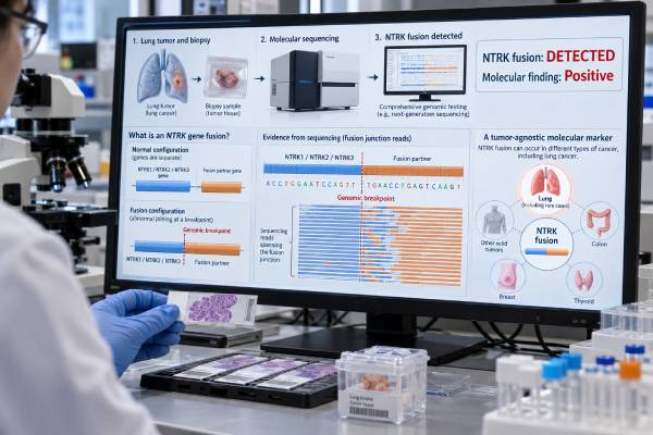
An NTRK fusion test looks for an abnormal joining of one of three genes—NTRK1, NTRK2, or NTRK3—to another gene. When the rearrangement creates an active TRK fusion protein, it can become the main growth driver of a cancer and can make the tumor eligible for TRK-targeted therapy. NTRK fusions are rare in non-small cell lung cancer, generally well below 1%, but they matter because treatment can be selected based on the fusion regardless of the organ where the cancer began. This is why NTRK is often described as a tumor-agnostic biomarker. RNA-based next-generation sequencing is especially effective for detecting these fusions and identifying their partners. DNA sequencing, pan-TRK immunohistochemistry, FISH, and RT-PCR can also be useful in selected settings. A positive result must represent a true, functional NTRK fusion; an NTRK point mutation, amplification, or isolated protein staining is not automatically equivalent.
- What a positive result means: A confirmed NTRK1, NTRK2, or NTRK3 fusion identifies a potentially actionable TRK-driven tumor.
- How common it is in lung cancer: NTRK fusions are uncommon in NSCLC, usually around 0.1%–0.3% in unselected cases and generally under 1%.
- Why it is tumor-agnostic: TRK inhibitors can be used based on the presence of an NTRK fusion across multiple solid tumor types, not only lung cancer.
- Preferred testing method: RNA-based NGS is particularly strong for fusion detection because it can show the expressed transcript and identify the fusion partner.
- Important limitation: A negative result is only as reliable as the specimen and assay; poor RNA quality, low tumor content, or incomplete fusion coverage can produce false-negative results.
Table of Contents
- What an NTRK Fusion Is
- Why NTRK Is a Tumor-Agnostic Marker
- Who Should Have NTRK Testing
- How NTRK Fusions Are Tested
- Understanding a Positive NTRK Fusion Result
- Negative, Equivocal, and Discordant Results
- Practical Next Steps After NTRK Testing
What an NTRK Fusion Is
The NTRK gene family contains NTRK1, NTRK2, and NTRK3, which encode the TRKA, TRKB, and TRKC receptor tyrosine kinases. These proteins normally participate in nervous-system development and signaling. In cancer, a chromosomal rearrangement can join the kinase-containing portion of an NTRK gene to a different partner gene.
The resulting fusion can remove normal regulatory controls and keep the TRK kinase continuously active. That persistent signal can drive pathways involved in cell growth, survival, and proliferation. When this happens, the fusion is considered an oncogenic driver rather than a passive genetic change.
Many partner genes have been reported. In lung cancer, examples include TPM3–NTRK1, SQSTM1–NTRK1, MPRIP–NTRK1, CD74–NTRK1, and less commonly fusions involving NTRK2 or NTRK3. The exact partner and breakpoint matter to the laboratory because a true driver fusion should retain the functional TRK kinase domain and usually be expressed as RNA.
NTRK fusions are rare in common adult cancers. In unselected NSCLC, estimates are usually around 0.1%–0.3%, although rates differ among cohorts and testing methods. Because the alteration is so uncommon, testing one gene at a time is inefficient. Broad molecular panels are a practical way to search for NTRK while also evaluating more frequent lung cancer drivers.
The rarity also affects how laboratory results should be judged. In a low-prevalence setting, even a test with good analytical performance can generate occasional false-positive screening results, which is why confirmatory molecular testing matters when pan-TRK IHC is used first. Conversely, a method with incomplete coverage can miss the very small number of true-positive cases. Efficient testing therefore depends on both sensitivity and specificity, not simply on whether a platform can technically report NTRK. Laboratories also need validation materials that represent different NTRK genes and fusion partners, because performance with one common fusion does not guarantee equal performance across all possible rearrangements.
A fusion is not the same as every other NTRK alteration. Molecular reports can also list NTRK point mutations, copy-number gains, or variants of uncertain significance. Those findings generally do not carry the same established treatment meaning as a confirmed activating NTRK gene fusion.
Why NTRK Is a Tumor-Agnostic Marker
NTRK fusions are called tumor-agnostic biomarkers because the molecular alteration can predict benefit from TRK inhibition across different solid tumors. In other words, the fusion itself can be therapeutically important whether it is found in lung cancer, thyroid cancer, sarcoma, salivary cancer, or another eligible malignancy.
This concept differs from traditional oncology, where treatment was selected mainly by the organ where the tumor began. Tumor-agnostic therapy focuses on a shared molecular driver that occurs across several tumor types.
The important qualifier is that the biomarker is a gene fusion, not simply the presence of the NTRK gene. Every person has normal NTRK genes. What matters is the abnormal fusion that creates a constitutively active TRK protein.
TRK inhibitors such as larotrectinib and entrectinib established the tumor-agnostic treatment model for NTRK fusion-positive solid tumors, and additional agents have expanded the treatment landscape. Treatment eligibility depends on the exact regulatory indication, age, disease setting, prior therapy, and current clinical guidance.
The tumor-agnostic label also does not erase the importance of the original cancer type. A patient with NTRK fusion-positive lung cancer is still staged and clinically managed as a lung cancer patient. Imaging, pathology, brain assessment, local treatment options, and other lung cancer biomarkers remain relevant. The fusion adds a powerful therapeutic target; it does not replace the rest of the diagnosis.
An integrated lung cancer biomarker panel is therefore useful because it identifies both common lung-specific drivers and rarer tumor-agnostic alterations within the same diagnostic process.
Who Should Have NTRK Testing
NTRK fusion testing is most efficiently performed as part of broad genomic profiling for patients with advanced or metastatic NSCLC when molecular testing is indicated. Because the fusion is rare, clinical features alone are not a reliable screening strategy.
NTRK-positive lung cancers are often adenocarcinomas and may occur in people with little or no smoking history, but those tendencies are not strict enough to select who gets tested. A patient who does not fit the “typical” profile can still harbor an NTRK fusion.
A lung cancer NGS test can evaluate NTRK1/2/3 together with EGFR, ALK, ROS1, RET, BRAF, KRAS, MET, HER2 and other genes. This reduces tissue use and avoids the delay of sequential single-gene testing.
Testing may also be important when prior molecular work was incomplete. For example, an older panel may have tested only EGFR, ALK, and ROS1 and omitted NTRK. Re-reviewing the original assay’s gene list and fusion design can show whether a newer broad panel would add useful information.
At progression, repeat molecular testing can sometimes identify a resistance mechanism. Tumors treated with first-generation TRK inhibitors may acquire mutations in the TRK kinase domain that interfere with drug binding. Tissue or plasma testing can help characterize resistance when the result could influence later therapy.
The key point is not to wait for a particular age, sex, smoking history, or metastatic pattern. NTRK testing is most reliable when incorporated into a systematic molecular workflow for eligible NSCLC rather than reserved for patients who “look” like they might have a rare fusion.
How NTRK Fusions Are Tested
NTRK fusions can be detected by several laboratory methods. RNA-based NGS is often favored because it can directly identify expressed fusion transcripts, but the best method depends on sample quality, available technology, and the tumor type.
RNA-based NGS
RNA sequencing is particularly effective for fusion detection because introns have already been removed from mature RNA. The assay can identify the exact 5′ partner and the NTRK kinase-containing 3′ portion of the transcript. Consensus recommendations emphasize careful RNA quality control, adequate tumor content, validated bioinformatics, and clear reporting of in-frame versus uncertain fusions.
The main weakness is specimen quality. Formalin fixation can degrade RNA, and a failed RNA assay should be reported as technically unsuccessful rather than simply negative.
DNA-based NGS
DNA panels are convenient because they can detect mutations, copy-number changes, and some fusions in one test. However, NTRK2 and NTRK3 in particular contain large intronic regions that can be difficult to cover comprehensively. A DNA panel may therefore miss a fusion that RNA testing could detect.
When DNA sequencing identifies an unusual structural rearrangement, RNA confirmation can show whether an expressed functional transcript is actually produced.
Pan-TRK immunohistochemistry
Pan-TRK IHC detects TRK protein expression and can serve as a screening tool. It is relatively fast and inexpensive, but it is not perfectly sensitive or specific. A 2022 meta-analysis found that pan-TRK IHC missed a meaningful fraction of molecularly confirmed fusions, with more false negatives among NTRK3 fusions.
For this reason, negative IHC cannot always exclude an NTRK fusion, and positive IHC generally requires molecular confirmation in low-prevalence tumors such as lung cancer.
FISH and RT-PCR
FISH can detect rearrangements involving NTRK loci, but separate assays may be needed for NTRK1, NTRK2, and NTRK3. It also does not necessarily show whether a rearrangement creates a functional transcript. RT-PCR can be highly sensitive for known fusion junctions but may miss novel partners or breakpoints that were not built into the assay.
| Method | Main strength | Main limitation |
|---|---|---|
| RNA NGS | Detects expressed fusions and identifies partners | Requires adequate-quality RNA |
| DNA NGS | Profiles many alteration types together | May miss fusions in difficult intronic regions |
| Pan-TRK IHC | Fast screening method | False positives and false negatives occur |
| FISH | Can show gene rearrangement without knowing the partner | May require separate gene assays and does not define the transcript |
| RT-PCR | Highly sensitive for targeted known fusions | Can miss unexpected partners or breakpoints |
Understanding a Positive NTRK Fusion Result
A strong positive result identifies a specific NTRK fusion that is predicted or known to create an active TRK kinase. A report may name the two genes, identify exons or breakpoints, state whether the fusion is in frame, and classify the finding as pathogenic, oncogenic, or therapeutically relevant.
A result such as TPM3::NTRK1 fusion detected is more informative than “NTRK altered.” The first describes a defined fusion. The second could refer to a mutation, copy-number change, or another event with different clinical meaning.
A confirmed fusion usually has three important implications:
- It defines a molecular driver. The tumor may depend heavily on abnormal TRK signaling.
- It can be therapeutically actionable. TRK inhibitors have demonstrated activity across NTRK fusion-positive cancers, including lung cancers.
- It can shape future resistance testing. If progression develops after TRK inhibition, molecular reassessment may identify kinase-domain resistance mutations or alternative signaling changes.
The fusion does not tell the cancer stage. A stage I lung cancer and a stage IV lung cancer could theoretically harbor the same molecular driver. Stage remains an anatomic assessment based on tumor size and invasion, lymph nodes, and metastatic spread.
The result is also usually somatic rather than inherited. Most NTRK fusions detected in lung tumors arose in the cancer cells. A routine tumor NTRK fusion does not by itself imply that family members need genetic testing.
Negative, Equivocal, and Discordant Results
A negative NTRK result means no reportable fusion was found by the assay. The strength of that conclusion depends on assay coverage and sample quality.
A high-quality RNA panel with adequate tumor content provides a strong negative result. A DNA-only panel with limited intronic coverage, an RNA test that barely passed quality metrics, or a plasma assay with very low tumor DNA provides less certainty.
A technically unsuccessful result should not be interpreted as negative. Common problems include insufficient tissue, too few tumor cells, severe RNA degradation, failed library preparation, and low sequencing depth. Another block, a new biopsy, or an alternative method may be appropriate if the result could change treatment.
Discordant results require method-specific reasoning. Pan-TRK IHC can be positive without a true fusion, while some real fusions can be IHC negative. FISH can identify a structural rearrangement that does not produce a functional transcript. DNA NGS can suggest a rearrangement that RNA sequencing either confirms or resolves as nonfunctional.
When a fusion result is labeled a variant of uncertain significance, clinicians should avoid assuming that it predicts TRK inhibitor benefit. The report should be reviewed for the genes involved, whether the TRK kinase domain is retained, whether the transcript is in frame, and whether clinical databases or published evidence support oncogenic function.
If NTRK is truly negative, the molecular workup should continue or be reviewed for other drivers. A ROS1 fusion, RET fusion, ALK fusion, EGFR mutation, MET exon 14 skipping alteration, KRAS G12C mutation, BRAF V600E, or another actionable event may provide the relevant target instead.
Practical Next Steps After NTRK Testing
NTRK testing itself does not require fasting or special preparation. The important practical step happens before the assay: obtaining and preserving enough tumor material for histology, fusion testing, other molecular biomarkers, and PD-L1 when needed.
After receiving the report, ask these questions:
- Which NTRK gene is involved—NTRK1, NTRK2, or NTRK3?
- Is this a confirmed gene fusion or another kind of NTRK alteration?
- Was the fusion detected at the RNA level, DNA level, by FISH, or only by IHC?
- Was the sample technically adequate and did quality controls pass?
- Is the fusion considered in frame and oncogenic?
- Has the full NSCLC biomarker profile been completed?
- Does the result make the patient eligible for an approved targeted therapy or a clinical trial in the current treatment setting?
If the result is positive, the oncology team reviews the full disease context rather than treating the fusion as an isolated laboratory fact. Stage, brain metastases, symptoms, prior therapies, organ function, drug interactions, and treatment access all influence the plan.
If the result is negative but the test was incomplete or technically weak, it can be worth repeating with RNA-based NGS. Because NTRK fusions are rare, missing even one due to assay design can deny a patient access to a highly relevant targeted treatment.
The molecular meaning of NTRK is unusually clear when the right alteration is present: a functional fusion links tumor growth to TRK signaling and creates a target that can be treated across cancer types. The challenge is not understanding why the fusion matters; it is detecting a rare event accurately and distinguishing it from NTRK findings that do not carry the same clinical significance.
References
- NTRK gene fusion testing and management in lung cancer 2024 (Review)
- Consensus Recommendations to Optimize the Detection and Reporting of NTRK Gene Fusions by RNA-Based Next-Generation Sequencing 2023 (Consensus)
- Japanese Society of Medical Oncology/Japan Society of Clinical Oncology/Japanese Society of Pediatric Hematology/Oncology-led clinical recommendations on the diagnosis and use of tropomyosin receptor kinase inhibitors in adult and pediatric patients with neurotrophic receptor tyrosine kinase fusion-positive advanced solid tumors 2023 (Guideline)
- Evaluating Targeted Next-Generation Sequencing Assays and Reference Materials for NTRK Fusion Detection 2022
- The sensitivity of pan-TRK immunohistochemistry in solid tumours: A meta-analysis 2022 (Meta-analysis)
- NTRK Fusion in Non-Small Cell Lung Cancer: Diagnosis, Therapy, and TRK Inhibitor Resistance 2022 (Review)
Disclaimer
NTRK results should be interpreted by qualified molecular pathology and oncology professionals using the complete tumor diagnosis and assay-quality information. A confirmed fusion can affect treatment, while a mutation, amplification, uncertain rearrangement, or technically limited negative result may have very different meaning. Do not change cancer treatment based on an NTRK report without specialist review.