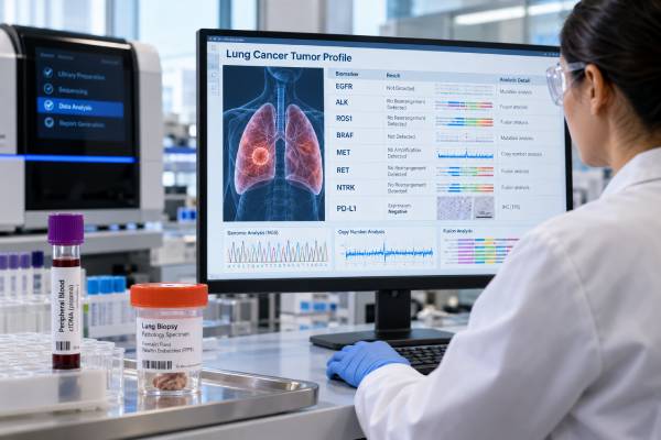
A lung cancer biomarker panel looks for molecular and protein features that can define the tumor more precisely than histology alone. In non-small cell lung cancer (NSCLC), especially adenocarcinoma, the panel may identify driver mutations, gene fusions, copy-number changes, and PD-L1 expression that influence treatment selection. The exact test menu varies by laboratory, so “biomarker panel” is not one universal assay. A high-quality workup usually combines broad genomic testing with methods that reliably detect fusions and with PD-L1 immunohistochemistry. Tissue remains the standard specimen when available, while plasma circulating tumor DNA (ctDNA) can add rapid, minimally invasive genomic information. A negative panel does not always mean the cancer has no targetable biology; specimen quality, assay coverage, tumor shedding, and emerging biomarkers matter. The final report should be interpreted with pathology, stage, prior therapy, and current guidelines before treatment decisions are made.
- Core purpose: identify actionable tumor drivers and PD-L1 expression before systemic treatment is selected.
- Common biomarkers: EGFR, ALK, ROS1, BRAF V600E, MET exon 14 skipping/amplification, RET, NTRK, KRAS G12C, HER2/ERBB2, and PD-L1 are commonly considered in modern NSCLC profiling.
- Best genomic approach: broad NGS, often with RNA fusion testing, reduces tissue use and can detect several alteration classes in one workflow.
- Blood can help: plasma ctDNA is useful when tissue is limited or rapid genotyping is needed, but a negative plasma result may require tissue testing.
- PD-L1 is different: PD-L1 is usually measured by immunohistochemistry and reported as a tumor proportion score rather than as a DNA mutation.
Table of Contents
- What a lung cancer biomarker panel is
- What EGFR, ALK, ROS1, BRAF, MET, RET, NTRK, and PD-L1 mean
- How the panel is performed
- How to interpret a biomarker report
- Tissue testing versus liquid biopsy
- How biomarker results shape treatment planning
- Limitations, missing biomarkers, and false negatives
- Practical next steps and questions to ask
What a lung cancer biomarker panel is
A biomarker panel is a coordinated set of tests that asks whether a lung tumor has molecular features linked to targeted therapy, immunotherapy, prognosis, resistance, or clinical-trial eligibility. It is most important in NSCLC because the number of clinically relevant drivers has expanded rapidly. Rather than testing EGFR first, then ALK, then ROS1, then another gene, many laboratories now use broad next-generation sequencing (NGS) so one specimen can answer several questions at once.
The word panel can be misleading because laboratories do not all test the same genes or alteration types. One panel may sequence hundreds of DNA genes but have weak fusion detection. Another may include both DNA and RNA, giving better coverage for ALK, ROS1, RET, and NTRK fusions. A third may be a small hotspot panel that covers only selected variants. Always review the assay menu, not just the marketing name.
A complete tumor profile also extends beyond DNA. PD-L1 is a protein-expression biomarker measured with immunohistochemistry (IHC), usually on tumor tissue. Histologic markers such as TTF-1, p40, and Napsin A help classify the tumor. Genomic and protein results are then integrated with stage and clinical features.
For advanced non-squamous NSCLC, broad molecular testing is generally favored because actionable alterations can be found even when the patient does not fit a classic demographic profile. Testing based only on smoking status, age, sex, or ancestry risks missing people who could benefit from targeted treatment.
What EGFR, ALK, ROS1, BRAF, MET, RET, NTRK, and PD-L1 mean
The most useful way to read a panel is by alteration class and clinical meaning.
| Biomarker | Typical alteration | What a positive result usually means |
|---|---|---|
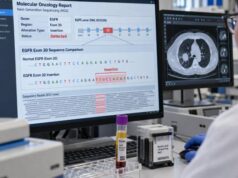
| EGFR | Exon 19 deletion, L858R, exon 20 insertion, uncommon kinase-domain mutations | Defines several distinct EGFR-driven subtypes; the exact variant determines drug sensitivity. |
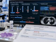
| ALK | Gene fusion, most often EML4-ALK | Identifies ALK-driven NSCLC and supports ALK-directed therapy. |
| ROS1 | Gene fusion | Identifies a ROS1-driven subtype with approved targeted options. |
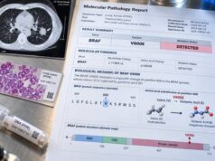
| BRAF | Most importantly V600E | V600E is an actionable MAPK-pathway driver; non-V600 variants require separate interpretation. |
| MET | Exon 14 skipping or amplification | Exon 14 skipping is a primary driver; high-level amplification can be a driver or a resistance mechanism depending on context. |
| RET | Gene fusion | Identifies RET fusion-positive NSCLC and supports RET-directed therapy. |
| NTRK1/2/3 | Gene fusion | Rare in lung cancer but actionable with tumor-agnostic targeted therapy. |
| PD-L1 | Protein expression by IHC | Helps estimate likelihood of benefit from immune-checkpoint therapy; it is not a gene mutation. |
Modern profiles also commonly include KRAS G12C and HER2/ERBB2 mutations. A broader NSCLC biomarker profile may include additional genes and emerging resistance markers.
The exact alteration matters more than the gene name alone. “MET positive,” for example, is incomplete unless the report says whether the finding is exon 14 skipping, amplification, overexpression, or something else. The same principle applies to HER2 mutation versus amplification versus protein overexpression, and to BRAF V600E versus non-V600 BRAF variants.
How the panel is performed
Most comprehensive workflows begin with a tissue biopsy or cytology specimen. A pathologist confirms the diagnosis, estimates tumor cellularity, and selects material for molecular testing. Tissue stewardship matters because a small biopsy may need to support morphology, IHC, PD-L1, DNA sequencing, and RNA analysis.
DNA NGS is strong for point mutations, small insertions/deletions, and many copy-number changes. It can detect EGFR, BRAF, KRAS, HER2, and MET exon 14–related variants, among many others. Some DNA panels also detect fusions, but complex intronic breakpoints can be difficult.
RNA NGS directly evaluates expressed fusion transcripts and is often especially valuable for ALK, ROS1, RET, and NTRK. A combined DNA/RNA approach therefore offers broad coverage across alteration classes. Targeted PCR, digital PCR, FISH, or IHC may be used when rapid single-marker testing is needed, when a comprehensive assay is unavailable, or when a result needs confirmation.
PD-L1 IHC is performed separately on tissue with a validated antibody assay. Results are commonly reported as tumor proportion score (TPS), the percentage of viable tumor cells showing membranous staining. PD-L1 does not belong in the DNA mutation table because it measures protein expression.
Turnaround time varies from a few days for targeted IHC or PCR to roughly one to several weeks for comprehensive testing depending on specimen transport, laboratory workflow, and whether tissue needs additional processing. The goal is not merely speed; it is getting a reliable complete profile early enough to guide the first major treatment decision.
How to interpret a biomarker report
Start by identifying findings labeled pathogenic, likely pathogenic, actionable, or an equivalent high-evidence category. Then read the exact variant, fusion partner, copy-number result, or protein score. A variant of uncertain significance should not be treated as if it were a validated driver.
For a mutation, the report may list variant allele frequency (VAF). VAF reflects the fraction of sequenced DNA molecules carrying the variant; it is influenced by tumor purity, copy number, clonal structure, and normal-DNA contamination. It is not a direct measure of how much cancer is in the body.
For a fusion, the report should ideally identify both genes and, when possible, the transcript or breakpoint. An RNA-confirmed expressed fusion can be especially reassuring when the DNA event is complex. For MET amplification, the magnitude and assay-specific definition matter because low-level copy gain and high-level focal amplification do not always have the same significance.
For PD-L1, the TPS category may affect immunotherapy options, but it should be interpreted alongside driver testing. A high PD-L1 score does not cancel an actionable oncogenic driver. Conversely, low PD-L1 does not mean immunotherapy can never be used; treatment decisions depend on regimen, stage, and the full clinical context.
A negative panel means no reportable alteration was found within the tested specimen and assay. It is more informative when the sample had adequate tumor, passed quality-control metrics, and the assay covered all recommended alteration classes. An “indeterminate” or “quantity not sufficient” report is not a biologic negative and often warrants another approach.
Tissue testing versus liquid biopsy
Tissue remains essential because it establishes histology and provides material for PD-L1 and other IHC tests. It can also reveal transformation at resistance, such as a change from adenocarcinoma to small-cell morphology. For many patients, tissue is therefore the anchor of the diagnostic workup.
Plasma ctDNA liquid biopsy can complement tissue. It is minimally invasive, can be collected quickly, and may capture DNA shed by multiple metastatic sites. Broad plasma NGS can detect mutations, some fusions, and copy-number changes, although performance varies by platform and alteration type.
The main limitation is tumor shedding. A blood test can be technically perfect and still be negative because too little tumor DNA entered the bloodstream. Low-volume disease, certain metastatic patterns, and treatment effects can reduce ctDNA. Therefore, a positive plasma driver result can be highly useful, while a negative result often needs tissue follow-up when an actionable driver remains plausible.
Liquid biopsy also cannot provide a PD-L1 TPS score or confirm histology. It should be viewed as a complementary genomic specimen rather than a universal replacement for tissue.
At progression, using tissue and plasma together can be powerful. Plasma may reveal heterogeneous resistance alterations across several sites, while tissue can show both molecular resistance and morphologic transformation. The choice should be driven by the question the team needs to answer.
How biomarker results shape treatment planning
The purpose of comprehensive testing is to avoid treating biologically different cancers as if they were the same. A confirmed EGFR mutation, ALK fusion, ROS1 fusion, BRAF V600E mutation, MET exon 14 skipping alteration, RET fusion, NTRK fusion, KRAS G12C mutation, or HER2 mutation can create a targeted-treatment pathway. The exact preferred therapy depends on stage, regulatory setting, prior treatment, brain metastases, comorbidities, and current guidelines.
This is why oncologists try to obtain biomarker results before starting first-line systemic therapy in advanced NSCLC whenever clinically feasible. Choosing treatment from PD-L1 alone before driver results are known can lead to a non-optimal sequence in some oncogene-driven tumors.
The panel can also identify resistance mechanisms. For example, MET amplification can emerge as a bypass mechanism after EGFR-targeted therapy, while ALK or EGFR can acquire on-target resistance mutations. A resistance mutation panel can be useful when the cancer progresses after a targeted drug.
Not every reported alteration has an approved matched therapy. Some findings support clinical-trial eligibility rather than standard care. The report may also list drugs approved in other tumor types; those annotations should not be assumed to apply to lung cancer without lung-cancer-specific evidence.
The most reliable treatment decision comes from integrating the molecular report with pathology, imaging, stage, prior therapy, and guideline evidence. Molecular tumor boards can help when the report contains rare variants, multiple drivers, uncertain amplification levels, or conflicting tissue and plasma findings.
Limitations, missing biomarkers, and false negatives
A panel can be “large” and still incomplete. Review whether it includes both DNA and RNA, whether it covers the relevant EGFR exons, whether it can detect MET exon 14 skipping and amplification, and whether fusions in ALK, ROS1, RET, and NTRK are robustly assessed. A hundreds-of-genes DNA panel is not automatically superior to a smaller assay with stronger coverage of clinically important alteration types.
Specimen quality is another major limitation. Low tumor content reduces sensitivity. Decalcified bone tissue may yield damaged nucleic acid. Tiny cytology samples can run out after diagnostic IHC. Older tissue may no longer represent the molecular state after several lines of therapy.
A negative plasma result has the additional problem of low ctDNA shedding. If the plasma panel is negative and tissue has not been comprehensively tested, tissue testing is often the next step when feasible. Conversely, if tissue failed because there was too little material, plasma may rescue useful information.
Panels also age. A test designed several years ago may omit biomarkers that are now clinically relevant. Ask for the current gene list and version date. As treatment options expand, retesting can be justified when an older limited panel no longer reflects current standards.
Finally, the report cannot replace clinical judgment. Biomarker prevalence statistics do not prove whether one person should or should not have a driver. A complete high-quality negative result narrows the molecular possibilities; it does not mean the cancer is biologically simple or that future testing will never add information.
Practical next steps and questions to ask
Keep the full pathology and biomarker reports. They should show the specimen source, collection date, tumor percentage when available, assay platform, genes tested, result categories, and PD-L1 method. These details matter if care later moves to another institution.
Before treatment begins, ask whether the panel has covered the full set of biomarkers recommended for the specific NSCLC subtype and stage. If a key result is missing, ask whether the existing tissue can support reflex testing or whether another specimen is needed.
Timing also matters. A specimen collected before any systemic therapy is often the clearest baseline for identifying the original driver, while a specimen collected after progression may contain new resistance alterations. If a report is several years old, verify whether its assay actually included biomarkers that are standard today. Patients with potentially resectable disease should also ask whether biomarker results have implications for perioperative or adjuvant treatment, because molecular testing is increasingly relevant outside metastatic disease. When tissue is scarce, the team can prioritize tests so essential pathology and PD-L1 are preserved while broad DNA/RNA profiling is completed efficiently.
A useful report should also distinguish a truly negative result from a technical failure. Terms such as “quantity not sufficient,” “low tumor fraction,” “failed quality control,” or “no result” signal that the clinical question may still be unanswered. In that situation, another tissue block, a new biopsy, or plasma ctDNA may be more appropriate than treating the report as a comprehensive negative panel.
Useful questions include:
- Was broad NGS performed, and did it include RNA fusion testing?
- Were EGFR, ALK, ROS1, BRAF, MET, RET, NTRK, KRAS, HER2/ERBB2, and PD-L1 assessed as appropriate?
- If MET was abnormal, was it exon 14 skipping, amplification, or another change?
- If HER2 was abnormal, was it a mutation, amplification, or protein-overexpression result?
- What is the PD-L1 TPS, and which assay was used?
- Did the sample pass quality control and contain enough tumor?
- If plasma was negative, has tissue been comprehensively tested?
- Are any findings variants of uncertain significance rather than established drivers?
- Would a molecular tumor board or clinical trial review help with a rare result?
A good biomarker workup is not simply a long gene list. It is a complete, technically reliable profile that answers the treatment-relevant questions with the least possible ambiguity and preserves enough tissue for pathology and future needs.
References
- Therapy for Stage IV Non-Small Cell Lung Cancer With Driver Alterations: ASCO Living Guideline, Version 2026.3.3. 2026 (Guideline)
- Oncogene-addicted metastatic non-small-cell lung cancer: ESMO Clinical Practice Guideline for diagnosis, treatment and follow-up 2023 (Guideline)
- New update to the guidelines on testing predictive biomarkers in non-small-cell lung cancer: a National Consensus of the Spanish Society of Pathology and the Spanish Society of Medical Oncology 2023 (Position Statement)
- The 2021 WHO Classification of Lung Tumors: Impact of Advances Since 2015 2022 (Review)
- The American Cancer Society National Lung Cancer Roundtable strategic plan: Advancing comprehensive biomarker testing in non-small cell lung cancer 2024 (Review)
- Updated Molecular Testing Guideline for the Selection of Lung Cancer Patients for Treatment With Targeted Tyrosine Kinase Inhibitors: Guideline From the College of American Pathologists, the International Association for the Study of Lung Cancer, and the Association for Molecular Pathology 2018 (Guideline)
Disclaimer
This article provides general information about lung cancer biomarker testing and is not a substitute for an oncologist’s or pathologist’s interpretation. Recommended biomarkers, assay methods, and treatment implications change over time and depend on tumor type, stage, specimen quality, prior therapy, and local regulatory guidance.