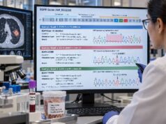
A MET amplification test checks whether lung cancer cells contain extra copies of the MET gene. MET encodes a receptor tyrosine kinase that can drive cancer growth when the gene is highly amplified. In non-small cell lung cancer (NSCLC), MET amplification may be present as a primary tumor alteration or appear later as an acquired resistance mechanism, especially after treatment of EGFR-mutant cancer with an EGFR tyrosine kinase inhibitor. The test can be performed with fluorescence in situ hybridization (FISH), next-generation sequencing (NGS), or other validated molecular methods. The result is not always a simple yes-or-no finding because copy number exists on a spectrum, laboratories use different thresholds, and chromosome 7 polysomy can raise MET copy number without producing the same biology as focal high-level amplification. A meaningful result therefore depends on the testing method, degree of amplification, tumor fraction, other genomic findings, treatment history, and the clinical question being asked.
- What it measures: MET amplification testing measures increased MET gene copy number, usually with FISH or NGS; it is different from MET exon 14 skipping and from c-MET protein overexpression.
- What a positive result means: High-level or focal MET amplification can act as an oncogenic driver or as a bypass resistance mechanism, but the clinical significance depends on the assay and threshold.
- Why thresholds matter: There is no single universal definition of MET amplification across all methods; studies have used different gene-copy and MET-to-chromosome-7 ratio cutoffs.
- When it is especially useful: Testing is often considered in advanced NSCLC molecular profiling and at progression on targeted therapy, particularly in EGFR-mutant cancer.
- What a negative result means: A negative result lowers the likelihood of detectable MET amplification in that specimen but may miss heterogeneous disease or low-level amplification, especially when plasma ctDNA is low.
Table of Contents
- What MET Amplification Is
- How MET Amplification Is Tested
- How to Read MET Amplification Results
- Primary vs Acquired MET Amplification
- MET Amplification vs Other MET Biomarkers
- What MET Amplification Can Mean for Treatment
- Limitations and Next Steps
What MET Amplification Is
The MET gene, located on chromosome 7, encodes the MET receptor. When activated normally, MET helps regulate cell growth, survival, movement, and tissue repair. Cancer cells can exploit this signaling system in several ways. One is gene amplification, in which extra copies of MET increase signaling enough to support tumor growth.
MET copy-number gain can arise through two biologically different processes. Focal amplification involves preferential duplication of the MET region and is more likely to represent true selection for MET signaling. Chromosome 7 polysomy means the cell contains extra copies of much or all of chromosome 7, so MET copy number rises along with other chromosome 7 genes. Those two patterns can look similar if a report lists only raw gene copy number.
This is why clinicians often care about both the absolute MET copy number and, when FISH is used, the ratio of MET signals to signals from the chromosome 7 centromere. A high ratio suggests that MET itself is selectively amplified rather than merely increased because the whole chromosome is duplicated.
MET amplification is one part of the broader NSCLC biomarker profile. It should be interpreted together with other drivers, co-mutations, pathology, and prior therapy rather than as an isolated laboratory number.
How MET Amplification Is Tested
There is no single testing method used everywhere. The two main approaches are FISH and NGS, with immunohistochemistry serving a different purpose because it measures protein expression rather than gene copy number.
FISH
Fluorescence in situ hybridization uses labeled DNA probes that bind to MET and usually to a reference region near the centromere of chromosome 7. A pathologist counts fluorescent signals in tumor-cell nuclei and calculates measures such as:
- average MET gene copy number per cell;
- MET-to-CEP7 ratio, where CEP7 represents the chromosome 7 centromere; and
- sometimes the percentage of cells meeting a high-copy threshold.
FISH can distinguish focal amplification from polysomy more directly than a simple copy-number estimate. Its disadvantages include manual interpretation, tumor-cell selection, specimen requirements, and the fact that thresholds vary across studies and laboratories.
Next-generation sequencing
A lung cancer NGS panel can estimate MET copy number while also testing many other genes. This is efficient because the same assay can identify EGFR, KRAS, BRAF, MET exon 14, gene fusions, and other alterations.
NGS copy-number estimates depend on sequencing depth, tumor purity, ploidy, bioinformatic algorithms, and assay calibration. Tissue NGS generally performs best when the sample contains enough tumor. Plasma NGS can detect amplification in circulating tumor DNA, but low tumor fraction can make copy-number gains difficult to call reliably.
Why one report may not match another
A patient can receive different-sounding results from FISH and NGS without either test necessarily being “wrong.” The methods measure amplification differently. One may call a sample MET amplified based on a ratio, another based on estimated copies, and another may reserve the term for only very high-level gains.
For a result that may affect treatment, the report should identify the method and threshold used. When the finding is borderline or unexpected, confirmatory testing with another validated method may sometimes help.
How to Read MET Amplification Results
The most important point is that MET amplification is a continuous variable, not a universally standardized binary marker. Higher and more focal amplification is generally more likely to represent true MET dependence than low-level copy-number gain.
Published studies have used a wide range of definitions. FISH-based trials have used MET-to-CEP7 ratios roughly from the high 1s to 3 or greater, while absolute gene-copy-number cutoffs have ranged from about 5 to 10 or more copies per cell. NGS studies have also used different thresholds. These values illustrate why the number printed on one laboratory report cannot automatically be transferred to another assay.
A report may use terms such as:
- copy-number gain;
- low-level amplification;
- intermediate amplification;
- high-level amplification; or
- focal amplification.
Those categories need the laboratory’s own definitions. A “copy number 6” result, for example, is not interpretable by itself without knowing whether it came from FISH or NGS, how tumor purity was handled, and what threshold the assay validated.
High-level focal amplification is more clinically compelling because it is more likely to function as a dominant oncogenic event. Low-level gain may reflect chromosome-level changes or a subclone rather than a cancer that is strongly dependent on MET.
A negative result also needs context. If a plasma sample contains little or no detectable ctDNA, the assay may miss a true amplification. Tissue confirmation can be important when suspicion is high.
Primary vs Acquired MET Amplification
MET amplification can occur in two main clinical settings.
Primary or de novo amplification
Primary MET amplification is present before targeted therapy and may act as an oncogenic driver. A 2024 systematic review found a median reported frequency of about 4.8% across several U.S. NSCLC studies, although prevalence varies according to assay and amplification definition.
High-level primary amplification is more likely to behave like a true driver than low-level gain. It may be detected during initial comprehensive molecular profiling, sometimes without another dominant actionable driver.
Acquired amplification after targeted therapy
MET amplification is also a well-recognized bypass resistance mechanism. It is particularly important in EGFR-mutant NSCLC progressing on EGFR inhibitors, including third-generation therapy. The MET pathway can reactivate downstream survival signaling even while EGFR remains inhibited.
The same 2024 review reported a median frequency around 15% for secondary MET amplification in EGFR-mutant NSCLC previously treated with an EGFR inhibitor, although individual studies varied widely because of different populations and definitions.
In this setting, MET amplification should be interpreted as part of a lung cancer resistance workup. The original EGFR driver is usually still relevant, and the new MET alteration may explain why EGFR blockade alone no longer controls the cancer.
MET amplification has also been described as an acquired resistance mechanism in ALK-, ROS1-, and RET-driven NSCLC. That broader pattern supports the idea that MET can function as a common bypass pathway when another oncogenic kinase is blocked.
MET Amplification vs Other MET Biomarkers
Several MET-related tests sound similar but measure different biological events. Confusing them can lead to incorrect interpretation.
| Biomarker | What it measures | Typical method |
|---|---|---|
| MET amplification | Extra copies of the MET gene | FISH or NGS |
| MET exon 14 skipping | Abnormal splicing that removes exon 14 from MET RNA | DNA and/or RNA sequencing |
| c-MET overexpression | Amount of MET protein in tumor cells | Immunohistochemistry |
| MET fusion | Rare rearrangement joining MET with another gene | NGS or other fusion assay |
A MET exon 14 skipping test is therefore not a substitute for an amplification test. Exon 14 skipping changes how MET is regulated by interfering with normal receptor degradation. Amplification increases the amount of MET gene material. Both can activate the pathway, but they are distinct biomarkers with different evidence bases.
Protein overexpression is also not interchangeable with gene amplification. Some tumors overexpress c-MET protein without high-level MET amplification, and some amplified tumors show different levels of protein expression. Treatment indications based on c-MET immunohistochemistry should be interpreted using the exact assay and criteria for that protein biomarker.
What MET Amplification Can Mean for Treatment
The treatment meaning depends on whether MET amplification is primary, high-level, acquired after another targeted therapy, or accompanied by additional drivers.
In primary MET-amplified NSCLC, responses to MET-directed tyrosine kinase inhibitors have generally been stronger in tumors with higher levels of amplification. This supports the concept that high-level amplification is more likely to represent true MET dependence. However, unlike MET exon 14 skipping, MET amplification has historically had less standardized testing thresholds and a more variable treatment evidence base.
In acquired EGFR resistance, the biology is different. The tumor may remain dependent on the original EGFR driver while also using MET as a bypass route. That provides the rationale for dual EGFR and MET inhibition rather than simply replacing one pathway with another. Clinical trials have evaluated several combinations in molecularly selected patients.
The existence of a MET amplification result does not mean that every MET-targeted drug is appropriate. Treatment depends on current regulatory approvals, guideline recommendations, disease setting, prior therapies, amplification level, co-alterations, and patient factors. Because the therapeutic landscape changes rapidly, the oncology team should match the exact biomarker definition used in the clinical evidence to the patient’s test result.
A high-quality molecular report may also identify whether the amplification is focal and whether another strong driver is present. Coexisting alterations can change how confidently MET is considered the main treatment target.
The specimen context can be just as important as the copy-number value. In a newly diagnosed tumor, a high-level MET amplification found before systemic therapy may represent a primary driver. In a repeat biopsy taken after months or years of EGFR-directed treatment, the same laboratory pattern may instead represent a newly selected resistant clone. The report cannot infer that history by itself; the oncology team has to connect the molecular finding with the treatment timeline.
Tumor percentage also affects interpretation. Tissue NGS compares sequencing read depth across genomic regions to estimate copy number, so a highly amplified gene can look less dramatic when the tumor is heavily mixed with normal cells. Modern bioinformatic methods try to correct for purity and ploidy, but the estimate still depends on specimen quality. FISH has a different advantage: the pathologist can visually count signals in individual tumor nuclei and compare MET with the chromosome 7 reference probe. That can help when focal amplification and polysomy need to be distinguished.
Plasma testing presents another challenge. A blood sample may contain ctDNA from several metastatic sites, which is useful when resistance is heterogeneous. But copy-number gains are harder to detect than many point mutations when the tumor fraction is low. A plasma report that identifies the original EGFR mutation but no MET amplification is more informative than a report in which no tumor-derived variants are detected at all. The latter may simply indicate that the sample contained too little ctDNA for a meaningful copy-number assessment. A lung cancer liquid biopsy should therefore be judged by both the reported alteration and evidence that tumor DNA was actually present.
Results can also change over time. An acquired MET-amplified clone may expand during an EGFR inhibitor and shrink after treatment is changed. If a later specimen is taken from a different lesion or after another therapy, the measured copy number may be lower or absent. This does not necessarily mean the earlier result was false; it may reflect clonal evolution. Serial testing is most useful when it is tied to a concrete decision, such as selecting therapy at progression, rather than performed simply to watch numbers change.
Finally, clinicians should distinguish a biomarker threshold from a biological law. Trial eligibility cutoffs are chosen to define study populations and enrich for likely responders. They do not create a sharp boundary where MET becomes biologically irrelevant one copy below the cutoff. Borderline values should therefore be interpreted cautiously, especially when the assay threshold differs from the threshold used in the evidence supporting a potential treatment.
Limitations and Next Steps
The central challenge in MET amplification testing is standardization, especially when a borderline result could directly influence treatment selection or trial eligibility. Different laboratories can use different specimens, platforms, thresholds, and reporting categories. That makes borderline results harder to compare than well-defined point mutations such as BRAF V600E.
Other limitations include tumor heterogeneity and specimen timing. An acquired MET-amplified clone may exist in only some metastatic sites. A biopsy of a non-amplified lesion can therefore miss resistance present elsewhere. Conversely, plasma can sample several lesions but may miss amplification when ctDNA shedding is low.
After receiving a result, useful questions include:
- Which method was used—FISH, tissue NGS, or plasma NGS?
- What threshold did the laboratory use to call amplification?
- Was the result low-level, intermediate, or high-level?
- Was focal amplification distinguished from chromosome 7 polysomy?
- Is another oncogenic driver present?
- Was this test done before treatment or after progression on a targeted drug?
- Does the treatment evidence use the same biomarker definition as this assay?
If the result is borderline and treatment would depend on it, confirmatory testing can be reasonable. If plasma is negative after targeted-therapy progression, tissue testing may provide better copy-number assessment and can also evaluate histologic transformation.
MET amplification is therefore best understood as a quantitative and contextual biomarker. The most meaningful result is not merely “more copies of MET,” but a well-characterized alteration measured by a validated assay and interpreted according to amplification level, tumor biology, and treatment history.
References
- Targeting MET in 2025: From Exon 14 Skipping to MET-Amplified Acquired Resistance in Non-Small Cell Lung Cancer 2026 (Review)
- Targeting Mesenchymal-Epidermal Transition (MET) Aberrations in Non-Small Cell Lung Cancer: Current Challenges and Therapeutic Advances 2026 (Review)
- Non-small cell lung cancer with MET amplification: review of epidemiology, associated disease characteristics, testing procedures, burden, and treatments 2024 (Systematic Review)
- Unraveling the Significance of MET Focal Amplification in Lung Cancer: Integrative NGS, FISH, and IHC Investigation 2024
- Overview of Molecular Detection Technologies for MET in Lung Cancer 2023 (Review)
Disclaimer
This article is for general education and does not replace personalized oncology advice. MET amplification results require interpretation using the specific assay, threshold, specimen, tumor type, and treatment history. Cancer treatment should not be started, stopped, or changed based on a copy-number result without review by the treating oncology team.