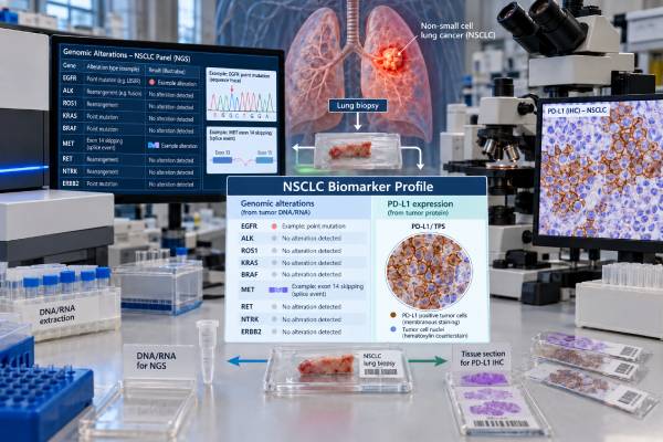
Non-small cell lung cancer (NSCLC) biomarker testing examines a tumor for molecular changes and protein markers that can influence treatment. In advanced NSCLC, broad testing can identify actionable alterations such as EGFR mutations, ALK or ROS1 fusions, KRAS G12C, BRAF V600E, MET exon 14 skipping, RET or NTRK fusions, and ERBB2/HER2 mutations. PD-L1 immunohistochemistry is usually assessed separately because it helps guide immunotherapy choices, especially when no targetable driver is found. Testing may use tumor tissue, a cytology cell block, blood-based circulating tumor DNA, or a combination. The most useful result is not simply “positive” or “negative”; it is a complete profile that shows which biomarkers were tested, which alteration was found, whether it is considered actionable, and whether the specimen was adequate. Results must also be interpreted with cancer stage, histologic subtype, prior treatments, and current drug approvals or guidelines.
- What it tests: NSCLC biomarker testing looks for actionable DNA mutations, gene fusions, splice changes, amplifications, and PD-L1 protein expression.
- Why broad testing matters: A targetable driver can make a matched targeted therapy more appropriate than starting treatment based only on PD-L1.
- How PD-L1 is reported: PD-L1 is commonly reported as tumor proportion score (TPS), including clinically important cut points such as 1% and 50%, but treatment rules depend on the setting and regimen.
- What a negative result means: “No alteration detected” does not prove the tumor lacks every driver; assay coverage, tumor amount, and specimen quality can limit detection.
- When retesting is useful: Repeat tissue or liquid biopsy may be considered when the first sample is inadequate, the disease progresses, or resistance biology could change treatment.
Table of Contents
- What NSCLC Biomarker Testing Measures
- Driver Mutations and Gene Fusions That Matter
- PD-L1 Testing and Result Meaning
- How NSCLC Biomarker Testing Is Performed
- How to Read an NSCLC Molecular Profile
- When Biomarkers Should Be Tested or Repeated
- Limitations, Discordant Results, and Next Steps
What NSCLC Biomarker Testing Measures
NSCLC biomarker testing is a group of tests, not one single assay. The goal is to identify biologic features of the cancer that can predict benefit from targeted therapy or immunotherapy and, in some settings, help with diagnosis, prognosis, or clinical-trial selection.
The two main categories are genomic biomarkers and protein biomarkers. Genomic testing examines DNA and often RNA for alterations that drive tumor growth. These include point mutations, small insertions or deletions, gene fusions, exon-skipping events, and copy-number changes. Protein testing uses immunohistochemistry (IHC) to measure markers such as PD-L1 directly in tumor tissue.
For advanced non-squamous NSCLC, comprehensive molecular profiling is generally preferred over a sequence of narrow single-gene tests because many actionable alterations are individually uncommon. A broad lung cancer NGS panel can evaluate numerous genes at once and may include both DNA and RNA analysis. RNA is especially useful for detecting fusions and splice alterations that can be difficult to capture with DNA-only methods.
Biomarker testing is different from hereditary genetic testing. Most NSCLC assays look for somatic changes acquired by cancer cells. A tumor result can occasionally raise concern for a germline variant, but confirmation requires a separate inherited-cancer evaluation using an appropriate non-tumor sample.
The final treatment decision also depends on factors that a biomarker panel cannot measure by itself, including cancer stage, performance status, other medical conditions, prior therapy, symptoms, and the location and pace of disease.
Driver Mutations and Gene Fusions That Matter
A driver alteration is a molecular change that helps the cancer grow and may create a treatment vulnerability. In contemporary NSCLC care, the most clinically established drivers include EGFR, ALK, ROS1, BRAF, KRAS, MET, RET, NTRK, and ERBB2/HER2. The exact list tested and considered actionable should follow current guidelines and drug availability in the patient’s country.
| Biomarker | Typical alteration | Why it matters |
|---|---|---|
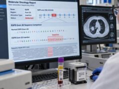
| EGFR | Exon 19 deletion, L858R, and other activating mutations | Can predict benefit from EGFR-targeted therapy |
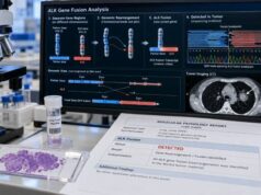
| ALK | Gene fusion or rearrangement | Can identify an ALK-driven tumor treatable with ALK inhibitors |
| ROS1 | Gene fusion | Can predict benefit from ROS1-directed therapy |
| KRAS | Especially KRAS G12C | G12C can be directly targetable in appropriate treatment settings |
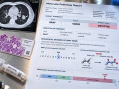
| BRAF | Especially V600E | Can support use of BRAF/MEK-directed therapy |
| MET | Exon 14 skipping; selected amplification settings | MET exon 14 skipping is an established target; amplification can also be relevant |
| RET | Gene fusion | Can identify tumors sensitive to selective RET inhibitors |
| NTRK1/2/3 | Gene fusion | Rare in NSCLC but can predict benefit from tumor-agnostic TRK therapy |
| ERBB2/HER2 | Activating mutation, usually in the kinase domain | Can influence selection of HER2-directed treatment |
EGFR illustrates why the exact variant matters. An EGFR mutation test should distinguish common sensitizing mutations from uncommon variants and resistance alterations rather than simply reporting “EGFR positive.” The same principle applies to BRAF, KRAS, and MET: gene name alone is not enough.
Fusions also require careful detection. ALK, ROS1, RET, and NTRK fusions often involve many possible partner genes. Assays that include RNA can improve confidence that a rearrangement creates an expressed fusion transcript. IHC or fluorescence in situ hybridization may still have a role as validated screening, companion, or confirmatory methods depending on the biomarker and laboratory.
Driver alterations are often treated as mutually exclusive dominant events, but co-alterations can occur. A report may therefore include additional changes in TP53, STK11, KEAP1, or other genes. These findings can provide biologic or prognostic context, but they do not automatically have an approved matched therapy.
PD-L1 Testing and Result Meaning
PD-L1 is a protein-expression biomarker used mainly to help select immunotherapy, not a driver mutation. It is measured by IHC on tumor tissue using a validated antibody assay.
In NSCLC, the most familiar reporting system is the tumor proportion score (TPS): the percentage of viable tumor cells showing membranous PD-L1 staining. A report may describe TPS as less than 1%, 1%–49%, or 50% or greater because these ranges are used in many treatment decisions. However, PD-L1 thresholds are not universal rules. The relevant cutoff depends on disease stage, treatment line, drug, regimen, regulatory approval, and whether the patient has a targetable driver.
A PD-L1 test for lung cancer can be clinically important even when broad molecular testing is also ordered. The two tests answer different questions:
- Molecular profiling asks whether the cancer contains a targetable genomic driver.
- PD-L1 asks how much of a specific immune-regulatory protein is expressed by tumor cells.
- A high PD-L1 TPS does not cancel an actionable driver alteration.
- A low or negative PD-L1 result does not mean immunotherapy can never be used; combination regimens and other clinical factors may still make it appropriate.
This is especially important in metastatic non-squamous NSCLC. If an actionable EGFR, ALK, ROS1, RET, MET, or another established driver is present, targeted therapy may take priority even when PD-L1 is high. Starting immunotherapy before driver results return can complicate sequencing and, for some targeted drugs, may increase toxicity concerns.
PD-L1 also has technical limitations. Expression can vary between tumor regions and can change over time or after therapy. Small biopsies may underrepresent heterogeneous staining. Different validated assays and treatment indications can use different scoring requirements, so the pathology report should identify the assay when clinically relevant.
How NSCLC Biomarker Testing Is Performed
Testing usually begins with the diagnostic specimen. Formalin-fixed, paraffin-embedded tissue from a core biopsy or surgical specimen is common, but cytology cell blocks can also work if they contain enough viable tumor. The pathology team must balance two competing needs: classify the cancer accurately and preserve enough material for molecular and PD-L1 testing.
For DNA and RNA profiling, the laboratory estimates tumor content, extracts nucleic acid, and runs a validated platform. Next-generation sequencing can detect many alteration types in parallel. Some panels are DNA-only; others pair DNA sequencing with RNA sequencing to improve fusion and exon-skipping detection.
A lung cancer liquid biopsy can analyze circulating tumor DNA (ctDNA) in plasma. Blood testing is valuable when tissue is unavailable, risky to obtain, or too slow to acquire, and it can shorten the time to an actionable result. However, a negative plasma test is not equivalent to a negative tumor test. Some cancers shed very little DNA into the bloodstream, especially when disease volume is low or confined to certain sites. If plasma is non-informative and tissue can be obtained, tissue testing is often still needed.
PD-L1 generally requires tumor cells and therefore is not replaced by standard ctDNA testing. The PD-L1 slide is scored by a pathologist, while sequencing results are interpreted through a molecular pathology workflow.
Turnaround time varies by laboratory and test. PD-L1 IHC can often return sooner than broad NGS, while comprehensive tissue sequencing may take roughly one to several weeks depending on specimen transport, tumor enrichment, assay complexity, and whether repeat testing is needed. Treatment planning should account for that timing when the patient is clinically stable enough to wait for the complete profile.
How to Read an NSCLC Molecular Profile
A useful molecular report should answer four questions: What was tested? What was found? Is the finding actionable? Was the assay technically adequate?
Pathogenic or likely pathogenic driver detected
This is the most straightforward result. The report names the gene and exact alteration, such as EGFR exon 19 deletion, KRAS G12C, BRAF V600E, MET exon 14 skipping, or an EML4-ALK fusion. It may also list approved therapies, guideline associations, or clinical trials. The oncology team then checks whether the treatment association applies to this cancer stage and prior-treatment setting.
No actionable alteration detected
This wording means the assay did not identify a qualifying target within its tested scope. It does not guarantee that no driver exists. Review the panel’s gene list and alteration types. A DNA-only panel may be less complete for some fusions, and a low-tumor specimen may reduce sensitivity. When the clinical picture strongly suggests an oncogene-driven tumor, RNA testing, repeat tissue, or plasma testing can sometimes resolve a negative result.
Variant of uncertain significance
A variant of uncertain significance, or VUS, is a DNA change whose clinical meaning is not established. A VUS should not be treated as though it were a proven driver simply because it appears in an important cancer gene. Classification can change as evidence grows, so laboratories may update interpretation over time.
PD-L1 result
PD-L1 should be read alongside the genomic profile, not in isolation. For example, “TPS 70%” indicates high expression by common cut-point conventions, but it does not mean a patient with an actionable EGFR mutation should automatically receive immunotherapy first.
The report may also contain tumor mutational burden, microsatellite instability, copy-number changes, or other exploratory biomarkers. A tumor mutational burden result can provide additional genomic context, but its role depends on the specific treatment indication and should not replace established driver testing or PD-L1 when those tests are required.
When Biomarkers Should Be Tested or Repeated
The best time to obtain a complete biomarker profile is before a treatment choice depends on it. In metastatic or otherwise advanced non-squamous NSCLC, broad genomic testing and PD-L1 assessment are commonly performed at diagnosis. Current precision-medicine guidance also supports broad testing across patients who could be eligible for matched therapy, rather than restricting testing based on age, sex, or smoking history.
Biomarkers increasingly matter in earlier-stage disease as well. EGFR and ALK status can affect adjuvant or perioperative treatment decisions in selected resected tumors, and PD-L1 or other testing may be required for particular perioperative regimens. The exact testing set therefore depends on stage and planned therapy, not only histology.
Repeat testing may be useful in several situations:
- The original sample is inadequate. Too few tumor cells, degraded nucleic acid, or test failure can leave the molecular profile incomplete.
- Only narrow testing was done. A patient may have had EGFR and ALK testing years ago but never received comprehensive profiling for newer targets.
- Cancer progresses on targeted therapy. Resistance can arise through new on-target mutations or bypass pathways. A lung cancer resistance mutation panel or broader profiling can identify mechanisms that affect the next treatment decision.
- The tumor biology appears to change. Rapid clinical change or a new lesion can occasionally reflect histologic transformation or a second cancer, situations that may require a new biopsy rather than blood testing alone.
At progression, plasma ctDNA is often attractive because it samples DNA released from multiple disease sites. Tissue remains valuable when plasma is negative, when transformation is suspected, or when morphology and protein markers need reassessment.
Limitations, Discordant Results, and Next Steps
No biomarker test is perfectly comprehensive. The main limitations are specimen quality, assay design, tumor heterogeneity, timing, and interpretation.
A small biopsy can fail because there is not enough tumor after diagnostic IHC. Bone specimens may be difficult if decalcification damages nucleic acids. Plasma can be falsely negative when ctDNA shedding is low. DNA sequencing can miss some RNA-level fusions or splice events. PD-L1 can vary across different parts of a tumor and between primary and metastatic sites.
Discordant results should be resolved by asking which method is most reliable for the alteration in question. For example, an unexpected fusion result may merit RNA-based confirmation. A plasma-negative result may justify tissue testing. A borderline copy-number result may need a method designed to assess amplification more directly. Laboratories should also distinguish true “negative” findings from “quantity not sufficient,” “test failed,” and “not assessed.”
After receiving the report, useful questions include:
- Was broad DNA and RNA testing performed, or only a limited panel?
- Were EGFR, ALK, ROS1, BRAF, KRAS, MET, RET, NTRK, and ERBB2/HER2 adequately assessed for the alteration types that matter?
- What is the PD-L1 TPS, and which assay produced it?
- Is any detected finding classified as actionable in this stage and treatment setting?
- Was there enough tumor for a reliable negative result?
- Would tissue or plasma retesting add information if no driver was found?
- If the cancer has progressed, is resistance testing needed before choosing the next therapy?
The practical goal is a profile that is complete enough to prevent a missed treatment opportunity. Molecular results, PD-L1, histology, stage, and clinical condition should be reviewed together. A complete report should also make technical gaps obvious, such as failed RNA sequencing, insufficient tumor, or an alteration type the panel does not cover. Those details can determine whether a second assay is worth pursuing. Because treatment approvals and biomarker recommendations change quickly, an oncology team should interpret the report using the most current guidance available at the time of the decision.
References
- Therapy for Stage IV Non-Small Cell Lung Cancer With Driver Alterations: ASCO Living Guideline, Version 2026.3.3 2026 (Guideline)
- Therapy for Stage IV Non–Small Cell Lung Cancer Without Driver Alterations: ASCO Living Guideline Update 2026 (Guideline)
- Prognostic and predictive biomarkers in non-small cell lung carcinoma 2024 (Review)
- Current status of molecular diagnostics for lung cancer 2024 (Review)
- Guidance for clinicians and patients with non-small cell lung cancer in the time of precision medicine 2023 (Review)
- Consensus Recommendations to Optimize Testing for New Targetable Alterations in Non-Small Cell Lung Cancer 2022 (Consensus)
Disclaimer
This article is for general education and does not replace individualized advice from an oncology, pathology, or molecular diagnostics team. Biomarker requirements, drug approvals, and preferred testing methods can change, and a result must be interpreted in the context of cancer stage, histology, prior treatment, specimen quality, and current guidelines. Do not start, stop, or delay cancer treatment based on a biomarker result without discussing it with the treating clinician.