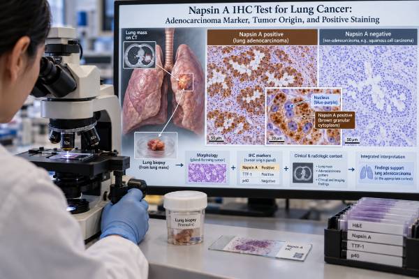
Napsin A immunohistochemistry (IHC) is a tissue stain used mainly to support the diagnosis of lung adenocarcinoma. Napsin A is an aspartic protease normally expressed in type II pneumocytes and some kidney cells. In a tumor biopsy, positive staining appears as granular cytoplasmic staining in cancer cells. A positive result can strongly support pulmonary adenocarcinoma when the tumor’s appearance and other markers fit, especially when Napsin A is interpreted together with TTF-1. It is not completely specific to the lung: some renal, gynecologic clear cell, thyroid, and other tumors can also express Napsin A. Likewise, some genuine lung adenocarcinomas are Napsin A-negative, particularly certain mucinous or poorly differentiated tumors. For these reasons, pathologists do not use Napsin A as a stand-alone “lung origin” test. They interpret it as part of a panel that may include TTF-1, p40, CK7, PAX8, and organ-specific markers chosen from the clinical and morphologic differential diagnosis.
- What it measures: Napsin A IHC detects Napsin A protein in tumor cells; positive staining is usually granular and cytoplasmic.
- What a positive result means: In the right morphologic setting, Napsin A positivity supports lung adenocarcinoma, especially when TTF-1 is also positive.
- What a negative result means: Negative staining does not rule out lung adenocarcinoma; a meaningful minority of pulmonary adenocarcinomas do not express Napsin A.
- What it cannot prove: Napsin A is not perfectly lung-specific because renal and some gynecologic or other carcinomas can be positive.
- Why panels matter: Pathologists combine morphology with Napsin A, TTF-1, p40, and other markers to subtype NSCLC while preserving tissue for molecular testing.
Table of Contents
- What Napsin A Is and Why It Is Used
- How the Napsin A IHC Test Is Performed
- How to Read Positive and Negative Staining
- Napsin A and TTF-1 in Lung Adenocarcinoma
- Can Napsin A Prove Tumor Origin?
- Common Pitfalls and Special Tumor Types
- What Happens After the Napsin A Result
What Napsin A Is and Why It Is Used
Napsin A is a protease involved in processing surfactant-related proteins in normal lung tissue. It is strongly expressed in type II pneumocytes and is also present in renal tissue. Many pulmonary adenocarcinomas retain Napsin A expression, which makes the protein useful as a marker of glandular differentiation in lung cancer.
The test is most valuable when a biopsy shows non-small cell carcinoma but the small sample does not clearly display gland formation or keratinization. Correctly separating adenocarcinoma from squamous cell carcinoma matters because the subtypes have different molecular-testing priorities and sometimes different treatment options.
Napsin A is therefore a diagnostic lineage marker, not a blood tumor marker and not a genomic biomarker. It does not measure a mutation, gene fusion, or treatment-resistance alteration. It is performed directly on cells in a tissue section or cytology cell block.
In routine practice, the pathologist first examines the hematoxylin-and-eosin (H&E) morphology. IHC is then used to resolve a specific diagnostic question. For a poorly differentiated NSCLC, Napsin A and TTF-1 can support adenocarcinoma, while p40 is a strong marker of squamous differentiation. A focused panel helps preserve limited biopsy material for a lung cancer NGS panel and other predictive tests.
How the Napsin A IHC Test Is Performed
Napsin A IHC is usually performed on formalin-fixed, paraffin-embedded tissue from a lung biopsy, lymph-node biopsy, resection specimen, or cytology cell block. The laboratory cuts a thin tissue section, applies an antibody directed against Napsin A, and uses a detection system that makes the antibody-binding sites visible under a microscope.
A pathologist evaluates three things:
- Location of staining: true Napsin A expression is cytoplasmic, often coarse or granular.
- Distribution: staining may be focal or diffuse across the tumor.
- Intensity: staining can be weak, moderate, or strong depending on the assay and tumor.
There is no universal patient-facing “normal range” or numeric cutoff comparable with a blood test. Laboratories validate their antibody clone, staining platform, controls, and interpretation criteria. Some studies have defined positivity using a percentage threshold, while routine diagnostic interpretation often relies on the combination of convincing cytoplasmic staining and morphology.
Normal lung cells can provide useful internal context. Type II pneumocytes may stain for Napsin A, and alveolar macrophages can show granular staining because they contain lysosomal material. The pathologist must identify which cells are actually tumor cells before calling a cancer positive.
Pre-analytic quality matters. Poor fixation, very small cell blocks, crush artifact, necrosis, or scant tumor can reduce staining quality. A technically inadequate result should not be treated the same as a true negative tumor.
How to Read Positive and Negative Staining
A Napsin A result is best interpreted as supporting evidence, not a diagnosis by itself.
Positive Napsin A staining
A positive result means tumor cells show convincing granular cytoplasmic Napsin A staining. In a lung mass with adenocarcinoma-like morphology, this finding supports pulmonary adenocarcinoma. Large studies have reported Napsin A positivity in roughly the mid-80% range for lung adenocarcinomas, with specificity that can be high when appropriate comparison groups are used.
The strength of the conclusion depends on context. A TTF-1-positive, Napsin A-positive tumor with classic lung adenocarcinoma morphology is more convincing than isolated weak Napsin A positivity in a patient with a known kidney or gynecologic cancer.
Negative Napsin A staining
A negative result does not rule out lung adenocarcinoma. Depending on the study and tumor subtype, approximately 10%–20% or more of lung adenocarcinomas can be negative. Sensitivity is lower in some mucinous tumors and poorly differentiated cancers.
When Napsin A is negative, the pathologist may rely on TTF-1, morphology, mucin stains, CK7, molecular findings, and other organ-specific markers. A tumor can be TTF-1 positive and Napsin A negative and still be a primary lung adenocarcinoma.
Focal or weak staining
Focal staining is not automatically meaningless, but it deserves caution. The pathologist considers whether the staining is in tumor cells, whether the pattern is appropriately granular and cytoplasmic, and whether other markers support the same lineage. Background macrophage staining can be a pitfall in small biopsies.
The final pathology report may not provide a percentage because the clinical purpose is usually classification rather than a treatment threshold. The key phrase may simply be “Napsin A positive” as part of an immunophenotypic summary.
Napsin A and TTF-1 in Lung Adenocarcinoma
Napsin A and TTF-1 are commonly used together because they provide complementary evidence. TTF-1 is a nuclear transcription factor, while Napsin A is a cytoplasmic enzyme. Their different staining patterns make the pair easy to evaluate on the same diagnostic problem.
A large 2024 tissue-microarray study found that, for distinguishing pulmonary adenocarcinoma, TTF-1 had high sensitivity while Napsin A had higher specificity; using both markers together increased specificity further. This does not mean every laboratory will reproduce identical percentages, but it illustrates why a two-marker interpretation is often stronger than either result alone.
| TTF-1 | Napsin A | Typical interpretation in the right morphology |
|---|---|---|
| Positive | Positive | Strong support for pulmonary adenocarcinoma, but still not absolute proof of origin |
| Positive | Negative | Lung adenocarcinoma remains possible; some pulmonary adenocarcinomas lack Napsin A |
| Negative | Positive | Can support adenocarcinoma, but extrapulmonary Napsin A-positive tumors must be considered |
| Negative | Negative | Does not exclude lung adenocarcinoma; morphology and additional markers become more important |
A focused TTF-1 IHC test can help clarify how nuclear TTF-1 staining differs from Napsin A and why neither marker should be used alone in difficult metastatic workups.
For squamous differentiation, p40 is usually more informative. A Napsin A-negative, p40-positive NSCLC supports squamous cell carcinoma, whereas Napsin A positivity favors glandular differentiation. Rare tumors can show mixed or unusual immunophenotypes, so morphology remains essential.
Can Napsin A Prove Tumor Origin?
No. Napsin A is highly useful but not lung-specific.
A large survey of human tumors found Napsin A expression not only in lung adenocarcinoma but also in substantial subsets of ovarian clear cell carcinoma, endometrial clear cell carcinoma, papillary renal cell carcinoma, and several less common tumor types. This is a major reason the marker cannot independently prove that a metastasis came from the lung.
The differential diagnosis depends on the patient’s history and tumor morphology. For example:
- A Napsin A-positive lung lesion in a patient with renal cancer may need PAX8 and other renal markers.
- A Napsin A-positive lesion in a woman with a pelvic mass may require PAX8 and gynecologic markers.
- A TTF-1/Napsin A double-positive pattern strongly favors lung adenocarcinoma in many settings, but unusual extrapulmonary tumors can still create exceptions.
Pathologists therefore build a panel around the clinical question. The right comparison is not “Napsin A positive = lung.” It is “Does the full morphology and immunophenotype fit a lung primary better than the plausible alternatives?”
This distinction is especially important when a biopsy comes from a lymph node, bone, liver, or another metastatic site. The pathologist may know the tissue is carcinoma but need to determine its most likely origin. Napsin A contributes evidence; it does not provide a standalone answer.
Common Pitfalls and Special Tumor Types
Several situations make Napsin A harder to interpret.
Mucinous lung adenocarcinoma
Invasive mucinous adenocarcinoma often has lower Napsin A and TTF-1 expression than conventional non-mucinous lung adenocarcinoma. Older studies reported Napsin A positivity in only about one-third of some mucin-producing lung adenocarcinomas. A negative stain therefore cannot be used to exclude pulmonary origin in a mucinous tumor.
Some antibody preparations have also produced problematic staining in extrapulmonary mucinous tumors. Marker clone, staining platform, and the complete panel matter.
Renal tumors
Normal kidney expresses Napsin A, and several renal cell carcinoma subtypes can be positive. This is one of the classic exceptions to the idea that Napsin A indicates lung origin. PAX8 is often useful in the differential because many renal tumors are PAX8 positive while primary pulmonary adenocarcinoma is generally PAX8 negative.
Gynecologic clear cell tumors
Ovarian and endometrial clear cell carcinomas can express Napsin A strongly. Clinical history, PAX8, other gynecologic markers, and morphology are critical when these tumors are possible.
Normal cells and macrophages
A small lung biopsy may contain many macrophages with granular cytoplasm. If the pathologist mistakes their staining for tumor-cell staining, the result can be overcalled. Proper cell identification under the microscope prevents this pitfall.
What Happens After the Napsin A Result
Napsin A usually helps answer a classification question, and that classification determines what comes next.
If morphology and IHC support lung adenocarcinoma, the remaining tissue may be directed to comprehensive molecular profiling for actionable drivers such as EGFR, ALK, ROS1, BRAF, KRAS, MET, RET, NTRK, and ERBB2/HER2. PD-L1 immunohistochemistry is commonly assessed separately. This broader NSCLC biomarker testing affects treatment far more directly than Napsin A itself.
If the staining pattern is unusual, the pathologist may add organ-specific markers or request clinical correlation. A Napsin A-positive tumor with PAX8 positivity, for instance, can shift the differential toward renal or gynecologic origin depending on morphology.
Patients reviewing a pathology report can ask:
- Was Napsin A staining present in the tumor cells or only background cells?
- Was TTF-1 also tested?
- Did morphology support adenocarcinoma?
- Were squamous markers such as p40 needed?
- Is there any clinical history that raises concern for a metastasis from another organ?
- Has enough tissue been preserved for molecular and PD-L1 testing?
Why tissue stewardship matters in a small lung biopsy
Small lung biopsies often contain only a few groups of viable tumor cells. Every additional IHC slide uses some of that material, and the same specimen may still be needed for PD-L1 testing and broad DNA/RNA profiling. Pathologists therefore try to use the smallest panel that can answer the classification question. A common approach in an undifferentiated NSCLC is to pair an adenocarcinoma marker such as TTF-1 or Napsin A with a strong squamous marker such as p40 rather than ordering a long list of stains at once.
This tissue-sparing strategy is clinically important because a precise subtype is only one part of modern lung-cancer diagnosis. If all tumor is exhausted during IHC, the patient may need another biopsy to obtain the molecular profile needed for treatment selection. The ideal workup gets enough diagnostic certainty while preserving tumor for the tests that directly guide therapy.
Napsin A can be especially useful when the morphology suggests adenocarcinoma but gland formation is subtle. Convincing tumor-cell staining may allow the pathologist to support glandular differentiation without adding many lower-yield stains. The exact panel still depends on the differential diagnosis and the amount of tissue available.
Napsin A is not a treatment-selection score
Unlike PD-L1, Napsin A does not have a clinically validated percentage threshold that selects immunotherapy. Unlike EGFR, ALK, ROS1, MET, or other genomic drivers, it does not identify a matched targeted drug. A report that says “Napsin A positive” is mainly helping establish what the tumor is, not which medicine should be prescribed.
That distinction can prevent a common misunderstanding. Strong or diffuse Napsin A staining does not mean the cancer is more targetable, and weak staining does not mean treatment will be less effective. Prognosis and treatment depend on stage, molecular alterations, PD-L1, the patient’s condition, and other tumor features. Napsin A contributes to the diagnostic foundation that makes those later decisions possible.
What if the stains disagree?
Discordant IHC patterns are not rare enough to ignore. A tumor may be TTF-1 positive but Napsin A negative, or it may have weak Napsin A with an unexpected p40 result. The pathologist does not resolve that conflict by counting positive markers. Instead, the team reviews morphology, staining location and intensity, internal controls, prior cancer history, imaging, and whether additional tissue is available.
If the pattern remains unusual, a broader panel or molecular result may clarify the diagnosis. Rare NSCLCs can show mixed differentiation, and metastatic cancers can imitate a pulmonary immunophenotype. In a difficult case, the final report may appropriately use language such as “favor adenocarcinoma” rather than claiming certainty that the evidence does not support.
The core interpretation is that Napsin A is a strong supporting marker for lung adenocarcinoma, not an absolute marker of lung origin. Its value comes from combining a characteristic cytoplasmic stain with the tumor’s morphology and a focused panel of complementary markers. When the clinical history or staining pattern is atypical, a carefully chosen second-line IHC panel is safer than forcing a lung-origin conclusion from one marker.
References
- Immunohistochemistry as a Cornerstone in Lung Cancer Diagnosis: Subtyping Based on the 2021 World Health Organization Classification and Its Practical Application 2026
- Diagnostic Utility of Immunohistochemistry in Lung Carcinoma Subtyping: A Clinicopathological Study From a Tertiary Care Center. 2026
- TTF-1 is a highly sensitive but not fully specific marker for pulmonary and thyroidal cancer: a tissue microarray study evaluating more than 17,000 tumors from 152 different tumor entities 2024
- Non-small cell lung carcinomas with diffuse coexpression of TTF1 and p40: clinicopathological and genomic features of 14 rare biphenotypic tumours 2022
- Napsin A Expression in Human Tumors and Normal Tissues 2021
- Napsin A, a new marker for lung adenocarcinoma, is complementary and more sensitive and specific than thyroid transcription factor 1 in the differential diagnosis of primary pulmonary carcinoma: evaluation of 1674 cases by tissue microarray 2012
Disclaimer
This article is for general education and does not replace interpretation by a qualified pathologist or oncology team. Napsin A staining must be read with morphology, controls, other IHC markers, clinical history, and specimen quality. A positive or negative Napsin A result should not be used alone to determine tumor origin or cancer treatment.