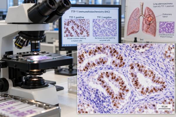
The TTF-1 immunohistochemistry test is a tissue stain that helps pathologists determine whether a tumor has features typical of lung adenocarcinoma or certain lung neuroendocrine cancers. TTF-1, also called NKX2-1, is a nuclear transcription factor normally expressed in lung and thyroid tissue. In a lung tumor, strong nuclear TTF-1 staining supports pulmonary adenocarcinoma when the microscopic appearance and other markers fit, but it does not prove lung origin by itself. About 15%–20% of lung adenocarcinomas can be TTF-1 negative, and some thyroid tumors and a smaller group of cancers from other organs can be positive. The test is therefore interpreted as part of an immunohistochemical panel rather than as a stand-alone answer. TTF-1 is especially useful in small biopsies, metastatic tumors of uncertain origin, and poorly differentiated non-small cell lung cancers where morphology alone does not clearly show glandular or squamous differentiation.
- What TTF-1 measures: The stain detects nuclear expression of the NKX2-1 protein in tumor cells; true positivity is primarily a nuclear staining pattern.
- What a positive result usually suggests: In the right clinical setting, TTF-1 positivity supports lung adenocarcinoma or a pulmonary neuroendocrine tumor, but thyroid origin and rare non-lung tumors must still be considered.
- What a negative result means: A negative stain does not rule out lung adenocarcinoma; roughly 15%–20% of cases may lack TTF-1 expression.
- How it is used: TTF-1 is commonly paired with Napsin A for adenocarcinoma and with squamous markers such as p40 when classifying non-small cell lung cancer.
- Why the panel matters: No single immunostain should determine tumor origin or subtype without morphology, clinical history, imaging, and complementary markers.
Table of Contents
- What TTF-1 Is and How the IHC Test Works
- What Positive TTF-1 Staining Means
- What Negative TTF-1 Staining Means
- TTF-1 in Lung Cancer Classification
- Using TTF-1 to Assess Tumor Origin
- Limits and Common Interpretation Pitfalls
- What Happens After the TTF-1 Result
What TTF-1 Is and How the IHC Test Works
TTF-1 stands for thyroid transcription factor 1. Its gene name is NKX2-1. The protein helps regulate development and specialized functions in the lung and thyroid, which is why pathologists can use its expression pattern as a clue to tissue lineage.
Unlike a blood tumor marker, TTF-1 immunohistochemistry is performed on cells from a biopsy, surgical specimen, cytology cell block, or another tumor sample. The tissue is exposed to an antibody that binds TTF-1. If the protein is present, the nuclei of tumor cells develop visible staining under the microscope.
The key feature is nuclear staining. Cytoplasmic color by itself is generally not interpreted as true TTF-1 positivity because the biologically relevant protein is a nuclear transcription factor. The pathologist evaluates the intensity, distribution, percentage of positive tumor cells, background staining, and whether internal controls behave as expected.
The specimen can be very small. Bronchoscopic biopsies, needle cores, pleural fluid cell blocks, and lymph-node aspirates may contain only a limited number of malignant cells, so pathologists try to use the smallest effective stain panel. Preserving tissue matters because the same sample may also be needed for DNA- and RNA-based molecular testing. When only a few tumor cells are present, the pathologist may prioritize stains that answer the immediate classification question and avoid unnecessary markers that consume tissue without adding much diagnostic value.
There is no universal patient-facing score comparable with a PD-L1 TPS. Reports may simply say “positive,” “negative,” “focal positive,” or describe the percentage and intensity. Different laboratories also use different antibody clones. Some clones are more sensitive than others, and that can slightly change how often unusual non-lung tumors or squamous carcinomas appear positive.
The TTF-1 result is therefore not interpreted in isolation. It is combined with the tumor’s shape and growth pattern, the specimen site, imaging findings, and other immunostains. In a small biopsy labeled only as non-small cell carcinoma, the Napsin A lung adenocarcinoma stain often complements TTF-1 because the two markers together can provide stronger evidence of glandular differentiation than either one alone.
What Positive TTF-1 Staining Means
A positive TTF-1 stain means that tumor cell nuclei contain detectable TTF-1 protein. In a suspicious lung mass, this finding often supports a diagnosis of primary lung adenocarcinoma, especially when the tumor also has adenocarcinoma morphology or Napsin A positivity.
Modern series suggest that around 80%–85% of lung adenocarcinomas are TTF-1 positive, although rates vary with specimen type, tumor subtype, antibody clone, staining threshold, and patient population. A 2026 multicenter analysis found TTF-1 negativity in about 15% of lung adenocarcinomas, which illustrates why positive staining is common but not universal.
TTF-1 positivity can also occur in lung neuroendocrine neoplasms, including many small cell lung cancers. That means a positive result does not automatically equal adenocarcinoma. The microscopic architecture, cell shape, proliferation pattern, and neuroendocrine markers determine whether the tumor belongs to a neuroendocrine category.
For a poorly differentiated tumor, a positive TTF-1 result can narrow the differential diagnosis but still needs context. Examples include:
- A lung mass with gland-forming tumor cells, TTF-1 positivity, and Napsin A positivity strongly supports pulmonary adenocarcinoma.
- A high-grade small blue cell tumor that is TTF-1 positive may be small cell lung cancer if neuroendocrine morphology and markers also fit.
- A neck or bone metastasis that is TTF-1 positive may be pulmonary in origin, but a thyroid primary must be excluded when clinically plausible.
- Rare non-lung tumors can show TTF-1 staining, so unexpected positivity should trigger a broader panel rather than an automatic lung diagnosis.
TTF-1 also has emerging prognostic associations. Recent studies have found that TTF-1-negative lung adenocarcinomas can have different genomic features and, in some treatment settings, worse outcomes than TTF-1-positive tumors. These observations are clinically interesting, but TTF-1 remains first and foremost a diagnostic lineage marker. It is not currently used alone to select a specific targeted therapy.
What Negative TTF-1 Staining Means
A negative TTF-1 result means that convincing nuclear staining was not detected in the tumor cells. The most important point is that TTF-1 negativity does not rule out primary lung adenocarcinoma.
Approximately 15%–20% of lung adenocarcinomas may be TTF-1 negative, and the proportion can be higher in certain histologic subtypes. Invasive mucinous adenocarcinoma, for example, often lacks TTF-1 and Napsin A. Some poorly differentiated tumors also lose lineage markers as they become less morphologically specialized.
When TTF-1 is negative, the pathologist usually asks whether other findings still support lung origin. Napsin A, cytokeratin patterns, mucin stains, and organ-specific markers can help. The exact panel depends on the main alternatives suggested by the patient’s history and imaging.
A TTF-1-negative lung tumor that is strongly positive for p40 may instead be squamous cell carcinoma. The p40 IHC test is one of the most specific commonly used markers of squamous differentiation in non-small cell lung cancer. This paired approach is useful in small specimens because it can classify many poorly differentiated NSCLCs while conserving tissue for molecular testing.
Negative TTF-1 staining can also be informative in a metastatic tumor. If a cancer involving the lung is TTF-1 negative and strongly expresses markers typical of colon, breast, kidney, prostate, or another organ, the overall profile may favor a metastasis rather than a new primary lung adenocarcinoma. But the absence of TTF-1 alone is never enough to make that conclusion.
A report may also describe a technically limited stain. Crush artifact, scant viable tumor, decalcification, poor fixation, or loss of tissue from a tiny biopsy can make a negative result less reliable. In that situation, the pathologist may repeat the stain on another block or recommend additional tissue if classification remains clinically important.
TTF-1 in Lung Cancer Classification
TTF-1 is especially useful when routine microscopy cannot confidently separate adenocarcinoma from squamous cell carcinoma. That distinction matters because tumor subtype influences molecular testing strategy, systemic treatment options, and how the diagnosis is communicated.
Current lung cancer pathology practice emphasizes conserving tissue while using a small, high-yield immunohistochemical panel. A common approach in a poorly differentiated NSCLC is to use TTF-1 as an adenocarcinoma marker and p40 as a squamous marker.
| TTF-1 | p40 | Pattern most commonly supported |
|---|---|---|
| Positive | Negative | Adenocarcinoma phenotype, if morphology and other findings agree |
| Negative | Diffuse positive | Squamous cell carcinoma phenotype |
| Negative | Negative | Unclassified or alternative lineage; additional workup may be needed |
| Both positive in separate populations or unusual patterns | Variable | Requires careful morphologic review and broader differential diagnosis |
These patterns are guides, not rigid rules. Adenosquamous carcinoma, for example, requires evidence of both glandular and squamous components and is generally diagnosed on a sufficiently large specimen rather than inferred from a few cells with mixed staining.
TTF-1 is also useful in distinguishing lung adenocarcinoma from mesothelioma and from metastatic adenocarcinomas, but those questions require their own marker panels. No pathologist should use TTF-1 as a universal “lung versus not lung” test because sensitivity and specificity are high but imperfect.
Once a tumor is classified as non-squamous NSCLC or lung adenocarcinoma, immunohistochemistry is only the beginning of biomarker workup. A modern lung cancer NGS panel can detect driver mutations and fusions that may determine targeted treatment. PD-L1 testing is performed separately because it measures immune-related protein expression, not lineage.
Using TTF-1 to Assess Tumor Origin
One of the most common reasons to order TTF-1 is to help determine whether an adenocarcinoma found in the lung, lymph node, pleura, bone, brain, or another site originated in the lung.
Strong nuclear TTF-1 positivity can be persuasive evidence of pulmonary origin, particularly when Napsin A is also positive and the clinical picture shows a dominant lung mass. Large tissue studies continue to show that TTF-1 is highly sensitive for pulmonary adenocarcinoma, but they also confirm that it is not fully specific.
The thyroid is the most important normal organ outside the lung that expresses TTF-1. Many thyroid cancers retain TTF-1, so a TTF-1-positive metastatic adenocarcinoma cannot be assigned to the lung without considering thyroid markers such as thyroglobulin or PAX8 when thyroid origin is plausible.
Rare positivity can also occur in tumors from other sites. The frequency depends partly on the antibody clone and the tumor type. For that reason, the pathologist chooses additional markers based on the differential diagnosis rather than ordering a fixed panel for every case.
Clinical information improves interpretation dramatically. A pathologist evaluating a brain metastasis with no history may view TTF-1 positivity as strong evidence for lung origin. The same stain in a patient with known thyroid carcinoma has a different meaning. Similarly, a TTF-1-negative liver lesion in someone with a lung mass and a colon mass may require gastrointestinal markers to establish which tumor is metastatic.
This is why pathology reports often use wording such as “immunophenotype supports lung primary” rather than stating that TTF-1 proves the origin. Immunohistochemistry produces a probability-weighted pattern. Final diagnosis integrates the pattern with morphology, imaging, prior cancers, and sometimes molecular findings.
Limits and Common Interpretation Pitfalls
The biggest interpretation error is treating TTF-1 as a binary lung-cancer detector. It is neither perfectly sensitive nor perfectly specific.
Several practical pitfalls matter:
- A negative result can still be lung adenocarcinoma. This is especially relevant in mucinous and poorly differentiated tumors.
- A positive result can be thyroid in origin. Thyroid lineage must be considered when the clinical scenario fits.
- Small cell lung cancer is often TTF-1 positive. Positive staining does not by itself distinguish adenocarcinoma from neuroendocrine carcinoma.
- Antibody clones differ. More sensitive clones may stain a wider range of tumors, potentially reducing specificity in unusual cases.
- Specimen quality matters. Crush artifact, necrosis, decalcification, poor fixation, or scant tumor can create weak or uninterpretable staining.
- Cytoplasmic staining is not the main diagnostic signal. The expected TTF-1 pattern is nuclear.
- Percent positivity is not a treatment score. Unlike PD-L1, TTF-1 does not have a broadly used therapeutic threshold such as 1% or 50% for lung cancer treatment selection.
Another mistake is assuming that TTF-1 positivity eliminates the need for molecular testing. It does not. Lung adenocarcinoma classification and molecular profiling answer different questions. A TTF-1-positive adenocarcinoma may still require testing for EGFR, ALK, ROS1, BRAF, KRAS G12C, MET exon 14 skipping, RET, NTRK, HER2, and other alterations according to current clinical standards and disease setting.
Conversely, the absence of TTF-1 does not mean molecular testing is unnecessary. A TTF-1-negative tumor can still harbor actionable changes, and treatment decisions should follow the full diagnosis and molecular profile rather than the IHC result alone.
What Happens After the TTF-1 Result
After the TTF-1 stain is reported, the next step depends on why it was ordered. If the goal was to classify a poorly differentiated lung tumor, the pathologist may integrate TTF-1 with p40, Napsin A, neuroendocrine markers, and the microscopic appearance to issue the final histologic diagnosis.
If the goal was to identify the origin of a metastasis, additional organ-specific stains may be added. The pathology report may state that the profile is consistent with, supports, or argues against lung origin rather than presenting TTF-1 as a definitive answer.
For confirmed NSCLC, the tumor may then proceed to predictive biomarker testing. A non-small cell lung cancer biomarker workup can include broad molecular profiling and PD-L1 testing, depending on stage, histology, tissue availability, and treatment context. Those results are much more directly connected to therapy selection than TTF-1 expression itself.
Patients reading a pathology report can ask several useful questions:
- Was the TTF-1 stain clearly positive, clearly negative, or only focal?
- Which other stains were used, and do they agree with the proposed diagnosis?
- Does the pathology favor a primary lung tumor or metastasis from another organ?
- Is there enough tissue remaining for molecular testing and PD-L1 testing?
- If the sample is limited, would another specimen materially change diagnosis or treatment planning?
A TTF-1 result should also be interpreted in the context of imaging. For example, a TTF-1-positive lymph node biopsy may strongly support lung origin if CT or PET imaging shows a dominant pulmonary mass and no thyroid lesion. If imaging shows several possible primaries, the pathologist may need a broader panel.
The most useful way to understand TTF-1 is as a lineage clue. Positive nuclear staining often points toward pulmonary adenocarcinoma, but the test works best when paired with morphology and complementary markers. Negative staining narrows some possibilities but does not eliminate lung cancer. The final diagnosis comes from the entire pattern, not from one brown stain on one slide.
References
- TTF-1 Expression in Lung Adenocarcinoma: Clinicopathologic, Genomic, and Immunophenotypic Correlates and Outcomes to Immunotherapy-Based Treatments and KRASG12C Inhibitors 2026
- TTF-1 is a highly sensitive but not fully specific marker for pulmonary and thyroidal cancer: a tissue microarray study evaluating more than 17,000 tumors from 152 different tumor entities 2024
- 2021 WHO Classification of Lung Cancer: Molecular Biology Research and Radiologic-Pathologic Correlation 2024 (Review)
- TTF-1 status in early-stage lung adenocarcinoma is an independent predictor of relapse and survival superior to tumor grading 2024
- The 2021 WHO Classification of Lung Tumors: Impact of Advances Since 2015 2022 (Review)
- Introduction to 2021 WHO Classification of Thoracic Tumors 2022
Disclaimer
TTF-1 immunohistochemistry is a pathology tool and cannot diagnose tumor origin or lung cancer subtype by itself. Results must be interpreted by a qualified pathologist with tumor morphology, other immunostains, clinical history, imaging, and molecular testing when appropriate. Treatment decisions should not be based on TTF-1 status alone.