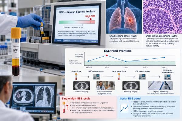
Neuron-specific enolase (NSE) is a blood biomarker that can be elevated in small cell lung cancer (SCLC) and other neuroendocrine conditions. The test measures forms of the enolase enzyme that contain the gamma subunit, which is abundant in neurons and neuroendocrine cells. In a person with confirmed SCLC, serial NSE results can provide supportive information about tumor burden, treatment response, and disease progression, especially when the marker was elevated before treatment. NSE cannot diagnose lung cancer by itself, and it is not recommended as a stand-alone screening test. A high result can also occur because of blood-sample hemolysis, neurologic injury, or other cancers and medical conditions. Laboratories use different assays and reference intervals, so there is no single universal “normal NSE” cutoff for every patient. The most useful interpretation compares the result with the laboratory’s own upper reference limit, specimen quality, imaging, pathology, symptoms, and previous NSE measurements.
- What it measures: NSE testing measures gamma-enolase–containing enzyme forms in serum or plasma and is most often used as a tumor marker in SCLC.
- What a high level means: Elevated NSE can support a high neuroendocrine tumor burden, but it is not specific for SCLC and cannot confirm cancer on its own.
- Normal range: Reference limits are assay-specific; interpret the number against the range printed by the testing laboratory rather than a universal cutoff.
- Important false-high cause: Even mild or visually hidden hemolysis can raise NSE because red blood cells and platelets contain NSE-related enzyme forms.
- Best use in monitoring: Trends are most informative when NSE was elevated at baseline; falling levels may accompany response, while a persistent rise can prompt clinical reassessment.
Table of Contents
- What NSE Is and Why It Is Measured
- NSE in Small Cell Lung Cancer
- How the NSE Blood Test Is Done
- How to Interpret High and Normal NSE
- NSE for Treatment Response and Recurrence Monitoring
- NSE Versus ProGRP and Other SCLC Markers
- Limitations, False Results, and Next Steps
What NSE Is and Why It Is Measured
NSE is an enzyme-related biomarker associated with neurons and neuroendocrine cells. Enolase helps cells process glucose for energy. Several enolase subunits exist, and clinical NSE immunoassays detect forms that contain the gamma subunit, especially gamma-gamma and alpha-gamma dimers.
Small cell lung cancer has neuroendocrine features, so many SCLC tumors release enough NSE for the concentration in blood to rise. This makes serum NSE useful as a supportive tumor marker, particularly after the diagnosis has already been established by tissue or cytology.
NSE is not a lung-specific protein. It is also present in nervous-system tissue, red blood cells, platelets, and other neuroendocrine tumors. For that reason, an abnormal blood result cannot tell a clinician where the signal came from. The diagnosis of SCLC still depends on the clinical picture, imaging, and microscopic examination of tumor cells, often with neuroendocrine IHC markers.
NSE is also different from genomic biomarkers. It does not detect a mutation, fusion, or treatment-resistance gene. Instead, the blood concentration can change as tumor activity changes. That makes it more analogous to a disease-burden marker than to a molecular target.
In practice, clinicians may order NSE with ProGRP testing or other laboratory markers. The value of the panel is contextual: one marker can be normal even when another is elevated, and neither replaces biopsy or imaging.
NSE in Small Cell Lung Cancer
NSE is most useful in SCLC when it is interpreted as one piece of evidence rather than a diagnostic yes-or-no test. SCLC often produces higher NSE concentrations than non-small cell lung cancer, and higher baseline levels tend to occur with greater disease burden or extensive-stage disease. However, sensitivity is incomplete, especially in limited-stage disease.
Older clinical studies established that NSE frequently falls when SCLC responds to chemotherapy and rises again with progression. More recent studies continue to support the value of serial tumor-marker measurements as prognostic and response-related information. In a 2024 prospective study of 232 patients with SCLC, ProGRP, NSE, and CYFRA 21-1 fell rapidly after the first chemotherapy cycle and correlated with radiologic response. A 2026 cohort study also found that dynamic NSE trajectories carried prognostic information in extensive-stage SCLC.
Baseline NSE can also have prognostic associations. A systematic review and meta-analysis of 18 studies involving 2,981 patients found that elevated pretreatment NSE was associated with worse overall survival and progression-free survival. Prognosis for an individual patient, however, cannot be calculated from NSE alone. Stage, performance status, treatment response, organ function, metastasis pattern, and many other factors contribute more directly to clinical decisions.
A small cell lung cancer biomarker panel may therefore include NSE as a monitoring marker while pathology and imaging establish diagnosis and stage. Emerging tissue biomarkers, such as DLL3 expression for particular therapies, answer different questions from serum NSE.
The most important practical distinction is diagnostic use versus monitoring use. A single high NSE result has limited specificity. A reproducible change over time in a patient with proven SCLC and an elevated baseline level can be much more informative.
How the NSE Blood Test Is Done
NSE is usually measured from a blood sample analyzed by an automated immunoassay. The laboratory may use serum or another validated specimen type depending on the platform. Most people do not need special preparation such as fasting unless other blood tests ordered at the same time require it. Follow the specific laboratory or oncology clinic instructions.
The blood-draw technique matters more for NSE than for many common tumor markers because hemolysis is a major source of false elevation. Hemolysis means red blood cells rupture and release their contents into the specimen. Red blood cells and platelets contain enolase forms that can react in NSE assays, so even mild hemolysis can raise the measured concentration.
This interference may be invisible. A tube can look normal to the eye while an automated hemolysis index detects enough free hemoglobin to affect the result. Laboratories therefore commonly evaluate specimen quality and may suppress, flag, repeat, or cautiously interpret an NSE result when hemolysis exceeds the assay’s validated limit.
Other pre-analytic factors can matter, including how long the sample sits before separation, storage temperature, transport, and repeated handling. A 2025 study of serum pre-analytical conditions emphasized that standardized collection and processing reduce false-positive NSE results.
There is no universal reference interval shared by all NSE assays. Different platforms, specimen types, calibration systems, and populations can produce different upper reference limits. Some laboratories use cutoffs in the approximate mid-teens to around 20 ng/mL (numerically equivalent to µg/L), but the correct cutoff is the one printed on the patient’s own report. Comparing a result with a cutoff from another assay can be misleading.
For longitudinal monitoring, consistency helps. If possible, serial NSE measurements should use the same laboratory and assay so that changes are less likely to reflect platform differences rather than biology.
How to Interpret High and Normal NSE
A result should be interpreted in relation to the laboratory’s upper reference limit, specimen quality, prior results, and the reason the test was ordered.
High NSE
A high NSE level means the assay detected more gamma-enolase–containing protein than expected for that laboratory’s reference population. In a patient with confirmed SCLC, a clearly elevated level can be consistent with active disease and may be higher when tumor burden is greater. It is not proof of progression unless the trend and the rest of the clinical evaluation agree.
High NSE can also occur in other neuroendocrine tumors and in some non-neuroendocrine cancers. Neurologic injury can increase NSE because neurons contain the enzyme; this is why NSE is also studied as a biomarker after cardiac arrest and brain injury. Hemolysis is especially important because it can create a false high result without any change in the cancer.
Normal NSE
A normal result does not rule out SCLC. Some patients, particularly those with limited disease or lower tumor burden, do not have an elevated marker at diagnosis. If a person’s NSE was normal before treatment, it is usually less useful as a personal monitoring marker because there is little baseline signal to follow.
Borderline or unexpected NSE
A small isolated increase deserves verification before it is interpreted as cancer growth. The clinician can review whether the sample was hemolyzed, whether the assay or laboratory changed, whether the patient had a recent neurologic event, and whether imaging or symptoms show a matching change.
A practical way to read the report is:
- Below the laboratory upper limit: not elevated, but SCLC can still be present.
- Modestly above the upper limit: nonspecific; repeat testing and clinical context may be important.
- Markedly elevated in known SCLC: may reflect substantial active disease, but confirm the specimen was not hemolyzed and correlate with imaging.
- Rising on serial tests: can raise concern for progression if the pattern is persistent and clinically concordant.
- Falling after treatment: can support response when the marker was elevated before therapy.
The number should never override stronger evidence. A CT scan showing clear progression is not dismissed because NSE remains normal, and an isolated high NSE does not establish relapse when imaging and the clinical picture are stable.
NSE for Treatment Response and Recurrence Monitoring
Serial NSE can be useful for monitoring SCLC, but it should complement imaging and clinical assessment rather than replace them. The marker is most informative when three conditions are met: SCLC is already confirmed, pretreatment NSE was elevated, and repeat samples are collected consistently without major hemolysis.
During effective systemic therapy, NSE often decreases. A rapid fall can support other evidence that tumor burden is shrinking. If the concentration does not fall as expected, remains persistently high, or begins to rise after an initial decrease, clinicians may look more closely for resistant or progressive disease. The timing of imaging and treatment decisions is still based on the full clinical plan, not on a marker threshold alone.
The direction and persistence of change matter more than a tiny one-time difference. For example, a sequence of 72, 38, and 18 ng/mL during treatment is qualitatively different from 18, 20, and 19 ng/mL around an assay’s reference boundary. The first pattern shows a large sustained decrease; the second may reflect ordinary analytic or biologic variation. Exact interpretation depends on the assay and clinical setting.
After a response, a sustained rise can sometimes precede radiographic or symptomatic progression. Historical studies observed NSE increases before clinically recognized relapse in some patients, but the lead time is inconsistent and false rises occur. Modern follow-up therefore does not substitute NSE testing for scheduled imaging.
Recent prospective evidence also supports looking at multiple markers over time. In patients with chemotherapy-induced remission, serial ProGRP, NSE, and CYFRA 21-1 carried prognostic information at several time points. In 2026 data from extensive-stage SCLC, persistent or rising marker trajectories separated higher-risk groups from patients with low, stable markers.
This is a useful way to think about NSE: it is a trend marker with clinical context, not a stand-alone recurrence detector. Any unexpected rise should trigger a review of specimen integrity and the broader disease assessment before it changes treatment.
NSE Versus ProGRP and Other SCLC Markers
NSE and ProGRP are commonly discussed together because both can be elevated in SCLC, but they measure different biology and have different strengths.
| Feature | NSE | ProGRP |
|---|---|---|
| What it reflects | Gamma-enolase–containing enzyme from neuroendocrine/neuronal and blood cells | Stable precursor fragment related to gastrin-releasing peptide biology |
| Common role | Supportive marker for burden, prognosis, and treatment trends | Supportive SCLC/neuroendocrine marker, often useful diagnostically |
| Major laboratory pitfall | Hemolysis can cause false elevation | Kidney dysfunction can influence concentration and interpretation |
| Can diagnose SCLC alone? | No | No |
Classic comparative studies found ProGRP more sensitive for SCLC diagnosis in some cohorts, while NSE had strong prognostic associations. More recent work supports using the markers as complementary rather than interchangeable. If one is normal at baseline, the other may still be elevated and useful for follow-up.
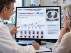
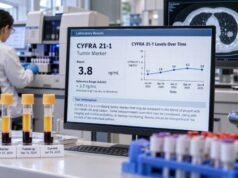
Other serum markers such as CYFRA 21-1 and CEA may also be measured in some centers, particularly when clinicians want broader prognostic information or the tumor has mixed features. None is specific enough to establish SCLC without tissue confirmation.
Pathology markers answer a different question. IHC for synaptophysin, chromogranin, INSM1, or other neuroendocrine markers helps classify tumor cells in the biopsy. Serum NSE cannot show tumor morphology or distinguish classic SCLC from every other neuroendocrine neoplasm.
Likewise, molecular testing is not replaced by NSE. If treatment decisions require genomic information, that information must come from an appropriate tissue or liquid-biopsy assay rather than a serum protein concentration.
Limitations, False Results, and Next Steps
The biggest limitation of NSE is lack of specificity. A high value can come from SCLC, another neuroendocrine cancer, neurologic tissue injury, or sample hemolysis. The test is therefore poorly suited to population screening or to diagnosing a lung mass by itself.
Hemolysis deserves special attention because it can produce a result that looks biologically meaningful. Studies have shown that even low-grade hemolysis increases measured NSE. Some laboratories use an automated hemolysis index, reject affected specimens, or apply validated correction approaches. Correction formulas are assay- and laboratory-dependent and should not be improvised by patients or clinicians from a published equation.
Other limitations include biological variability and assay differences. If serial results come from different laboratories, a modest change may reflect calibration rather than cancer behavior. A value close to the upper reference limit is also less persuasive than a large, reproducible change that tracks imaging and symptoms.
When an NSE result is unexpectedly high, reasonable next steps include:
- Check the laboratory comment and hemolysis index. A flagged specimen may need recollection.
- Compare with the patient’s baseline and previous values. The trend matters more than one isolated number.
- Review the clinical context. Recent neurologic injury, another neuroendocrine tumor, or other causes may change interpretation.
- Correlate with imaging and symptoms. Tumor-marker changes should fit the overall disease picture before treatment is changed.
- Consider complementary markers. ProGRP or other tests may add information when clinically appropriate.
- Use pathology for diagnosis. A suspicious lung lesion still requires the appropriate diagnostic workup; NSE cannot determine histologic subtype on its own.
For patients already being treated for SCLC, the most useful question is usually not “Is my NSE high?” but “How has my NSE changed compared with my pretreatment value, and does that change match the scans and my clinical course?” That framing avoids overreacting to assay noise and keeps the marker in its proper role as supportive evidence.
References
- Association between dynamic trajectories of serum tumor markers and survival outcomes in patients with extensive-stage small cell lung cancer: a retrospective cohort study 2026
- Improving the screening ability of neuron-specific enolase on small cell lung cancer 2025
- Neuron-Specific Enolase-What Are We Measuring? 2024 (Review)
- Managing hemolysis in serum neuron-specific enolase measurements – an automated algorithm for routine practice. 2024
- Prognostic value of tumor markers ProGRP, NSE and CYFRA 21-1 in patients with small cell lung cancer and chemotherapy-induced remission 2024
- Prognostic value of neuron-specific enolase for small cell lung cancer: a systematic review and meta-analysis 2020 (Systematic Review and Meta-analysis)
Disclaimer
This article is for general education and does not replace diagnosis or monitoring by an oncology team. NSE reference limits and hemolysis rules differ by laboratory, and a high or rising result should be interpreted with specimen quality, pathology, imaging, symptoms, and other clinical findings. Do not change cancer treatment based on an NSE value alone.