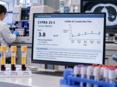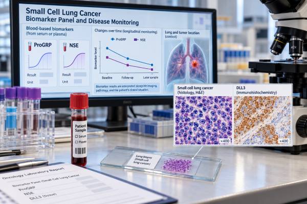
A small cell lung cancer (SCLC) biomarker panel may combine blood markers such as progastrin-releasing peptide (ProGRP) and neuron-specific enolase (NSE) with tumor markers such as delta-like ligand 3 (DLL3). These markers answer different questions. ProGRP and NSE can support the clinical picture and, when elevated at baseline, may help track change during treatment. DLL3 is a protein found on the surface of many SCLC cells and has become important because it can be targeted by newer therapies. None of these markers can diagnose SCLC by itself, and there is no universal three-marker score that replaces biopsy, pathology, imaging, or clinical assessment. The most useful interpretation comes from knowing which specimen was tested, which assay was used, whether the marker was abnormal before treatment, and how the result changes over time. Kidney function, sample hemolysis, tumor heterogeneity, and treatment-related biological changes can all affect interpretation.
- ProGRP: A blood marker often associated with SCLC; serial changes can be useful when the level is elevated before treatment, but kidney dysfunction can raise it.
- NSE: A blood marker of neuroendocrine cell injury and tumor activity; hemolysis can falsely increase the result, so sample quality matters.
- DLL3: A tumor-cell surface protein expressed in many SCLCs and a therapeutic target; it is not a routine blood marker for tracking tumor burden.
- No single cutoff diagnoses SCLC: Results must be interpreted with tissue pathology, imaging, symptoms, stage, and the laboratory’s assay-specific reference range.
- For monitoring, trends matter more than one value: Falling or rising ProGRP or NSE can add useful information, but treatment decisions should not be based on marker movement alone.
Table of Contents
- What the SCLC biomarker panel measures
- ProGRP in small cell lung cancer
- NSE in small cell lung cancer
- DLL3 expression and what it means
- How biomarkers are used for disease monitoring
- Limits and common interpretation problems
- Questions to ask about SCLC biomarker results
What the SCLC biomarker panel measures
A ProGRP, NSE, and DLL3 “panel” is best understood as a group of complementary biomarkers, not as one standardized laboratory test. ProGRP and NSE are generally measured in blood. DLL3 is usually assessed in tumor tissue or studied through other specialized methods because it is a cell-surface protein. The three markers therefore describe different parts of SCLC biology.
ProGRP is the stable precursor of gastrin-releasing peptide. SCLC cells often show neuroendocrine differentiation and can release ProGRP-related products. A high ProGRP concentration can support suspicion for SCLC in the right setting and may provide a measurable baseline for later follow-up.
NSE is an enzyme found in neurons and neuroendocrine cells. Blood NSE can rise when neuroendocrine tumor cells are abundant or damaged. It can also rise for reasons unrelated to SCLC, particularly if red blood cells break open in the sample.
DLL3 is different. It is an inhibitory ligand in the Notch signaling pathway and is abnormally displayed on the surface of many SCLC cells while having limited expression on most normal cell surfaces. That pattern made DLL3 attractive as a drug target. Modern SCLC research also places DLL3 within a broader biology that includes transcriptional subtypes and changing surface-antigen expression.
These markers should not be confused with the stains used to establish the tissue diagnosis. Pathologists commonly use tumor shape and growth pattern together with neuroendocrine immunohistochemical markers. A blood result cannot replace that process. Likewise, a ProGRP test for lung cancer or an NSE test for lung cancer is most useful when interpreted in the context of a confirmed or strongly suspected diagnosis.
There is also no broadly accepted formula that combines the three results into “low-risk,” “medium-risk,” and “high-risk” panel categories. A report should be read marker by marker, with attention to specimen type and assay method.
ProGRP in small cell lung cancer
ProGRP is one of the more SCLC-associated serum or plasma biomarkers in clinical use. It tends to have greater specificity for SCLC than NSE, although neither marker is accurate enough to establish a diagnosis alone. A markedly elevated result can strengthen the case for SCLC when imaging and pathology findings point in the same direction, but it can also create a reason to recheck the diagnosis when the clinical picture does not fit.
Laboratories use different analyzers, specimen types, and reference intervals, so there is no single universal “normal” cutoff to apply to every ProGRP report. The value should be compared with the reference range printed by the laboratory that performed the test. For follow-up, using the same assay and preferably the same laboratory reduces unnecessary variation.
Why kidney function matters
ProGRP is affected by renal clearance. Reduced kidney function can raise the blood concentration even when tumor burden has not increased. This is one of the most important reasons not to interpret an isolated high value as direct evidence of progression. A clinician may review creatinine, estimated glomerular filtration rate, recent dehydration, and the previous ProGRP pattern before deciding what a change means.
What a falling or rising ProGRP can suggest
If ProGRP is clearly elevated before treatment and then falls during effective chemotherapy or chemoradiotherapy, the direction of change can support evidence that tumor activity is decreasing. A later sustained rise may raise concern for recurrence or progression, especially if it is accompanied by new symptoms or imaging findings.
The reverse is also important: some people with SCLC never have a clearly elevated ProGRP. In that situation, repeatedly measuring a marker that was normal at baseline may add little. Monitoring is usually more informative when the patient has a measurable pretreatment abnormality that tracks with the disease.
A single increase should rarely trigger a major treatment decision without confirmation. Biological variation, kidney function, assay variation, and the timing of the sample can all matter.
NSE in small cell lung cancer
NSE is another long-used blood biomarker for SCLC and other neuroendocrine tumors. It reflects neuroendocrine biology less specifically than ProGRP. An elevated NSE can occur with SCLC, but it can also appear in other cancers, neurological injury, and conditions that damage blood cells. Its main practical strength is often serial measurement in a patient whose NSE was elevated when active SCLC was documented.
The laboratory reference interval again matters more than a generic cutoff found online. Different assays are not perfectly interchangeable. When possible, compare results produced by the same method over time.
Hemolysis is a major source of false elevation
Red blood cells contain enolase. If they rupture during or after the blood draw, they can release intracellular material and raise measured NSE. This process is called hemolysis. A difficult blood draw, rough specimen handling, delayed processing, or other pre-analytic problems can contribute.
That means an unexpected NSE spike deserves a basic quality check before it is interpreted as cancer growth. The laboratory may flag a hemolyzed specimen. Depending on the degree of interference and the clinical setting, repeating the blood draw can be more informative than reacting to the number.
Using NSE as a trend marker
When baseline NSE is high, a substantial decline during treatment can accompany response. Persistent elevation or a renewed rise can occur with active disease. However, the marker is not a substitute for response assessment by imaging. SCLC can progress in a way that is not mirrored cleanly by the blood value, and NSE can fluctuate for non-cancer reasons.
ProGRP and NSE can sometimes provide complementary information because they are affected by different confounders. For example, kidney dysfunction may complicate ProGRP interpretation while hemolysis may complicate NSE. Looking at both trends can be helpful, but two abnormal blood markers still do not equal a tissue diagnosis.
DLL3 expression and what it means
DLL3 is a tumor-associated surface protein that has moved from a research biomarker to a clinically important therapeutic target. Studies commonly report DLL3 expression in a large majority of SCLC tumors, often roughly 70% to 80%, although the exact percentage depends on the assay, threshold, tumor sample, and patient population.
Unlike ProGRP and NSE, DLL3 is not normally followed as a routine serum concentration. It is generally studied on tumor cells, most often with tissue-based methods in research or clinical development settings. The result may be reported as positive or negative, as a percentage of tumor cells showing staining, or with another assay-specific scoring system.
Why DLL3 matters clinically
DLL3 is located on the surface of many SCLC cells, allowing therapies to bind the cancer cell while engaging another killing mechanism. Tarlatamab, for example, is a bispecific T-cell engager that connects DLL3 on tumor cells with CD3 on T cells. This approach established DLL3 as more than a descriptive marker: it is a validated therapeutic target in SCLC.
A DLL3 result should not automatically be treated like an EGFR mutation or another classic driver alteration. The treatment relevance depends on the specific drug, regulatory indication, local practice, and whether a companion diagnostic is required. A patient should not assume that a certain staining percentage is necessary for a DLL3-directed treatment unless the current treatment label or clinical-trial protocol says so.
SCLC biology also changes under treatment pressure. Molecular studies describe major SCLC states associated with transcription factors such as ASCL1, NEUROD1, and POU2F3, plus an inflamed phenotype. DLL3 expression is often strongest in neuroendocrine-rich states, but tumors can contain more than one state and can shift over time. That heterogeneity helps explain why one tissue sample may not capture every cancer cell.
For broader context, a lung cancer biomarker panel used in non-small cell lung cancer answers a different molecular question. SCLC usually does not follow the same driver-mutation testing pathway as lung adenocarcinoma.
How biomarkers are used for disease monitoring
Disease monitoring in SCLC is built around the patient’s symptoms, physical findings, laboratory data, and imaging. Biomarkers can add another signal, but they should support that framework rather than replace it.
A practical monitoring approach starts before treatment. If ProGRP and NSE are going to be followed, the most useful baseline sample is obtained when active disease is known to be present. The clinician can then compare later values with that starting point. The exact testing schedule varies with stage, treatment, symptoms, and local practice; there is no universal rule that every patient needs both markers at every treatment cycle.
A useful way to read serial results is:
- Confirm the baseline. Was ProGRP, NSE, or both clearly above the laboratory reference range before therapy?
- Check confounders. Review kidney function for ProGRP and specimen hemolysis for NSE.
- Look for a sustained direction. Several results moving in the same direction are usually more meaningful than one small fluctuation.
- Compare with the clinical picture. Marker decline is more convincing when symptoms and imaging also improve.
- Investigate discordance. A rising marker with stable imaging, or worsening scans with stable markers, should prompt clinical review rather than automatic conclusions.
For example, suppose ProGRP is markedly elevated at diagnosis, falls steadily after treatment begins, and then remains low while imaging shows tumor shrinkage. The concordant pattern can provide additional confidence that the marker is useful for that patient. Months later, if ProGRP rises repeatedly, the change may justify closer evaluation, but imaging is still needed to establish whether disease has returned or progressed.
DLL3 is not usually used this way. A tissue DLL3 result describes target expression at a particular place and time. Repeated biopsies solely to track DLL3 are not routine for most patients. Research is exploring blood-based and imaging approaches for dynamic target assessment, but these are not the same as standard ProGRP or NSE monitoring.
If clinicians need to look for broader molecular changes in a lung cancer, lung cancer liquid biopsy uses circulating tumor DNA and answers a different question from protein tumor markers.
Limits and common interpretation problems
The biggest mistake is treating a biomarker result as if it were a stand-alone diagnosis. SCLC is diagnosed through pathology in the proper clinical setting. Biomarkers can support, refine, or monitor that diagnosis, but they do not override tissue findings by themselves.
Other common interpretation problems include:
- Using a universal cutoff from another laboratory. ProGRP and NSE assays can differ, so the report’s own reference interval and method matter.
- Ignoring renal function. A ProGRP rise may reflect reduced clearance rather than a larger tumor burden.
- Ignoring hemolysis. NSE is especially vulnerable to false elevation from a damaged blood sample.
- Comparing different assay platforms as though they are identical. Small method-related shifts can look like biological change.
- Expecting every SCLC to release blood markers. Some tumors do not produce a useful baseline elevation.
- Calling DLL3 a blood tumor marker. DLL3 is primarily a cell-surface target assessed on tumor tissue or by specialized investigational methods.
- Assuming DLL3 expression is uniform. Intratumoral heterogeneity and phenotype changes can create differences between samples or over time.
- Using markers to replace imaging. A reassuring blood level cannot rule out progression, and a high value cannot localize disease.
Another important distinction is between prognostic and predictive information. A prognostic biomarker is associated with how the disease may behave overall. A predictive biomarker helps identify the chance of benefit from a particular treatment. ProGRP and NSE have been studied for prognosis and monitoring, but they are not established stand-alone treatment-selection tests. DLL3 is a therapeutic target, yet the need for formal DLL3 testing depends on the specific therapy and current clinical requirements.
Finally, SCLC itself is biologically diverse. Newer research on molecular subtypes, circulating tumor cells, immune markers, and surface antigens may eventually produce more sophisticated panels. At present, however, these emerging tools should not be mixed with established routine testing without explaining which results are standard care and which remain investigational.
Questions to ask about SCLC biomarker results
The most useful result is one that can be connected to a clear clinical question. Patients and caregivers can ask the oncology team what the test was meant to answer and what action, if any, the result changes.
For ProGRP or NSE, useful questions include:
- Was this marker elevated before treatment, making it suitable for serial monitoring?
- What is this laboratory’s reference range, and has the same assay been used each time?
- Could kidney function explain the ProGRP value?
- Was the NSE specimen hemolyzed or otherwise flagged by the laboratory?
- Is the change large and sustained enough to be meaningful, or could it be normal analytical variation?
- Do the marker trend, symptoms, and imaging tell the same story?
For DLL3, ask what specimen was tested, what method and scoring system were used, and whether the result is required for a specific treatment decision. If a report gives a percentage or staining category, the relevant threshold should come from the validated assay or treatment protocol, not from a general internet range.
It is also reasonable to ask whether another biopsy is needed when the disease behaves differently from expected. In selected situations, repeat tissue can clarify histology or biological change, but the decision depends on safety, accessibility, and whether the result would alter care.
The core principle is simple: treat the biomarker as one piece of evidence. ProGRP and NSE can be useful longitudinal signals when they start high and are measured consistently. DLL3 provides information about an important SCLC surface target. Pathology confirms what the tumor is, imaging shows where it is and how it changes, and the oncology team integrates all of those data before changing treatment.
References
- Decoding Small Cell Lung Cancer: Molecular Subtypes, Surface Antigens, and the Target-Modality Problem 2026 (Review)
- Associations between progastrin-releasing peptide (ProGRP) and neuron-specific enolase (NSE) and survival in patients with limited-stage small cell lung cancer (LS SCLC) receiving chemoradiotherapy (CRT) 2025
- Harnessing delta-like ligand 3: bridging biomarker discovery to next-generation immunotherapies in refractory small cell lung cancer 2025 (Review)
- Taking a Bite Out of Small Cell Lung Cancer By Leveraging Precision-Directed Delta-Like Ligand-3 Therapies 2025 (Review)
- DLL3-guided therapies in small-cell lung cancer: from antibody-drug conjugate to precision immunotherapy and radioimmunotherapy 2024 (Review)
- Evaluating the diagnostic and prognostic value of serum TuM2-PK, NSE, and ProGRP in small cell lung cancer 2023
Disclaimer
SCLC biomarker results cannot diagnose, stage, or monitor lung cancer reliably without the rest of the clinical evaluation. ProGRP and NSE can be affected by non-cancer factors, and DLL3 testing requirements depend on the specific treatment and assay. Decisions about biopsy, imaging, treatment, or suspected progression should be made with the oncology team using the complete clinical record.