The PD-L1 test for lung cancer is an immunohistochemistry test that measures programmed death-ligand 1 protein on tumor cells and helps guide the use of immune checkpoint inhibitors in non-small cell lung cancer. The most common lung cancer score is the tumor proportion score (TPS), reported from 0% to 100% as the percentage of viable tumor cells showing qualifying membrane staining. Common interpretation bands are less than 1%, 1%–49%, and 50% or higher, but the treatment meaning depends on the exact drug, disease stage, assay, and clinical setting. PD-L1 is an imperfect predictive biomarker: a high score increases the likelihood of benefit from some immunotherapy approaches but does not guarantee response, while a low or negative score does not mean immunotherapy can never work. Molecular driver results, tumor histology, stage, prior therapy, and patient health must be considered alongside the PD-L1 report.
- What TPS measures: TPS is the percentage of viable tumor cells with partial or complete PD-L1 membrane staining, expressed from 0% to 100%.
- Common result groups: TPS below 1% is often called negative, 1%–49% intermediate or positive, and 50% or higher high expression, although treatment thresholds are indication-specific.
- What a high score means: High PD-L1 can support immunotherapy selection in some NSCLC settings, but it is not a guarantee that treatment will work.
- What a low score means: Low or absent PD-L1 does not rule out benefit from checkpoint inhibitors, especially when they are used in combination regimens.
- Why assay details matter: Different validated PD-L1 antibody assays and scoring systems are linked to specific clinical evidence, so the clone, platform, specimen quality, and scoring method should be reviewed.
Table of Contents
- What PD-L1 Is and Why It Is Tested
- How the TPS Score Is Calculated
- What PD-L1 Results Mean
- How the PD-L1 Test Is Performed
- Limits and Sources of Variation
- PD-L1 and Other Lung Cancer Biomarkers
- Practical Next Steps After a PD-L1 Result
What PD-L1 Is and Why It Is Tested
PD-L1 is a protein that can be expressed on the surface of cancer cells and some immune cells. Its normal biologic role is to help regulate immune responses. When PD-L1 binds the PD-1 receptor on activated T cells, it can send an inhibitory signal that reduces immune activity.
Some tumors use this pathway to avoid immune attack. Immune checkpoint inhibitors can block PD-1 or PD-L1 and restore part of the immune response against cancer. Because tumors with higher PD-L1 expression are more likely to benefit from certain checkpoint-inhibitor strategies, PD-L1 immunohistochemistry became a clinically important predictive test in NSCLC.
The test is predictive, not diagnostic. PD-L1 does not establish that a lung mass is cancer and does not determine whether the tumor is adenocarcinoma or squamous cell carcinoma. Pathology establishes the diagnosis first; PD-L1 helps with treatment planning after the cancer type and clinical setting are known.
PD-L1 is also not a gene mutation. The assay detects protein expression in tissue rather than sequencing DNA. That distinction matters because lung cancer treatment planning often combines PD-L1 with molecular profiling. A lung cancer biomarker panel may identify actionable driver alterations, while PD-L1 provides separate information about the tumor-immune interface.
The amount of PD-L1 can vary among different areas of the same tumor and between a primary tumor and metastases. It can also change over time or after treatment. The result is therefore a snapshot from a particular specimen, not a permanent property of every cancer cell.
This biologic variability helps explain why two specimens from the same patient can produce different percentages without either laboratory being wrong. A small bronchoscopic biopsy may capture one local immune environment, while a later metastatic biopsy samples another. When the difference could materially affect treatment, clinicians may ask the pathologist whether repeat testing on a newer or larger specimen is reasonable. The decision should be purposeful, because repeating PD-L1 on every available block can use tissue needed for more informative molecular studies and may not resolve tumor heterogeneity completely.
How the TPS Score Is Calculated
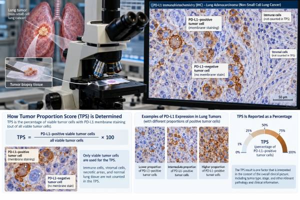
For many NSCLC indications, PD-L1 is reported as the tumor proportion score, or TPS. The pathologist estimates the percentage of viable tumor cells that show qualifying partial or complete membrane staining at any intensity.
The simplified formula is:
TPS = PD-L1-positive viable tumor cells ÷ all viable tumor cells × 100
If about 60 of every 100 viable tumor cells show appropriate membrane staining, the TPS is approximately 60%. If staining is seen in fewer than 1 of every 100 tumor cells, the report may be TPS less than 1%.
Only tumor-cell membrane staining counts for TPS. Cytoplasmic staining alone does not count. Immune cells may show PD-L1 staining, but they are not included in the TPS numerator. Some other tumor types and some lung cancer assay frameworks use different scoring systems that include immune cells, so it is important not to apply a CPS or immune-cell score as though it were a TPS.
Common TPS bands are:
| TPS result | Common description | General meaning |
|---|---|---|
| <1% | Negative or very low expression | Little or no qualifying tumor-cell PD-L1 staining; does not exclude immunotherapy benefit in all regimens |
| 1%–49% | Positive/intermediate expression | PD-L1 is present, but whether it changes therapy depends on the specific indication and treatment strategy |
| ≥50% | High expression | Can support checkpoint-inhibitor monotherapy in some advanced NSCLC settings when other clinical requirements are met |
These bands are useful for orientation but should not be treated as universal treatment rules. Regulatory indications and clinical guidelines can use different cutoffs, assays, stages, and combination strategies. A TPS of 50% is not inherently “good,” and 0% is not inherently “bad.” The number predicts probability of benefit in specific treatment contexts rather than measuring how aggressive the cancer is.
What PD-L1 Results Mean
A high PD-L1 TPS means a larger proportion of the sampled tumor cells express PD-L1 on their membranes. In advanced NSCLC without a more dominant treatment-defining molecular driver, high expression can make single-agent checkpoint inhibition an option in some settings. It can also remain relevant when immunotherapy is used with chemotherapy.
However, PD-L1 is not a yes-or-no response test. Some patients with TPS of 50% or higher do not respond to immunotherapy, while some patients with TPS below 1% respond—particularly when checkpoint inhibitors are combined with chemotherapy or other agents.
Several reasons explain this imperfect prediction. The immune response depends on more than PD-L1. Tumor genetics, antigen presentation, T-cell infiltration, co-mutations, prior treatments, steroid exposure, organ function, microbiome influences, and other factors can all affect outcome.
A low or negative TPS therefore means low measured expression in that specimen, not “immunotherapy cannot work.” The oncology team interprets the result within the evidence for the proposed regimen.
Likewise, a very high TPS does not override an actionable molecular driver. A patient with an EGFR mutation, ALK fusion, ROS1 fusion, or another targetable alteration may have a treatment sequence centered on targeted therapy even if PD-L1 is high. That is why molecular results should be available whenever possible before committing to first-line systemic therapy in advanced NSCLC.
PD-L1 also does not measure tumor burden. A TPS of 80% does not mean 80% of the body is cancer, and it does not correspond to stage IV disease. A small early-stage tumor can have high PD-L1, while an extensive metastatic tumor can have low expression.
How the PD-L1 Test Is Performed
PD-L1 testing is performed on a tumor specimen, usually formalin-fixed and paraffin-embedded tissue from a biopsy or surgical resection. Validated cytology cell blocks can also be used in many laboratories when they contain enough viable tumor cells.
The laboratory cuts a thin tissue section and performs immunohistochemistry with a validated PD-L1 antibody assay. Common antibody clones used in lung cancer include 22C3, 28-8, SP263, and SP142. These assays were developed on different platforms and with different clinical trial programs.
Studies have shown relatively strong tumor-cell agreement among 22C3, 28-8, and SP263 in many settings, while SP142 tends to stain fewer tumor cells and uses distinct scoring approaches for some indications. Because treatment evidence and regulatory approvals are assay-linked, laboratories should use appropriately validated methods rather than assuming all clones are freely interchangeable.
The pathologist first confirms that there is enough viable tumor to score. Necrotic tissue, crushed cells, poor fixation, scant tumor, or old unstained sections can reduce reliability. The pathologist then estimates the percentage of viable tumor cells with qualifying membrane staining.
No fasting or blood preparation is needed because PD-L1 is not a blood test. The patient’s practical preparation is the same as for the biopsy or procedure used to obtain tissue. If tissue already exists from a prior biopsy, PD-L1 can often be performed on the stored block without another procedure, provided sufficient suitable material remains.
For small samples, tissue conservation is important. The same specimen may need diagnostic IHC, PD-L1, DNA sequencing, and RNA fusion testing. Coordinated pathology workflows reduce the risk of exhausting the block before all necessary biomarkers are completed.
Limits and Sources of Variation
PD-L1 is a useful biomarker, but it is biologically and technically variable. The 2024 multidisciplinary lung cancer guideline from the College of American Pathologists and partner organizations emphasizes use of validated assays and careful attention to the clinical indication.
The major limitations include:
- Tumor heterogeneity: One biopsy can sample a high-expression area while another part of the tumor has lower expression.
- Primary-versus-metastatic differences: PD-L1 can differ between tumor sites.
- Time and treatment effects: Chemotherapy, radiation, targeted therapy, and evolving tumor biology can change expression.
- Specimen age and handling: Poor fixation, old blocks, or stored unstained slides can reduce antigen detection in some settings.
- Small samples: A tiny biopsy may contain too few viable tumor cells to represent the whole tumor reliably.
- Assay differences: Antibody clones, staining platforms, and scoring algorithms are not identical.
- Observer variation: Borderline cases near a cutoff can be scored differently by experienced pathologists.
These limitations are most important near treatment thresholds. A TPS of 48% and a TPS of 52% may not represent a meaningful biologic difference, even though a cutoff at 50% can place the results into different regulatory categories. Clinicians should interpret borderline values with the complete clinical context rather than treating the cutoff as a natural biologic boundary.
Cytology specimens can provide useful PD-L1 results when properly prepared and validated. A 2023 systematic review and meta-analysis found good overall diagnostic performance compared with matched histology, but adequacy and laboratory validation still matter.
Repeat testing can be reasonable when the original specimen was inadequate, when a new biopsy represents a later disease state, or when a prior result is clinically discordant with the current treatment question. Repeat testing is not automatically required every time the disease is reassessed.
PD-L1 and Other Lung Cancer Biomarkers
PD-L1 answers a different question from driver-gene testing. It estimates expression of an immune checkpoint ligand; molecular tests identify DNA or RNA alterations that can directly drive tumor growth.
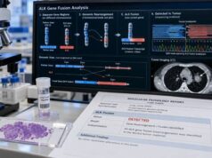
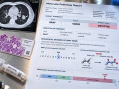
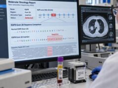
For example, the EGFR mutation test can identify sensitizing EGFR alterations with established targeted-therapy implications. ALK, ROS1, RET, and NTRK fusion tests look for kinase rearrangements. MET exon 14 skipping, BRAF V600E, KRAS G12C, and HER2 alterations define other molecular subgroups.
A high PD-L1 result should therefore be read beside, not instead of, the molecular profile. This is especially important in metastatic non-squamous NSCLC, where an actionable driver can change first-line treatment even when TPS is high.
Tumor mutational burden is another immune-related biomarker, but it is not the same as PD-L1. The TMB test in lung cancer estimates the number of somatic mutations per megabase of sequenced DNA. PD-L1 measures protein expression by IHC. The two biomarkers correlate imperfectly and should not be substituted for each other.
Histologic markers such as TTF-1 and p40 also serve a separate role. They help classify the tumor as adenocarcinoma, squamous cell carcinoma, or another type. A tumor can be TTF-1 positive and have any PD-L1 TPS; one result does not predict the other with enough accuracy to replace testing.
This layered approach—diagnosis, histologic classification, molecular profiling, and immune-biomarker testing—explains why a modern lung cancer pathology report can contain several very different kinds of “positive” or “negative” results.
Practical Next Steps After a PD-L1 Result
When reviewing a PD-L1 report, first confirm the scoring system. In NSCLC, look for a line such as PD-L1 TPS: 70% rather than assuming that any percentage on the report is TPS.
Then review five practical points:
- Which assay was used? The report may list 22C3, 28-8, SP263, SP142, or another validated method.
- Was the specimen adequate? Look for comments about limited tumor, necrosis, decalcification, or technical limitations.
- What is the exact TPS? The actual percentage is more informative than a vague “positive” label.
- Are molecular driver results complete? Treatment decisions in advanced NSCLC often require both PD-L1 and genomic information.
- Which treatment setting applies? Stage, prior therapy, histology, performance status, autoimmune disease, organ transplantation, and other clinical factors can change the significance of the score.
If the TPS is below 1%, the discussion does not end with “no immunotherapy.” Combination regimens may still be appropriate depending on the cancer and treatment setting. If TPS is 50% or higher, the discussion does not end with “immunotherapy alone.” A targetable driver, urgent disease burden, symptoms, and other factors may point to another strategy.
Patients should also know that PD-L1 does not predict immune-related side effects. A high TPS does not mean a person is more likely to develop thyroiditis, colitis, pneumonitis, hepatitis, or other checkpoint-inhibitor toxicities. Risk assessment for those complications is separate.
The most useful interpretation is therefore treatment-specific: What does this TPS mean for the therapies being considered for this exact cancer today? That question keeps the number connected to its true purpose. PD-L1 is valuable because it changes probabilities and eligibility in defined settings—not because it supplies a universal answer about whether immunotherapy will succeed.
References
- Programmed Death Ligand-1 and Tumor Mutation Burden Testing of Patients With Lung Cancer for Selection of Immune Checkpoint Inhibitor Therapies: Guideline From the College of American Pathologists, Association for Molecular Pathology, International Association for the Study of Lung Cancer, Pulmonary Pathology Society, and LUNGevity Foundation 2024 (Guideline)
- Pathways to Precision: Guideline for Programmed Death Ligand-1 and Tumor Mutation Burden Testing to Support the Selection of Immune Checkpoint Therapies in Lung Cancer 2024 (Practice Guideline)
- Improving practice in PD-L1 testing of non-small cell lung cancer in the UK: current problems and potential solutions 2024
- Accuracy of Cytologic vs Histologic Specimens for Assessment of Programmed Cell Death Ligand-1 Expression in Non-Small Cell Lung Cancer: A Systematic Review and Meta-Analysis 2023 (Systematic Review)
- PD-L1 immunohistochemistry: Clones, cutoffs, and controversies 2022 (Review)
- Atlas of PD-L1 for Pathologists: Indications, Scores, Diagnostic Platforms and Reporting Systems 2022 (Review)
Disclaimer
PD-L1 is a predictive biomarker, not a stand-alone diagnosis or guarantee of immunotherapy response. The TPS must be interpreted with the assay used, specimen quality, lung cancer stage and histology, molecular driver results, treatment history, and current clinical guidance. Do not start, stop, or change immunotherapy based on a PD-L1 percentage without oncology review.