Sleep apnea is more than loud snoring. When breathing stalls during sleep, oxygen dips and the body surges with stress signals. Over months and years this pattern can strain blood vessels, elevate blood pressure, fragment deep sleep, and erode attention, memory, and mood. The good news: diagnosing sleep apnea is far simpler than it used to be, and treatments have become quieter, more comfortable, and easier to personalize. In this guide, you will learn the key signs, who is at risk, what to expect from home sleep tests versus lab studies, how to read your report, and how to choose and adapt therapy so it actually works. For a broader view on how sleep fits into recovery and healthy aging, see our condensed primer on sleep, stress, and recovery strategies. Use this article as a practical map—from suspicion to diagnosis to confident, sustainable treatment.
Table of Contents
- Common Signs: Snoring, Pauses, Morning Headaches, and Fatigue
- Risk Factors in Midlife and Older Adults
- Home Sleep Test Versus Lab Study: What to Expect
- AHI and Oxygen Desaturation: Understanding Your Report
- Treatment Options: CPAP, Oral Appliances, and Positional Therapy
- Adherence Tips, Mask Fit, and Dryness Solutions
- When to See ENT or Consider Further Evaluation
Common Signs: Snoring, Pauses, Morning Headaches, and Fatigue
Obstructive sleep apnea (OSA) occurs when the upper airway narrows or collapses during sleep. Airflow drops, oxygen falls, and the brain briefly wakes you to reopen the airway. These micro-arousals may be too short to remember, yet they fragment sleep architecture and raise nighttime blood pressure and heart rate. The pattern repeats in cycles across the night.
Typical clues show up in three places—at night, on waking, and through the day.
Nighttime clues
- Loud, habitual snoring that worsens in the supine (back-sleeping) position.
- Witnessed pauses, gasps, or choking reported by a bed partner; some people startle themselves awake.
- Restless sleep with frequent awakenings, dry mouth, or nocturia (nighttime urination).
- Reflux flare-ups in the early morning hours, often overlooked but common with airway instability.
Morning clues
- Non-refreshing sleep even after 7–8 hours in bed.
- Morning headaches, often dull and bilateral, that ease within a few hours.
- Sore throat or dryness, a sign of mouth breathing.
- Brain fog on waking; some people feel “hungover” without alcohol.
Daytime clues
- Sleepiness in passive situations: reading, TV, meetings, as a passenger in a car.
- Irritability or low mood and reduced stress tolerance.
- Attention lapses and memory drift, especially for names and recent details.
- Performance drops in tasks that require sustained focus or reaction time.
Not everyone snores loudly, and women often present differently—insomnia, fatigue, and morning headaches may be more prominent than obvious apneas. In older adults, the signal can be subtle: falls in attention, worse balance, or worsening blood pressure despite treatment.
If two or more clusters resonate—nighttime symptoms plus daytime sleepiness or resistant hypertension—screening is reasonable. A validated questionnaire (such as STOP-BANG) can estimate risk in clinic, but testing is what confirms or rules out OSA. The decision to test should not hinge on weight alone; airway anatomy, hormones, and arousal threshold all matter.
Finally, remember that symptom severity does not always track with measured severity. Some people with “mild” apnea by numbers feel wrecked; others with “severe” apnea are asymptomatic. Treat the person and their goals, not just the label.
Risk Factors in Midlife and Older Adults
Sleep apnea reflects a mix of anatomy, neuromuscular control, arousal threshold, and ventilatory stability. Risk rises with age—not only due to weight gain but because tissues lose tone and craniofacial structures change. Post-menopausal status increases risk independent of body mass; progesterone withdrawal and fat redistribution can destabilize the airway. In men, andropause may bring central weight gain and reduced upper-airway responsiveness.
Core risk factors include:
- Neck circumference ≥40 cm; fat around the tongue base and lateral pharyngeal walls narrows airflow.
- Craniofacial crowding (retrognathia, high-arched palate, midface deficiency), often under-recognized.
- Nasal obstruction from allergies, deviated septum, turbinate hypertrophy, or chronic rhinitis.
- Alcohol near bedtime (relaxes airway dilator muscles), opioids (depress ventilatory drive), and sedatives (raise arousal threshold in unhelpful ways).
- Supine sleeping and late, heavy meals, which promote reflux and positional airway collapse.
- Comorbidities: hypertension (especially resistant), atrial fibrillation, type 2 diabetes, heart failure, and stroke history.
Patterns differ by sex and age:
- Women may report insomnia, fatigue, and headaches more than snoring or witnessed apneas—yet have significant disease. Menopause often unmasks apnea; hormone therapy may influence symptoms but is not a treatment for OSA.
- Older adults exhibit more arousals, lighter sleep, and reduced arousal threshold; even “mild” apnea can disrupt cognition and stability.
- Lean individuals can have OSA due to anatomy, nasal resistance, or low arousal threshold; never assume low BMI rules it out.
Lifestyle and circadian habits also tilt risk. Irregular schedules blunt sleep pressure and fragment slow-wave sleep; late caffeine and evening bright light push bedtimes later, compressing recovery sleep. Alcohol within three hours of bedtime increases snoring and events even in people without baseline OSA.
For hormone-related sleep changes that can amplify risk, see a concise overview of menopause and midlife sleep adjustments in hormone-related sleep strategies.
Red flags warranting earlier testing:
- Resistant hypertension despite two or more medications.
- Atrial fibrillation recurrence after cardioversion or ablation.
- Labile or nocturnal angina, or morning headaches most days.
- Sleepiness that endangers safety (drowsy driving, near-misses).
The takeaway: risk is multifactorial. If your history, body cues, and comorbidities line up, move from suspicion to objective testing—home or lab—to quantify the problem and match treatment to physiology.
Home Sleep Test Versus Lab Study: What to Expect
Today, most adults with a high likelihood of moderate to severe OSA can be diagnosed at home. A home sleep apnea test (HSAT) is typically a type III device that records airflow (nasal cannula), respiratory effort (chest/abdominal belts), oxygen saturation (finger oximeter), and heart rate. Some kits also estimate snoring and body position. You take the kit home, wear it for one or more nights, and return it for analysis by a sleep technologist and interpretation by a sleep physician.
Strengths of HSAT
- Measures the signals most relevant to obstructive events.
- Convenient, usually lower cost, and more representative of typical sleep at home.
- Good at confirming moderate–severe OSA in uncomplicated adults.
Limitations of HSAT
- Does not directly measure sleep stages, arousals, or limb movements.
- Cannot rule in central sleep apnea, hypoventilation, or complex parasomnias.
- A negative or inconclusive result in a high-risk person should be followed by an in-lab study.
In-lab polysomnography (PSG) records brainwaves (EEG), eye movements, chin and leg muscle tone, airflow, respiratory effort, oxygen, ECG, snoring, position, and sometimes CO₂. It is the standard for complex cases, for evaluating coexisting sleep disorders (e.g., periodic limb movements), and for titrating positive airway pressure (PAP) when needed. A split-night study combines diagnosis in the first half with PAP titration in the second if criteria are met.
Who should go straight to the lab?
- Significant cardiorespiratory disease, suspected hypoventilation, neuromuscular disorders.
- Chronic opioid therapy or suspicion of central sleep apnea.
- Prior stroke, severe insomnia that may distort HSAT, or inconclusive HSAT despite high suspicion.
- Unusual symptoms that suggest narcolepsy, REM behavior disorder, or nocturnal seizures.
What to expect logistically
- For HSAT: a brief setup visit or video instruction; one or two nights of recording; result interpretation within days.
- For PSG: arrive in the evening; sensors are applied; sleep in a quiet, dark room; leave in the early morning; results typically ready within one to two weeks.
After diagnosis
- If OSA is confirmed, many providers start auto-adjusting PAP (APAP) at home or schedule a titration night. Oral appliance therapy requires a referral to a qualified dentist trained in sleep medicine for a custom mandibular advancement device (MAD).
Curious where consumer devices fit? Wearables estimate sleep and breathing irregularity but are not diagnostic. For a practical lens on which metrics help and which to ignore, see wearable data that actually informs care.
AHI and Oxygen Desaturation: Understanding Your Report
Your sleep study report translates many hours of signals into a handful of numbers. Understanding the key metrics helps you and your clinician choose the right therapy and track progress.
Apnea–Hypopnea Index (AHI)
- Counts the average number of apneas (complete airflow pauses) and hypopneas (partial reductions with arousal or desaturation) per hour of sleep.
- Severity bands (adults): mild 5–14, moderate 15–29, severe ≥30 events/hour.
- AHI can vary night to night and shift with body position and REM sleep.
Oxygen metrics (often more predictive of cardiovascular stress)
- Oxygen Desaturation Index (ODI): number of ≥3–4% drops per hour; correlates with event burden.
- Nadir SpO₂: lowest oxygen saturation recorded; dangerous if very low (e.g., mid-70s to low-80s).
- T90: percent of sleep time with oxygen saturation <90%; sustained hypoxemia is a red flag even when AHI is modest.
- Hypoxic burden: an integrated measure of depth × duration of desaturations; increasingly used in research to assess risk beyond AHI.
Sleep architecture and arousals
- In lab studies, the report lists percentages of N1, N2, N3 (deep) and REM sleep, plus the arousal index (brief awakenings per hour). Fragmentation, even with moderate AHI, can explain deep fatigue and cognitive fog.
Positional and REM effects
- Positional OSA: AHI is far higher when supine. Positional therapy can help when non-supine AHI is low.
- REM-predominant OSA: events cluster in REM, often with deeper desaturations. This pattern matters because REM tends to occur later in the night; cutting sleep short can hide the problem.
Interpreting “mild”
- “Mild” by AHI can still be clinically important if ODI is high, T90 is elevated, symptoms are significant, or comorbidities exist (resistant hypertension, atrial fibrillation, diabetes). On the other hand, asymptomatic mild OSA with minimal hypoxemia may be reasonable to treat with targeted lifestyle changes or an oral appliance rather than PAP, depending on goals.
Tracking improvement
- On PAP, expect AHI on therapy to drop into the 1–5 range most nights; leaks and mouth breathing can falsely elevate reported AHI. Focus on consistent usage (≥4 hours on ≥70% of nights as a minimum; aim for full-night use), symptom relief, and normalized oxygen metrics.
If you are setting targets for deeper, more restorative sleep alongside OSA treatment, review practical stage balance in deep and REM sleep goals. It helps align oxygen and arousal improvements with next-day performance.
Treatment Options: CPAP, Oral Appliances, and Positional Therapy
The best treatment matches your physiology, preferences, and life context. The main options—positive airway pressure, mandibular advancement devices, positional therapy, and targeted lifestyle changes—can be combined.
Positive Airway Pressure (PAP)
- What it does: A gentle air “splint” keeps the airway open during inhalation and exhalation.
- Modes:
- CPAP (continuous pressure): one fixed pressure set by titration or derived from APAP data.
- APAP (auto-adjusting): pressure varies as events and flow limitation appear.
- Bi-level (different inhale/exhale pressures): reserved for specific needs (e.g., hypoventilation, pressure intolerance).
- Starting path: Many adults begin with APAP at home to accelerate therapy and refine settings. Expect the clinician to review 1–4 weeks of usage data and adjust ranges.
Mandibular Advancement Devices (MAD)
- What they do: Gently advance the lower jaw to expand the airway and stabilize the tongue base.
- Who benefits: Snoring and mild to moderate OSA, especially with retrognathia, normal BMI, or PAP intolerance. Custom, titratable devices made by a qualified dentist outperform boil-and-bite trays.
- Expectations: A repeat HSAT or PSG with the adjusted device verifies efficacy. Mild dental occlusion changes can occur over years; regular follow-up is key.
Positional Therapy
- When to use: If OSA is supine-predominant (non-supine AHI is low), train side-sleeping. Options include soft backpacks, positional belts, vibratory trainers, or body pillows. Recheck efficacy because positional habits may drift.
Weight, nasal airflow, and alcohol
- Weight reduction improves collapsibility in many patients. Even 5–10% loss can lower AHI meaningfully. Bariatric surgery can be considered when indicated, though OSA may persist and still require therapy.
- Nasal health (saline rinses, allergy control, nasal steroids when appropriate) reduces resistance and improves comfort on PAP or with MAD.
- Alcohol timing: Avoid within 3–4 hours of bedtime to reduce events and arousals.
Hypoglossal Nerve Stimulation (HGNS)
- A pacemaker-like device stimulates the tongue protrusor muscle during breathing. Candidates typically have moderate–severe OSA, PAP intolerance, BMI below program cutoffs, and specific airway patterns on endoscopy. Requires ENT evaluation and surgery.
Choosing among options
- Severe OSA (especially with hypoxemia): PAP first, unless anatomic surgery is clearly indicated.
- Mild–moderate, symptomatic: PAP or MAD; choose what you will use consistently.
- Positional pattern: Add positional therapy or consider it as a primary approach if non-supine disease is minimal.
- Nasal obstruction: Treat aggressively; it is a force multiplier for every modality.
If nasal breathing is chronically difficult, adjunct steps in nasal breathing and airway care can improve comfort—and treatment success.
Adherence Tips, Mask Fit, and Dryness Solutions
The most effective therapy is the one you use all night, most nights. Early experience sets the tone: small adjustments in the first two weeks often decide long-term success.
Mask selection and fit
- Match the interface to your breathing pattern.
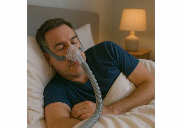
- If you breathe mostly through the nose, start with a nasal mask or nasal pillows—lighter and often more comfortable.
- If mouth leaks wake you or the device reports high leak, try a soft chin strap or switch to a hybrid/full-face mask.
- Sizing matters. Use manufacturer fitting guides; a mask that is too tight leaks more by distorting the seal. Aim for a gentle, even seal with the machine on at your typical pressure.
- Positioning: Adjust headgear while lying in your sleeping position; side sleepers may benefit from a PAP-friendly pillow that offloads the mask.
Dryness, congestion, and pressure intolerance
- Heated humidification reduces nasal dryness and congestion. If condensation (“rainout”) forms in the tube, raise tube temperature, use a heated hose, or lower room humidity.
- Nasal hygiene matters: nightly saline rinses, allergen control, and—when appropriate—topical nasal steroids.
- Pressure comfort: Use ramp features to fall asleep at a lower pressure; ask about exhalation pressure relief. If aerophagia (air swallowing) or bloating occurs, review pressures and sleep position.
Behavioral anchors
- Build a cue ritual: mask on, two minutes of relaxed belly breathing, lights out.
- Set a minimum use rule: even on tough nights, use PAP for the first half of the night—when apnea is often worst and deep sleep occurs. Aim to extend to full-night use.
- Track how you feel: energy, mood, and focus often improve before objective metrics look perfect.
Problem-solving
- Claustrophobia: practice when awake—mask on while reading for 10 minutes, then with the device on for 10 minutes, then lights out. Increase gradually.
- Skin irritation: ensure a gentle fit; consider mask liners; clean oils off seal surfaces daily.
- Leaks: reseat the mask in bed with the device on; check for mouth leaks if using a nasal interface.
- Persistent awakenings: confirm pressure range and investigate coexisting insomnia or periodic limb movements.
If insomnia coexists, PAP adaption improves markedly when insomnia is addressed directly. A brief, structured approach like cognitive behavioral therapy for insomnia (CBT-I) can cut awakenings and make therapy stick; see a step-by-step primer on insomnia strategies that pair with PAP.
Set expectations
- Most users feel clearer and less sleepy within 1–2 weeks, with steady gains over a month. Blood pressure often improves within weeks when adherence is solid. Give yourself time to dial in the mask, humidity, and pressure.
When to See ENT or Consider Further Evaluation
Sometimes the best next step is a specialist evaluation. Ear, nose, and throat (ENT) surgeons—often called “sleep surgeons” in this context—assess structural contributors to OSA and tailor interventions that complement or substitute for PAP when appropriate.
Signs you may benefit from ENT input
- Persistent PAP intolerance after attentive troubleshooting (fit, humidity, pressure adjustments) and coaching.
- Clear anatomic obstacles: marked nasal septal deviation, turbinate hypertrophy, large tonsils, adenoid regrowth, tongue base crowding, or retrognathia.
- Positional OSA that resists conservative measures.
- Interest in alternatives such as hypoglossal nerve stimulation or maxillomandibular advancement, where candidacy hinges on airway anatomy and BMI thresholds.
What the evaluation includes
- Focused nasal and oropharyngeal exam; often flexible nasopharyngoscopy.
- Review of your sleep study (look for REM-predominant or positional patterns, hypoxemia severity, and arousal burden).
- Consideration of drug-induced sleep endoscopy (DISE) to visualize collapse patterns before surgical or neurostimulation therapies.
Surgical and procedural options
- Nasal surgery (septoplasty, turbinate reduction) to reduce resistance and improve PAP or MAD tolerance.
- Palatal procedures (uvelectomy variants, expansion sphincter pharyngoplasty) for specific collapse patterns.
- Tongue-base reduction or lingual tonsillectomy when posterior tongue bulk dominates.
- Maxillomandibular advancement (MMA) in severe craniofacial crowding.
- Hypoglossal nerve stimulation (HGNS) for eligible patients with moderate–severe OSA and PAP intolerance.
When to repeat testing or broaden evaluation
- Major weight change (gain or loss ≥10%) since the last study.
- PAP on-treatment AHI persistently elevated (>5–10) despite good mask seal.
- New or worsening symptoms: witnessed central apneas, progressive morning headaches, or unexplained nocturnal awakenings.
- Comorbidity shifts (e.g., new heart failure, stroke) that may change the apnea pattern or indicate central contributions.
Shared decision-making
- Align interventions with your goals: alertness, blood pressure control, bed-partner harmony, and long-term vascular health.
- Expect a staged plan: optimize nasal airflow and position, ensure a fair PAP trial, and then layer in device-based or surgical options when benefits outweigh burdens.
The common path: diagnose accurately, start with the simplest effective therapy, and escalate thoughtfully when barriers persist. With this approach, most people achieve stable, restorative sleep and meaningful daytime gains.
References
- Treatment of Adult Obstructive Sleep Apnea with Positive Airway Pressure: An American Academy of Sleep Medicine Clinical Practice Guideline (2019) (Guideline)
- Clinical Practice Guideline for Diagnostic Testing for Adult Obstructive Sleep Apnea: An American Academy of Sleep Medicine Clinical Practice Guideline (2017) (Guideline)
- Obstructive Sleep Apnea in Adults: Screening (2022) (Guideline)
- Referral of adults with obstructive sleep apnea for surgical consultation: an American Academy of Sleep Medicine clinical practice guideline (2021) (Guideline)
- Mandibular advancement devices in obstructive sleep apnea: an updated review (2022) (Review)
Disclaimer
This article provides general educational information and is not a substitute for personalized medical advice, diagnosis, or treatment. Always seek the guidance of your physician or a qualified sleep specialist regarding symptoms, testing, or changes to your therapy. If you have severe sleepiness, drowsy driving, or signs of low oxygen (e.g., blue lips, chest pain), seek urgent care.
If this guide helped you, please consider sharing it with a friend or on Facebook or X. Your support helps us continue creating practical, evidence-informed resources for healthy aging.

