
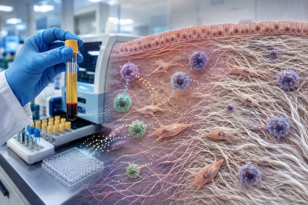
A transforming growth factor-beta test measures a TGF-beta protein in blood, most often TGF-beta 1. TGF-beta regulates immune tolerance, wound repair, extracellular-matrix production, blood-vessel biology, cell growth, and tissue scarring. Despite its central role in fibrosis and cancer biology, a circulating TGF-beta result is not a validated stand-alone test for liver, lung, kidney, or heart fibrosis, immune suppression, autoimmune disease, or cancer. Interpretation is difficult because most TGF-beta circulates in an inactive latent complex, assays may measure total or active protein, and platelets contain large amounts of TGF-beta 1. Platelet activation during collection or clotting can make serum and inadequately prepared plasma results appear high. Reliable plasma testing may require rapid double centrifugation to produce platelet-free plasma and immediate freezing. Laboratory methods, specimen types, activation steps, and units differ, so values are not interchangeable. The test is used mainly in research or selected specialist evaluations. When fibrosis is suspected, organ-specific imaging, function tests, validated fibrosis scores, and sometimes biopsy are usually more clinically meaningful than one blood cytokine concentration.
- TGF-beta 1 is commonly measured, but TGF-beta 2 and TGF-beta 3 are distinct isoforms with different distributions.
- Most secreted TGF-beta is latent and must be activated before it can signal.
- Platelets are a major blood source, making collection and platelet removal critical.
- High blood TGF-beta is nonspecific and does not prove active tissue fibrosis or cancer.
- A normal concentration cannot exclude strong local TGF-beta signaling inside an affected organ.
Table of Contents
- TGF-Beta Biology and Signaling
- What the Blood Test Measures
- Platelets, Specimen Handling, and False Elevation
- TGF-Beta and Fibrosis
- Immune Regulation, Wound Healing, and Cancer
- Meaning of High or Low Results
- Clinical Limitations and Common Misinterpretations
- Follow-Up Testing and Practical Interpretation
TGF-Beta Biology and Signaling
Transforming growth factor-beta is a family of signaling proteins with three mammalian isoforms: TGF-beta 1, TGF-beta 2, and TGF-beta 3. They share receptors and many downstream pathways but differ in where they are produced, how they are regulated, and their roles during development and tissue repair. Most clinical blood assays focus on TGF-beta 1.
TGF-beta is produced by platelets, macrophages, T cells, regulatory T cells, fibroblasts, endothelial cells, epithelial cells, cancer cells, and many other tissues. It is secreted in an inactive form bound to latency-associated peptide. This small latent complex can also attach to latent TGF-beta binding proteins in the extracellular matrix.
Activation releases mature TGF-beta so it can bind type II and type I receptors. The receptor complex phosphorylates SMAD proteins, which move to the nucleus and change gene expression. TGF-beta also signals through non-SMAD pathways that affect cell survival, migration, metabolism, and cytoskeletal behavior.
Latent activation is tightly controlled. Integrins, proteases, reactive oxygen species, mechanical force, acidic conditions, and tissue injury can expose active TGF-beta at a particular site. This local control means a tissue can have intense signaling while the measurable circulating concentration remains low or unchanged.
The biological effect depends on the cell and context. TGF-beta can suppress proliferation and inflammatory cytokine production in some immune cells, support regulatory T-cell differentiation, and limit tissue-damaging responses. In other settings, it can promote type 17 responses, fibroblast activation, extracellular-matrix deposition, epithelial-to-mesenchymal changes, and immune escape.
This dual behavior is often described as context-dependent rather than simply anti-inflammatory or pro-fibrotic. A blood result cannot reveal which receptor pathway is active, which tissue is responding, or whether the net effect is protective repair or harmful scarring.
What the Blood Test Measures
The test may be performed by enzyme-linked immunoassay, chemiluminescent immunoassay, or another antibody-based method. The report should specify the isoform, specimen, and whether the assay measures active TGF-beta, total TGF-beta after activation, or both.
Most circulating TGF-beta 1 is latent. To measure total TGF-beta 1, laboratories commonly acidify the specimen or use another activation step that releases mature protein from the latent complex. The result then includes protein that was not biologically active in the patient’s circulation at the moment of collection.
An active-only assay attempts to quantify the small fraction already capable of receptor binding. Active TGF-beta is unstable, present at low concentrations, and easily altered during collection and storage. Active and total results therefore answer different questions and cannot be compared directly.
Serum and plasma are also different. Serum is produced after blood clots, a process that activates platelets and releases abundant TGF-beta 1. Serum concentrations can largely reflect clotting-related platelet release rather than freely circulating protein. Plasma avoids deliberate clotting but remains vulnerable to platelet contamination and activation.
Results may be reported in picograms per milliliter, nanograms per milliliter, or another unit. Reference intervals depend on the antibody pair, calibration standard, activation procedure, and specimen preparation. A cutoff from one laboratory or research paper should not be applied to another method.
Some cytokine panels include TGF-beta, while others omit it because the specimen preparation needed for accurate platelet-poor measurement differs from routine cytokines. A general cytokine panel should not be assumed to handle TGF-beta optimally unless the laboratory states its validated process.
The assay measures concentration, not signaling activity. It does not measure receptor phosphorylation, SMAD activation, tissue matrix production, or the balance between active ligand and inhibitors. Those endpoints require tissue, cell-based, molecular, or imaging approaches.
Platelets, Specimen Handling, and False Elevation
Platelets contain exceptionally large stores of latent TGF-beta 1 in their granules. When they are activated by clotting, difficult venipuncture, prolonged tourniquet use, temperature change, centrifugation, or handling, they release this protein. Even a small number of residual platelets can substantially increase a plasma result.
For platelet-free plasma, laboratories may require EDTA blood to be centrifuged promptly, followed by a second higher-speed centrifugation of the separated plasma. The upper portion is transferred without disturbing the platelet-rich lower layer and frozen quickly. Exact speeds, times, tube types, and temperature instructions vary and must be followed.
A routine single spin that is adequate for many chemistry tests may leave enough platelets to invalidate TGF-beta interpretation. Visible hemolysis is not the only concern; the specimen can look clear while containing activated or residual platelets.
Serum generally has much higher TGF-beta 1 because clot formation releases platelet stores. A serum reference interval may account for this, but the biological meaning differs from carefully prepared platelet-free plasma. A serum result and a plasma result should not be trended as though they are the same test.
Platelet count and activation state can influence measurements. Thrombocytosis may increase available platelet-derived protein, while thrombocytopenia may lower it. Antiplatelet drugs, inflammation, surgery, exercise, and sample trauma may also affect release, although their impact depends on the method.
Some laboratories recommend co-measuring a platelet-release marker or documenting residual platelet count in research. Routine clinical reports may not provide this information. When a value is unexpectedly high, preanalytic contamination should be considered before attributing it to systemic fibrosis. Platelet factor 4 or another granule-release marker can sometimes reveal platelet activation, while a residual platelet count can show whether the plasma was truly platelet poor. These checks are method dependent and are not universal clinical standards, but they illustrate why collection quality is part of the result rather than a minor technical detail. Repeated freezing and thawing, delayed separation, and transfer close to the cell layer can also change the measured concentration.
The collection site should coordinate with the performing laboratory before drawing the sample. TGF-beta is a poor test for an unscheduled collection when staff are unfamiliar with the required double-spin protocol. Repeating an improperly prepared specimen with the same process is unlikely to clarify the result. A properly documented redraw is especially important when an unexpected result might otherwise trigger expensive imaging, referral, or an incorrect fibrosis label for the patient.
TGF-Beta and Fibrosis
TGF-beta 1 is a major driver of fibrogenesis. After persistent injury, it activates fibroblasts and tissue-specific precursor cells, promotes their transformation into matrix-producing myofibroblasts, increases collagen and fibronectin synthesis, reduces matrix breakdown, and changes tissue architecture.
In the liver, TGF-beta signaling activates hepatic stellate cells and contributes to fibrosis from metabolic liver disease, viral hepatitis, alcohol, autoimmune injury, and cholestatic disease. In the lungs, it participates in fibroblast activation and extracellular-matrix accumulation. Similar pathways operate in kidney, heart, skin, and other organs.
Biological importance does not make circulating TGF-beta 1 a reliable fibrosis stage test. Much of the signal is local, platelet contamination is substantial, and blood concentrations overlap among healthy people and patients with different diseases. The amount in plasma does not directly equal the amount of activated signaling in an organ.
Liver fibrosis is evaluated with established tools such as liver enzymes, platelet count, FIB-4 or other validated scores, elastography, imaging, and sometimes biopsy. Pulmonary fibrosis assessment relies on high-resolution imaging, pulmonary function, oxygenation, clinical trajectory, and multidisciplinary review. Kidney and heart fibrosis use their own function tests and imaging.
A high TGF-beta result cannot identify which organ is fibrotic, distinguish active injury from established scar, or determine reversibility. It also cannot separate fibrosis from wound healing, platelet activation, cancer-associated signaling, or systemic inflammation.
A normal result does not exclude fibrosis. Active ligand may be concentrated in tissue and rapidly bound to receptors or matrix. Fibrosis can continue through other pathways even if circulating TGF-beta is not elevated. Conversely, a high circulating value does not prove that canonical SMAD signaling is active in a particular organ. Receptor abundance, integrin-mediated activation, matrix binding, inhibitory proteins, and cross-talk with inflammatory and mechanical pathways all affect the tissue response. For this reason, clinicians do not use a routine TGF-beta blood result to stage fibrosis or monitor response to approved antifibrotic treatment. Organ function, imaging, validated fibrosis measures, symptoms, and treatment-specific endpoints remain more informative.
TGF-beta pathway inhibitors are being studied for fibrotic disease, but broad blockade can interfere with immune tolerance, vascular health, wound repair, and tumor suppression. Treatment development increasingly aims to target an isoform, activation mechanism, cell type, or tissue rather than suppressing all TGF-beta biology. A routine blood level does not determine eligibility for these therapies outside a defined protocol.
Immune Regulation, Wound Healing, and Cancer
TGF-beta helps limit excessive immune activation. It can reduce T-cell proliferation, macrophage activation, and inflammatory cytokine production. Together with other signals, it supports regulatory T-cell development and tissue tolerance. However, it can also participate in type 17 differentiation and chronic inflammation, depending on the surrounding cytokines.
In wound healing, TGF-beta recruits fibroblasts, promotes matrix formation, influences blood vessels, and helps close damaged tissue. These effects are necessary in a controlled repair response. Persistent activation can turn repair into pathologic scarring or contracture.
Cancer biology is similarly dual. Early in tumor development, TGF-beta can suppress cell proliferation and maintain tissue organization. Later, cancer cells may evade those growth-inhibitory effects while using TGF-beta to promote invasion, metastasis, stromal remodeling, angiogenesis, and immune suppression.
Tumors can create a TGF-beta-rich microenvironment that excludes or weakens cytotoxic T cells and natural killer cells while supporting regulatory cells and fibroblasts. This tissue-localized signaling is a major therapeutic research target. A blood concentration cannot establish that a tumor uses this mechanism.
Elevated TGF-beta has been reported in many cancers, autoimmune diseases, infections, cardiovascular disorders, pregnancy complications, and inflammatory states. Associations at the group level do not create an accurate diagnostic test for an individual. A high result should not be interpreted as evidence of cancer without standard clinical evaluation.
Low TGF-beta signaling can also be harmful. Genetic defects in receptors or downstream pathways can affect vascular development, immune regulation, skeletal features, and connective tissue. These disorders are diagnosed through phenotype and genetic testing, not a low circulating cytokine level.
Because TGF-beta influences many systems, nonspecific symptoms cannot be reliably assigned to a high or low blood value. Fatigue, pain, cognitive symptoms, and inflammation require a broader differential diagnosis and organ-specific assessment.
Meaning of High or Low Results
A high TGF-beta 1 result first raises a technical question: was the specimen serum, ordinary plasma, platelet-poor plasma, or platelet-free plasma? Was it processed within the required time, double-spun, transferred carefully, and frozen? Without that information, biological interpretation may be impossible.
If collection was valid, elevation can reflect platelet activation, wound healing, inflammation, tissue injury, fibrosis, malignancy, pregnancy-related changes, cardiovascular disease, kidney or liver dysfunction, or another process. The test cannot distinguish among them.
The magnitude does not map to a universal severity scale. A person with advanced organ fibrosis may have a modest or normal circulating value, while a poorly prepared specimen from a healthy person can be very high. Trends are meaningful only when collection, processing, specimen type, and assay are identical.
A low value may reflect true lower concentration, reduced platelet contribution, assay variation, sample dilution, or differences in activation of latent protein. There is no widely accepted clinical “TGF-beta deficiency” threshold for routine diagnosis.
Inherited defects in TGF-beta signaling, such as receptor or SMAD pathway disorders, can exist despite a normal or high ligand concentration. The problem is how cells respond, not necessarily how much cytokine circulates. Genetic and pathway-specific testing are required.
Medicines can influence production or signaling. Corticosteroids, immunosuppressants, antifibrotic therapy, chemotherapy, antiplatelet drugs, and investigational pathway inhibitors may change the result or the clinical context. Treatment should not be started, stopped, or adjusted solely because of a TGF-beta concentration.
A result outside range is best described as nonspecific and method-dependent. It may justify checking specimen integrity and investigating the clinical problem that prompted testing, but it does not define an organ diagnosis.
Clinical Limitations and Common Misinterpretations
The largest limitation is preanalytic variation from platelets. Another is the difference between total and active protein. A total assay that acid-activates latent complexes measures potential ligand, not the amount signaling in vivo.
Circulating concentration also ignores tissue compartmentalization. Receptors, integrins, matrix proteins, proteases, and mechanical forces determine local activation. Two people with the same plasma value can have very different tissue signaling.
There is no universal reference range or disease cutoff. Research studies use different matrices, preparation protocols, antibodies, standards, and patient populations. Comparing a personal result with a published graph can be misleading.
TGF-beta testing is sometimes included in nonstandard panels marketed for chronic inflammatory symptoms, environmental exposure, mold-related illness, immune imbalance, or generalized fibrosis. A high result does not identify a toxin, prove a specific exposure syndrome, or establish that TGF-beta is causing symptoms. Claims should be compared with validated diagnostic criteria and established organ testing.
The test is not a cancer screen. It cannot locate a tumor, distinguish benign from malignant disease, or predict future cancer in an otherwise healthy person. Standard screening follows age, risk, and evidence-based guidelines.
It is also not a direct autoimmune test. TGF-beta may participate in tolerance and inflammation, but autoimmunity is diagnosed through symptoms, examination, disease-specific antibodies, imaging, pathology, and organ function.
Serial monitoring can create false confidence. A lower number may result from fewer residual platelets rather than clinical improvement. A higher number may reflect collection trauma rather than disease progression. Standardized processing is mandatory before interpreting change.
Finally, the test does not replace biopsy or validated noninvasive fibrosis assessment. Its strongest use remains mechanistic research, clinical trials, and selected specialist investigations where the assay method and decision pathway are predefined.
Follow-Up Testing and Practical Interpretation
Begin with the laboratory report. Identify TGF-beta 1, 2, or 3; active or total measurement; serum or plasma; units; reference interval; and specimen instructions. Ask whether platelet-free preparation was required and documented.
If the result conflicts with the clinical picture, contact the laboratory before repeating it. The most useful repeat is one collected under a validated protocol, not simply another draw. Use the same laboratory if a trend is necessary. Record the tube type, time from collection to each centrifugation, spin conditions, transfer technique, freezing time, storage temperature, and number of freeze-thaw cycles when the result will influence research or specialized care. Do not compare serum with platelet-free plasma, or one laboratory’s total assay with another laboratory’s active assay, as though the numbers form a continuous trend. A change between such methods may reflect the specimen and assay rather than the patient.
For suspected liver fibrosis, evaluate the cause, liver enzymes, platelet count, validated serum scores, ultrasound or magnetic-resonance elastography, and imaging. For lung fibrosis, use high-resolution computed tomography, pulmonary function, oxygen assessment, exposure history, autoimmune testing, and multidisciplinary review. Other organs require similarly focused pathways.
When immune dysregulation is the concern, complete blood counts, lymphocyte subsets, immunoglobulins, inflammatory markers, autoantibodies, infection testing, and cell-function studies are often more actionable. A Treg panel measures cells, not circulating TGF-beta, and still does not provide a complete tolerance assessment.
When cancer is suspected, evaluation depends on the site and symptoms: examination, imaging, endoscopy, blood counts, tumor-specific markers where validated, pathology, and molecular studies. TGF-beta should not delay standard diagnostic work.
A useful interpretation states the limitation directly: the concentration is elevated or reduced on a specified assay, platelet contribution and latent-protein activation may influence the value, and the result is not diagnostic of fibrosis, malignancy, or immune disease. It should then name the organ-specific next step.
Urgent care is guided by symptoms, not this cytokine value. Breathing difficulty, jaundice, rapidly worsening swelling, chest pain, neurologic deficits, severe bleeding, or other signs of organ failure require immediate assessment regardless of whether TGF-beta is normal or high.
The central question is not simply how much TGF-beta is present in blood. It is where the pathway is active, what triggered it, which cells are responding, whether repair has become harmful fibrosis, and whether a validated test shows organ damage. A blood concentration alone cannot answer those questions.
References
- The pivotal role of TGF-β/Smad pathway in fibrosis and its therapeutic potential 2025 (Review)
- TGF-β inhibitors: the future for prevention and treatment of liver fibrosis 2025 (Review)
- Selective blockade of latent TGF-β1 activation suppresses fibrosis and cancer in preclinical models 2026 (Research Article)
- Blood Platelets as an Important but Underrated Circulating Source of TGFβ 2021 (Review)
- Human Transforming Growth Factor beta 1 (TGF-b1) 2026 (Laboratory Test Guide)
- Transforming Growth Factor beta1, Plasma 2026 (Laboratory Test Guide)
Disclaimer
This article is for general education and does not diagnose fibrosis, autoimmune disease, cancer, environmental illness, or a TGF-beta signaling disorder. TGF-beta results require method-specific interpretation with specimen type, platelet removal, active-versus-total measurement, medicines, symptoms, and validated organ testing. Do not change treatment based on a cytokine concentration without qualified medical guidance.





