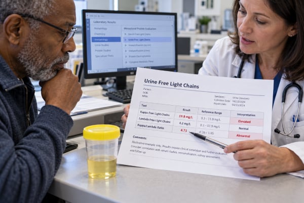
A urine free light chain test measures free kappa and free lambda immunoglobulin light chains that have passed into urine. These proteins are made by plasma cells as part of normal antibody production, but a clonal plasma cell or B-cell disorder can produce a large excess of one light-chain type. That excess may appear in urine as monoclonal light chains, historically called Bence Jones proteins, and can be associated with multiple myeloma, AL amyloidosis, or monoclonal gammopathy of renal significance (MGRS). The test can also be influenced by kidney filtration and tubular reabsorption, so urine kappa, lambda, and their ratio are not interpreted like serum free light chains and do not have one universal “normal” range across laboratories. In modern practice, serum free light-chain testing, serum protein electrophoresis, and serum immunofixation are central to monoclonal gammopathy evaluation, while urine electrophoresis and immunofixation remain important in selected diagnostic and monitoring situations. A urine free light chain result should therefore be interpreted as one part of a coordinated hematology and kidney workup.
- Urine free light chains are kappa or lambda antibody fragments that can spill into urine when production is high or kidney handling is abnormal.
- A strong excess of one light-chain type can suggest a monoclonal protein, but a urine kappa/lambda ratio alone does not diagnose multiple myeloma.
- Urine reference ranges are assay- and laboratory-specific because kidney filtration, tubular reabsorption, urine concentration, and collection method affect results.
- Serum free light chains, serum electrophoresis, and serum immunofixation are usually interpreted alongside urine studies when a plasma cell disorder is suspected.
- Kidney injury with unexplained proteinuria, declining eGFR, anemia, hypercalcemia, bone pain, or a monoclonal protein may require prompt hematology and nephrology evaluation.
Table of Contents
- What kappa and lambda free light chains are
- Why free light chains appear in urine
- Urine kappa, lambda, and ratio results
- Myeloma and other monoclonal disorders
- How kidney disease affects the test
- Collection and companion tests
- How abnormal results are interpreted
What kappa and lambda free light chains are
Antibodies are built from heavy chains and light chains. Each intact immunoglobulin contains either kappa or lambda light chains. Plasma cells normally produce a slight excess of light chains during antibody synthesis, and these unbound fragments circulate as free light chains.
Free light chains are small enough to pass through the kidney’s glomerular filter. Under normal conditions, proximal tubule cells reabsorb and break down most of the filtered light chains, so only small amounts reach the final urine. If production rises sharply or tubular reabsorption is impaired, more free light chains appear in urine.
A clonal plasma cell population usually produces a single immunoglobulin pattern. If the clone makes mainly kappa light chains, kappa may become disproportionately high; if it makes lambda, lambda may predominate. The monoclonal urinary light chains historically known as Bence Jones proteins are an important clue in plasma cell disorders.
A direct quantitative urine free light-chain assay measures kappa and lambda concentrations and may calculate a ratio. This should not be confused with urine immunofixation, which looks for a discrete monoclonal band and helps identify its type, or with UPEP, which separates proteins and can quantify a monoclonal component.
Why free light chains appear in urine
There are two major reasons free light chains increase in urine: excess production and abnormal kidney handling.
With excess production, a plasma cell or B-cell clone releases more of one light-chain type than the kidneys can reabsorb. Multiple myeloma is the best-known cause, but it is not the only one. Light-chain MGUS, AL amyloidosis, some lymphomas, Waldenström macroglobulinemia, and MGRS can also produce monoclonal light chains.
With kidney tubular injury, reabsorption of filtered light chains can fall even when production is not massively increased. This can raise both kappa and lambda in urine. Therefore, simply seeing elevated urinary light chains does not prove a monoclonal disorder.
Polyclonal immune activation can also raise free light-chain production. Infections, autoimmune disease, and inflammation can increase both kappa and lambda. A balanced rise is different from a clearly restricted monoclonal pattern, but the distinction is not always reliable from a urine ratio alone.
The kidneys are not passive in this process. Light chains are filtered, reabsorbed, metabolized, and sometimes directly nephrotoxic. Certain monoclonal light chains can form casts, deposit in kidney tissue, crystallize in proximal tubules, or contribute to amyloid. This explains why hematologic and renal evaluation often need to proceed together.
Urine kappa, lambda, and ratio results
There is no single universal urine free kappa, lambda, or kappa/lambda ratio range that can be safely applied to every laboratory. Assays differ, and urine results vary with collection method, concentration, kidney filtration, tubular function, and total urine volume.
Some reports provide concentrations in mg/L, while timed collections may report mg/24 hours. A spot sample can be highly affected by hydration unless the laboratory uses an additional creatinine correction. Reference intervals printed on the specific report should therefore take priority over ranges found online.
The ratio is conceptually simple: kappa concentration divided by lambda concentration. A strongly skewed ratio may support clonal excess of one light-chain type, but several cautions matter:
- A urine ratio is not interchangeable with the serum free kappa/lambda ratio.
- Reduced kidney filtration can change serum free light-chain concentrations and serum ratio interpretation.
- Tubular dysfunction can change how much of each light chain reaches urine.
- Very dilute urine may lower measured concentrations.
- Assay sensitivity near the lower detection limit can make a ratio unstable.
For these reasons, a urine free light-chain ratio is generally interpreted together with serum free light chains and monoclonal protein studies rather than used as a stand-alone screening threshold.
If the goal is to prove whether a urine light chain is monoclonal, urine protein electrophoresis and immunofixation often provide more direct information about the protein pattern.
Myeloma and other monoclonal disorders
A monoclonal free light-chain finding raises a differential diagnosis, not a single diagnosis. Multiple myeloma requires evidence of a clonal plasma cell disorder plus defined myeloma features or biomarkers; a urine result alone is not enough.
Multiple myeloma can cause anemia, bone lesions or bone pain, hypercalcemia, kidney injury, recurrent infections, and other complications. Some cases produce an intact monoclonal immunoglobulin; others are light-chain predominant. Light chains can contribute to kidney injury by forming obstructive casts in distal tubules, among other mechanisms.
MGUS is a smaller clonal condition that does not meet criteria for myeloma or another malignancy and lacks attributable organ damage. However, the label “MGUS” should not be used when the monoclonal protein itself is causing kidney disease. That situation may fall under MGRS.
MGRS includes kidney lesions caused by a nephrotoxic monoclonal immunoglobulin produced by a clone that may be too small to meet conventional criteria for multiple myeloma or lymphoma. The concept is important because even a low-level monoclonal protein can cause major renal damage. Evaluation often includes serum and urine electrophoresis, serum and urine immunofixation, serum free light chains, bone marrow assessment, and kidney biopsy when indicated.
AL amyloidosis is another important disorder in which monoclonal light chains misfold and form amyloid deposits in organs. Kidney involvement often causes substantial albuminuria or nephrotic syndrome. The diagnostic workup is broader than a urine free light-chain measurement.
A Bence Jones protein urine test refers to the same general clinical problem of monoclonal light chains in urine, although modern methods are more specific than the historical heat-based tests.
How kidney disease affects the test
Kidney function changes both the amount of free light chain in blood and the amount that appears in urine. As GFR falls, serum free light chains accumulate because renal clearance decreases. This can raise both kappa and lambda and can widen the expected serum ratio range in CKD.
Urine behavior is more complicated. Filtered light chains are normally reabsorbed in proximal tubules. If tubules are injured, more light chain may escape into urine even when serum production is not dramatically abnormal. Conversely, severe loss of filtration may reduce the filtered load into urine despite high blood concentrations.
This is why urine light-chain concentration does not simply track serum concentration. A patient with advanced CKD can have high serum free light chains from reduced clearance, while another patient with tubular damage may excrete more urinary light chains despite a different serum pattern.
Monoclonal light chains can also cause kidney disease themselves. Cast nephropathy is most strongly associated with very high circulating involved free light chains in myeloma. Other monoclonal proteins cause light-chain deposition disease, AL amyloidosis, proliferative glomerulonephritis with monoclonal deposits, or light-chain proximal tubulopathy.
If kidney injury is unexplained, the combination of proteinuria pattern, uACR, UPCR, serum creatinine, serum free light chains, and immunofixation can be more informative than any one urine concentration. A large gap between total protein and albumin may be a clue to non-albumin protein such as light chains.
Collection and companion tests
Urine free light chains may be measured in a spot sample or a timed collection depending on the laboratory and clinical purpose. Historically, 24-hour urine collections were commonly used in myeloma because they allow measurement of total monoclonal protein excretion. Collection accuracy matters: missing voids can substantially underestimate daily excretion.
No special diet is usually required. Normal hydration is appropriate unless the clinician gives different instructions. If a 24-hour collection is requested, follow the start and stop times exactly and keep the specimen as directed by the laboratory.
The companion blood and urine tests are often more important than the quantitative urine light-chain value itself. A typical monoclonal protein evaluation can include:
- serum protein electrophoresis;
- serum immunofixation;
- serum free kappa and lambda light chains with serum ratio;
- urine protein electrophoresis;
- urine immunofixation;
- complete blood count, calcium, creatinine, and other metabolic testing.
Serum screening is now highly sensitive, and many guidelines use serum electrophoresis, serum immunofixation, and serum free light chains as the initial laboratory panel for suspected monoclonal gammopathy. Urine studies still add value in light-chain disease, AL amyloidosis evaluation, renal presentations, and selected monitoring settings.
How abnormal results are interpreted
A useful interpretation asks three questions: Is one light-chain type disproportionately increased? Is there direct evidence of a monoclonal protein? Is there organ involvement?
A modest increase in both urinary kappa and lambda with a non-restricted pattern may reflect reduced kidney handling or polyclonal immune activation. A marked excess of one type, particularly when urine immunofixation identifies a matching monoclonal band and serum studies support it, is more concerning for a clonal disorder.
The size of the abnormality matters, but there is no urine ratio cutoff that independently proves myeloma. The diagnosis depends on bone marrow or tissue evidence, monoclonal protein studies, imaging, and myeloma-defining clinical or biomarker features.
Kidney findings can alter urgency. A patient with rapidly rising creatinine, heavy proteinuria, and a newly detected monoclonal light chain may need urgent hematology and nephrology assessment. Kidney biopsy can be essential when MGRS is suspected because the same monoclonal protein can cause several distinct renal lesions that require different interpretation.
If the urine test is negative but clinical suspicion remains high, the workup should not stop automatically. Serum free light chains and serum immunofixation may detect disease that is not obvious in urine. The opposite is also possible: urine immunofixation may reveal small monoclonal free light chains that add diagnostic information.
The safest way to use a urine free light-chain result is as a pattern-finding test within a larger monoclonal protein evaluation. Its value comes from integration, not from treating a kappa, lambda, or ratio number as a diagnosis by itself.
Examples of light-chain patterns and common pitfalls
Consider a patient whose urine free kappa and lambda concentrations are both mildly elevated, with no discrete monoclonal band on immunofixation and a serum free light-chain pattern compatible with reduced kidney clearance. That combination can occur in CKD and does not automatically indicate a plasma cell cancer. The balanced elevation and absence of clear clonality are important context.
A different pattern is a markedly elevated urine kappa concentration, a strongly skewed urine ratio, a monoclonal free kappa band on urine immunofixation, and an abnormal serum involved free light chain. That constellation is much more concerning for a clonal process and would usually lead to a formal plasma cell or B-cell evaluation. Even then, the final diagnosis could range from a precursor condition to active myeloma or MGRS depending on marrow findings and organ involvement.
A third pattern is heavy total urine protein with only modest albumin and a new acute kidney injury. If serum free light chains are very high, clinicians may worry about light-chain cast nephropathy. Because kidney injury can progress rapidly, this is not a situation to interpret from a urine ratio alone or to delay while repeating outpatient samples.
One common pitfall is applying the familiar serum kappa/lambda reference interval to urine. The physiology is different, and laboratories may use different assays and reference limits. Another is assuming that a normal urine result excludes myeloma. Serum free light chains and serum immunofixation can detect abnormalities that urine testing misses, especially with low-level or non-urinary secretion.
Patients sometimes focus on whether kappa is “high” or lambda is “high” without considering clonality. Both can rise in inflammation or kidney disease. The diagnostic question is whether one type is disproportionately produced by a clone and whether that clone is causing organ damage.
Finally, serial results should ideally be followed using the same assay platform and interpreted with the same companion tests. Switching methods can create apparent changes that reflect analytical differences rather than biology. Hematology laboratories often standardize the monitoring panel once a monoclonal disorder has been established.
Age and coexisting inflammation can further complicate interpretation. Older adults are more likely to have both reduced kidney function and incidental monoclonal gammopathy, so an abnormal light-chain result does not automatically reveal which process is causing symptoms. Sometimes a kidney biopsy is the only way to prove that the monoclonal protein is responsible for renal injury.
Treatment monitoring also differs from initial diagnosis. Once a specific clone is identified, clinicians usually follow the marker that best reflects that disease—often serum involved free light chain, serum M-protein, urine M-protein, or a combination. Repeating every possible test at every visit is not always necessary; the monitoring plan is tailored to the disease and baseline secretion pattern.
A final practical point is that “Bence Jones protein negative” and “urine free light chains normal” are not always equivalent statements because laboratories may use different methods. Read the actual method and companion immunofixation result rather than relying only on a shorthand label.
References
- Multiple myeloma: 2024 update on diagnosis, risk-stratification, and management 2024 (Review)
- Diagnosis and Management of Multiple Myeloma: A Review 2022 (Review)
- A review of clinical guidelines, laboratory recommendations and external quality assurance programs for monoclonal gammopathy testing 2023 (Review)
- Recommendations for the study of monoclonal gammopathies in the clinical laboratory. A consensus of the Spanish Society of Laboratory Medicine and the Spanish Society of Hematology and Hemotherapy. Part I: Update on laboratory tests for the study of monoclonal gammopathies 2023 (Position Statement)
- Monoclonal gammopathies of renal significance 2023 (Review)
Disclaimer
This article is for general education and cannot diagnose multiple myeloma, MGUS, amyloidosis, or MGRS. Urine free light-chain results are assay-specific and must be interpreted with serum testing, kidney function, urine immunofixation or electrophoresis, symptoms, and the broader clinical evaluation. Seek prompt medical care for rapidly worsening kidney function, severe weakness, confusion, dehydration, major swelling, or other acute symptoms.