
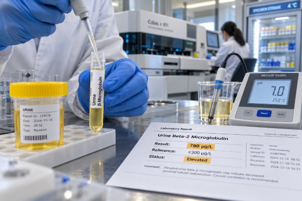
A beta-2 microglobulin urine test is a specialized marker of proximal kidney tubule function. Beta-2 microglobulin is a small protein released from the surface of most nucleated cells. It circulates in blood, passes easily through the glomerular filter, and is normally reabsorbed and broken down almost completely by proximal tubular cells. When those tubules are injured or cannot reabsorb filtered proteins efficiently, urinary beta-2 microglobulin rises. This creates a pattern of low-molecular-weight, or tubular, proteinuria. The test is not the standard way to screen the general population for chronic kidney disease; uACR and eGFR are more widely recommended for that purpose. Beta-2 microglobulin is most useful when a clinician specifically suspects proximal tubular damage or wants to characterize non-albumin proteinuria. Sample handling matters because beta-2 microglobulin is unstable in acidic urine, so collection and preservation instructions from the laboratory are important for avoiding falsely low results.
- High urinary beta-2 microglobulin usually suggests impaired proximal tubular reabsorption and tubular proteinuria.
- A low or undetectable urine level is usually expected and is not a sign of a beta-2 microglobulin deficiency.
- Urinary beta-2 microglobulin can degrade in acidic urine, especially around pH below 5.5–6, so sample handling can materially affect the result.
- There is no single universal reference range; results may be reported as mcg/L, mg/L, or a creatinine-adjusted ratio, and the laboratory’s interval should be used.
- The urine test reflects tubular handling, while serum beta-2 microglobulin is influenced strongly by kidney filtration and by conditions that increase immune-cell turnover.
Table of Contents
- What Urine Beta-2 Microglobulin Measures
- Why High Levels Suggest Tubular Injury
- When the Test Is Used
- Sample Collection and pH Stability
- Causes of High Urinary Beta-2 Microglobulin
- How to Interpret the Result
- Follow-Up and Next Steps
What Urine Beta-2 Microglobulin Measures
Beta-2 microglobulin, often abbreviated β2M or B2M, is a low-molecular-weight protein that forms part of major histocompatibility complex class I molecules on the surface of most nucleated cells. As cells normally turn over, beta-2 microglobulin enters the bloodstream.
Because the protein is small, it is freely filtered through the kidney glomeruli. Healthy proximal tubules then reabsorb and catabolize nearly all of the filtered beta-2 microglobulin. Only a small fraction reaches final urine. A rise in urinary B2M therefore indicates that the proximal tubules are not handling filtered low-molecular-weight proteins normally.
This mechanism is different from albuminuria. Albumin is larger and high urine albumin often reflects increased permeability of the glomerular filtration barrier. Beta-2 microglobulin usually crosses the glomerulus but is then reclaimed. A high urine B2M level therefore points more toward post-filtration tubular dysfunction.
The test may be performed on a random urine sample or as part of a timed collection, depending on the laboratory. Results can be reported as a concentration or normalized to urine creatinine. A spot urine creatinine adjustment helps reduce the effect of dilution but introduces its own dependence on creatinine production.
Urinary B2M is one member of a group of low-molecular-weight markers that includes alpha-1 microglobulin and retinol-binding protein. The broader tubular proteinuria pattern may be recognized by elevations in one or more of these proteins.
Why High Levels Suggest Tubular Injury
Proximal tubular cells perform extensive reabsorptive work. They reclaim glucose, amino acids, phosphate, bicarbonate, uric acid, electrolytes, and filtered low-molecular-weight proteins. When these cells are injured, reabsorption becomes less efficient and beta-2 microglobulin spills into urine.
Studies comparing urinary B2M with kidney biopsy markers have shown that higher urine levels correlate with proximal tubular injury. This makes B2M useful as a functional indicator of tubular protein handling, although it does not identify the exact cause of injury.
A high urine B2M can occur even when a standard protein dipstick is not strikingly abnormal. Dipsticks are most sensitive to albumin and are less reliable for low-molecular-weight proteins. Quantitative total protein and specific tubular markers can therefore reveal protein loss that routine dipstick screening underestimates.
The relationship also works in the other direction: a high albumin level does not necessarily mean B2M will be high. Purely glomerular disease can initially produce albumin-rich proteinuria with relatively preserved tubular reabsorption. In advanced or heavy glomerular disease, however, proximal tubules can become secondarily stressed, creating a mixed glomerular-tubular pattern.
The distinction between the urine and blood test is important. Serum beta-2 microglobulin tends to rise when kidney filtration falls because less B2M is cleared from the blood. It can also rise when production increases, as in inflammatory states and some blood cancers. Urinary B2M is more specifically tied to what the proximal tubules do with the filtered protein, although very high serum levels can increase the filtered load and complicate interpretation.
When the Test Is Used
Urine beta-2 microglobulin is a targeted test, not a universal kidney screen. Clinicians may order it when the main question involves proximal tubular damage, low-molecular-weight proteinuria, or a disease known to affect tubular function.
Possible uses include:
- evaluation of suspected tubulointerstitial nephritis;
- assessment of drug- or toxin-related tubular injury;
- investigation of Fanconi-type proximal tubular dysfunction;
- selected cases of acute kidney injury;
- evaluation of unusual proteinuria when albumin does not account for total protein;
- monitoring in certain inherited metabolic diseases that affect the kidneys;
- selected transplant, infectious, or inflammatory kidney evaluations;
- research or specialist monitoring of tubular injury and CKD progression.
The test usually sits beside other kidney markers rather than replacing them. Serum creatinine and eGFR assess overall filtration. uACR measures albumin leakage. Urinalysis and microscopy can reveal blood, white cells, casts, glucose, and other clues. Serum electrolytes can show whether proximal tubular dysfunction is causing clinically important losses.
For example, if a patient has glucose in the urine despite a normal blood glucose level, low serum phosphate, metabolic acidosis, and high urinary B2M, the pattern supports proximal tubular dysfunction more strongly than B2M alone. Conversely, a high B2M with no other abnormalities may need confirmation before a specific diagnosis is assigned.
Sample Collection and pH Stability
Sample handling is unusually important for beta-2 microglobulin because the protein is unstable in acidic urine. At low pH, urinary enzymes can degrade B2M after the urine has been produced. If degradation occurs before analysis, the laboratory may report a falsely low value.
Classic laboratory studies found substantial instability below about pH 5.5 and, in some pathological urine samples, instability even around pH 6 depending on temperature and enzymatic activity. For this reason, laboratories may specify a minimum urine pH, require prompt processing, or provide instructions for alkalinization or preservation.
Do not attempt to change urine pH yourself unless the laboratory specifically provides a collection procedure. Adding household substances to a urine specimen can invalidate the test. Follow the supplied container and transport instructions exactly.
The pH issue is one reason urine alpha-1 microglobulin is sometimes preferred for tubular screening. Alpha-1 microglobulin is more stable in acidic urine and answers a similar proximal reabsorption question.
Urine concentration also matters. A very dilute specimen can produce a low concentration even when daily excretion is increased, while dehydration can increase concentration. Creatinine-adjusted reporting partly corrects this, and some laboratories may prefer a timed collection. No approach completely eliminates biological and pre-analytical variation.
Causes of High Urinary Beta-2 Microglobulin
A high urine B2M result most commonly indicates proximal tubular dysfunction, but many diseases and exposures can produce that mechanism.
Acute tubular injury from ischemia, severe illness, or nephrotoxins can reduce reabsorptive capacity. In critical illness, urinary low-molecular-weight proteins may rise before or alongside conventional acute kidney injury markers. B2M has been studied in settings such as severe infection, hyperbilirubinemia, and acute respiratory distress syndrome.
Tubulointerstitial nephritis can raise B2M when inflammation involves the tubular compartment. Causes include medications, autoimmune disease, infections, and other inflammatory conditions. Urinalysis may show white blood cells or casts, but findings vary.
Toxins and drugs can injure proximal tubules. Heavy metals and some medications are recognized causes of low-molecular-weight proteinuria. The relevant exposure history is critical because a B2M result cannot identify which toxin or drug is responsible.
Inherited or metabolic disorders can damage proximal tubules over time. Recent studies have used urinary B2M to monitor kidney involvement in disorders of amino acid and cobalamin metabolism. In these conditions, B2M is a marker of tubular damage rather than the primary disease itself.
Chronic kidney diseases can produce mixed injury. Polycystic kidney disease, diabetic kidney disease, chronic interstitial disease, and other conditions may involve tubular pathways in addition to changes in eGFR or albuminuria.
Urinary tract and kidney infections can sometimes increase tubular markers when the upper urinary tract or renal parenchyma is involved. A B2M result does not diagnose infection and should be interpreted with symptoms, urinalysis, and culture when infection is suspected.
How to Interpret the Result
There is no single universal normal range for urinary beta-2 microglobulin. Laboratories may use different assays, sample types, units, creatinine corrections, and reference populations. The result should therefore be compared with the reference interval on the same report.
A high value is most convincing when it fits other evidence of tubular dysfunction. Useful companion findings include low-molecular-weight proteinuria, inappropriate urinary glucose, phosphate or bicarbonate wasting, abnormal urine sediment, a relevant drug or toxin exposure, and changes in kidney function.
A normal result should also be viewed critically if the urine was strongly acidic or sample handling was uncertain. A falsely low result caused by degradation is a known limitation. Repeating the test with correct handling or using a more acid-stable tubular marker may be appropriate.
High serum B2M can complicate interpretation. When blood concentrations are increased because of reduced GFR, inflammation, or malignancy, more protein may be presented to the glomerular filter. Urinary excretion reflects both the filtered load and the ability of proximal tubules to reclaim it. This is another reason the urine value should not be interpreted in isolation.
The magnitude of the elevation is not a direct measure of remaining kidney function. A person with marked tubular proteinuria can have a relatively preserved eGFR, while someone with advanced CKD may have high serum B2M for reasons related to reduced filtration. Different kidney markers answer different physiologic questions.
Follow-Up and Next Steps
After an elevated urine beta-2 microglobulin result, the next step is usually to identify the source and clinical significance of tubular dysfunction. A clinician may confirm the abnormality, review sample pH and handling, and compare it with other urine and blood findings.
Potential follow-up tests include uACR, total urine protein, serum creatinine and eGFR, electrolytes, bicarbonate, phosphate, uric acid, and urinalysis with microscopy. When Fanconi syndrome is suspected, clinicians may also assess urine glucose in relation to blood glucose and measure other solute losses.
Medication review is important. A temporal relationship between a new drug and tubular abnormalities may change management. Occupational and environmental exposures should also be considered when heavy metals or solvents are plausible.
Kidney imaging may be useful if obstruction or structural disease is suspected. Nephrology referral is appropriate for persistent unexplained tubular proteinuria, worsening kidney function, major electrolyte or acid-base abnormalities, or a pattern suggesting a systemic or inherited disorder.
Seek urgent medical attention if suspected tubular injury occurs with rapidly rising creatinine, very low urine output, severe dehydration, dangerous electrolyte abnormalities, significant poisoning, or symptoms of severe systemic illness. In those situations, immediate clinical stabilization matters more than any single biomarker value.
For stable abnormalities, trends using the same laboratory method can help show whether tubular dysfunction is improving, persistent, or worsening. The result is most useful when it answers a focused clinical question and is interpreted with the rest of the kidney evaluation.
Distinguishing a True Elevation From a Sample Problem
Urine beta-2 microglobulin is unusually sensitive to preanalytical conditions, especially urine acidity. Beta-2 microglobulin can break down in acidic urine after it has entered the bladder or collection container. If degradation occurs, the laboratory may report a falsely low value even when proximal tubular reabsorption is impaired. This limitation is one reason the test is less straightforward than routine uACR.
Collection instructions therefore matter. Some laboratories require a fresh specimen, rapid processing, refrigeration, or adjustment of urine pH according to their validated method. Patients should follow the specific laboratory instructions rather than trying to alkalinize or preserve urine on their own. If the sample pH was too low for the assay’s stability requirements, a normal result may not be reliable and recollection can be appropriate.
A true high result should also fit the surrounding pattern. Proximal tubular dysfunction may be accompanied by non-albumin proteinuria, urinary glucose despite a normal blood glucose level, phosphate wasting, bicarbonate loss, uric-acid wasting, or changes in other low-molecular-weight proteins. Serum creatinine may be normal early because eGFR reflects overall filtration and can remain preserved despite substantial tubular dysfunction. Conversely, serum beta-2 microglobulin can rise when GFR falls, which is a different phenomenon from increased urinary beta-2 microglobulin caused by failed tubular reabsorption.
Comparing beta-2 microglobulin with albumin helps localize the process. Predominantly albumin-rich proteinuria points more strongly toward glomerular disease. Elevated total protein with relatively little albumin and a high tubular marker supports a tubular pattern. Mixed injury is common, particularly in acute kidney injury, drug toxicity, systemic illness, and advanced CKD, so the categories are not mutually exclusive.
Magnitude and trend matter more than a barely abnormal isolated number. Reference intervals vary by assay, collection type, and reporting unit. A repeat result collected correctly is often useful when an unexpected elevation has no clinical explanation. If the value is persistently high, medication review, exposure history, electrolyte studies, urinalysis, and kidney function testing help determine whether there is a reversible cause.
The test is therefore best viewed as a localization marker. It can support evidence that proximal tubules are injured, but it cannot name the exact disease, establish chronicity, or measure overall kidney filtration by itself. Those questions require the rest of the clinical and laboratory evaluation.
When Another Tubular Marker May Be Preferable
Beta-2 microglobulin is not the only low-molecular-weight protein used to assess proximal tubular function. Alpha-1 microglobulin, retinol-binding protein, and other biomarkers may be chosen depending on the laboratory, suspected disease, and specimen conditions. Alpha-1 microglobulin is generally more stable in acidic urine, which can make it more practical when sample pH control is difficult.
The choice of marker should match the question. If the main concern is glomerular kidney risk in diabetes or hypertension, uACR has far stronger guideline support than urine beta-2 microglobulin. If the concern is a proximal tubular toxic exposure or Fanconi-type process, a low-molecular-weight protein can add information that uACR may miss. No single marker replaces urinalysis, serum creatinine, electrolytes, and exposure history.
Interpreting a Normal Result
A normal urine beta-2 microglobulin result lowers the likelihood of substantial proximal tubular failure only when the specimen was collected and handled correctly. It does not exclude early or focal tubular injury, and it does not exclude glomerular disease. If suspicion remains high because of glycosuria without hyperglycemia, phosphate wasting, acidosis, or another tubular abnormality, clinicians may repeat the specimen or use a different tubular marker.
This distinction is especially important when urine was acidic or processing was delayed. In that setting, a low value can reflect degradation rather than healthy tubular reabsorption.
References
– Urinary biomarkers of tubular injury and inflammation in ADPKD patients under tolvaptan therapy 2026 – Acute hyperbilirubinemia determines an early subclinical renal damage: Evaluation of tubular biomarkers in cholemic nephropathy 2024 – Prospective Multicenter Study on Early Proximal Tubular Injury in COVID-19-Related Acute Respiratory Distress Syndrome 2024 – Kidney urinary biomarkers in patients with branched-chain amino acid and cobalamin metabolism defects 2023 – Urinary β2-Microglobulin Is a Good Indicator of Proximal Tubule Injury: A Correlative Study with Renal Biopsies 2014
Disclaimer
This article is for general education and does not diagnose tubular kidney disease. Urine beta-2 microglobulin is sensitive to sample pH and laboratory handling, and reference ranges vary by method. Discuss abnormal results with a qualified healthcare professional, especially when kidney function is changing or electrolyte abnormalities are present.