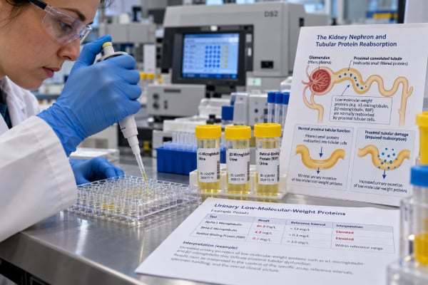
Tubular proteinuria occurs when the kidney’s proximal tubules fail to reabsorb small proteins that normally pass through the glomerular filter and are almost completely reclaimed. The result is increased urinary loss of low-molecular-weight proteins such as alpha-1 microglobulin, beta-2 microglobulin, retinol-binding protein, and cystatin C. This pattern differs from classic glomerular proteinuria, in which albumin is the dominant protein because the glomerular filtration barrier has become abnormally permeable. Tubular proteinuria may appear with acute tubular injury, tubulointerstitial disease, Fanconi syndrome, inherited proximal tubule disorders, monoclonal light-chain injury, and some medication or toxin exposures. A routine dipstick can miss or underestimate the problem because it is most sensitive to albumin. Diagnosis therefore relies on quantitative total protein, albumin, targeted low-molecular-weight proteins, kidney function, and other evidence of proximal tubular dysfunction.
- Tubular proteinuria means small filtered proteins are not being reabsorbed normally by the proximal tubules.
- Common markers include alpha-1 microglobulin, beta-2 microglobulin, retinol-binding protein, and urinary cystatin C.
- A standard protein dipstick may be weakly positive or negative despite tubular protein loss because it mainly detects albumin.
- Total protein that is high relative to urine albumin can suggest a non-albumin pattern and prompt tubular or monoclonal protein testing.
- Follow-up often includes kidney function, urine glucose, phosphate, bicarbonate-related testing, uric acid, medication review, and tests for the underlying cause.
Table of Contents
- What Tubular Proteinuria Means
- How Low-Molecular-Weight Proteins Are Handled
- Tests Used to Detect Tubular Proteinuria
- How to Interpret the Protein Pattern
- Common Causes of Tubular Proteinuria
- Tubular Versus Glomerular and Overflow Proteinuria
- Follow-Up, Treatment, and When to Worry
What Tubular Proteinuria Means
The glomeruli allow many small proteins to enter the initial filtrate. In a healthy kidney, the proximal tubules recover most of them before urine leaves the nephron. Tubular proteinuria develops when this recovery process is impaired.
The proteins involved are much smaller than albumin and include several molecules used as tubular biomarkers. Because they are normally reabsorbed so efficiently, even a modest loss of proximal tubular function can increase their urinary excretion.
This is different from albuminuria, which usually reflects increased permeability of the glomerular barrier. Tubular proteinuria can contain some albumin, but albumin often makes up a smaller fraction of total urine protein than it does in glomerular disease.
The total amount of protein in isolated tubular proteinuria is often lower than in severe glomerular disease. It may range from a mild increase to roughly 1–2 g/day in some disorders, although mixed disease can produce higher levels. A very large nephrotic-range protein loss is more typical of a glomerular process than of isolated tubular dysfunction.
The term “tubular proteinuria test” therefore refers less to one universal assay and more to a testing strategy that identifies low-molecular-weight proteins and shows that the proximal tubule is not handling them normally.
How Low-Molecular-Weight Proteins Are Handled
The proximal tubule contains a highly developed endocytic system. Filtered proteins bind receptors such as megalin and cubilin at the apical surface of tubular cells, are internalized, and are processed within endosomes and lysosomes. This system recovers useful amino acids and prevents large losses of filtered proteins.
When proximal tubular cells are injured, receptor-mediated uptake can fail. Low-molecular-weight proteins then remain in tubular fluid and appear in urine. The same injury can interfere with reabsorption of glucose, phosphate, bicarbonate, amino acids, and uric acid.
This explains why tubular proteinuria may be one clue within a broader proximal tubular syndrome. For example, Fanconi syndrome can produce low-molecular-weight proteinuria together with glycosuria despite normal blood glucose, phosphate wasting, metabolic acidosis from bicarbonate loss, aminoaciduria, and uric acid wasting.
Tubular handling can also be disrupted by ischemia, inflammation, nephrotoxins, inherited receptor or endosomal disorders, and toxic monoclonal light chains. The protein pattern tells clinicians that the proximal tubule is involved, but other tests are needed to identify the cause.
Tests Used to Detect Tubular Proteinuria
There is no single test panel used everywhere. The exact workup depends on local laboratory availability and the suspected disorder.
Total protein and albumin
A spot UPCR measures total urine protein, while uACR measures albumin specifically. Comparing the two can be helpful. If total protein is clearly elevated but albumin accounts for only a small portion, non-albumin proteins become more likely.
This comparison is not a stand-alone diagnostic formula because the assays and units differ, but a marked mismatch can prompt more specific testing.
Low-molecular-weight protein assays
Common markers include:
- Alpha-1 microglobulin: a stable low-molecular-weight protein often used to detect proximal tubular dysfunction.
- Beta-2 microglobulin: another tubular marker, although it is less stable in acidic urine and can degrade when urine pH is low.
- Retinol-binding protein: a sensitive marker of proximal tubular reabsorptive failure with relatively good urinary stability.
- Urinary cystatin C: can rise when proximal tubular uptake is impaired and is used in some kidney injury settings.
A urinary retinol-binding protein test is particularly useful when Fanconi syndrome or other proximal tubule disorders are suspected.
Electrophoresis and immunofixation
Urine protein electrophoresis can help separate albumin from other protein fractions. Immunofixation is used when monoclonal immunoglobulin light chains are a concern. This distinction matters because light-chain overflow can mimic or directly cause tubular proteinuria.
How to Interpret the Protein Pattern
The interpretation depends on the combination of total protein, albumin, low-molecular-weight proteins, urine sediment, and kidney function.
| Pattern | Likely source | Helpful next tests |
|---|---|---|
| Albumin dominates total protein | Glomerular proteinuria | uACR, urine microscopy, glomerular disease evaluation |
| Total protein elevated, albumin relatively low, low-molecular-weight proteins high | Tubular proteinuria | Alpha-1 microglobulin, RBP, beta-2 microglobulin, proximal tubular function tests |
| Total protein elevated with monoclonal light chains | Overflow proteinuria | Serum free light chains, electrophoresis, immunofixation |
| Albumin and tubular proteins both high | Mixed glomerular and tubular injury | eGFR, urine sediment, underlying disease evaluation |
Reference ranges vary widely between tubular markers and assays. Results may be reported as concentration or as a marker-to-creatinine ratio. A value should therefore be judged using the laboratory’s own reference interval rather than a universal cutoff.
Urine pH can affect some proteins. Beta-2 microglobulin is less stable in acidic urine, which may produce a falsely low result if sample handling is poor. RBP and alpha-1 microglobulin are often favored when sample stability is a concern.
The magnitude of tubular proteinuria also needs context. A small elevation can occur in early or limited tubular injury, while very high low-molecular-weight protein excretion strongly supports substantial proximal tubular dysfunction. A falling eGFR, electrolyte wasting, or metabolic acidosis increases the clinical significance of the finding.
Common Causes of Tubular Proteinuria
Tubular proteinuria can result from inherited, acquired, acute, or chronic conditions.
Fanconi syndrome is a classic cause. It represents generalized proximal tubular dysfunction rather than one disease. Inherited causes include cystinosis, Dent disease, Lowe syndrome, and other rare disorders. Acquired Fanconi syndrome can occur with certain drugs, monoclonal gammopathies, toxins, and other kidney diseases.
Medication-related tubular injury may occur with selected antivirals, chemotherapies, antibiotics, and other nephrotoxic agents. Risk is influenced by dose, exposure duration, kidney function, age, and other medications. A medication should never be stopped solely on the basis of an online description; the prescribing clinician must weigh risks and alternatives.
Heavy metals and toxins can damage proximal tubular cells. Cadmium is a classic example. Occupational, environmental, and supplement exposure histories can therefore be relevant.
Tubulointerstitial nephritis can impair tubular function through inflammation. Depending on the cause, there may also be white blood cells in urine, reduced kidney function, fever, rash, or eosinophilia, though these features are not always present.
Acute tubular injury from ischemia, sepsis, or nephrotoxins can increase urinary tubular proteins. Research has explored many such markers as early indicators of kidney injury before serum creatinine changes fully develop.
Monoclonal light-chain disorders can both create overflow proteinuria and directly injure proximal tubular cells. A person can therefore have a mixed pattern of light-chain excess and tubular reabsorptive failure.
Tubular Versus Glomerular and Overflow Proteinuria
Distinguishing the mechanism matters because the underlying diseases and treatments differ.
Glomerular proteinuria is usually dominated by albumin. It is common in diabetes, primary glomerulopathies, lupus nephritis, and many other diseases affecting the filtration barrier. Heavy glomerular proteinuria can become nephrotic range and lead to edema and hypoalbuminemia.
Tubular proteinuria is dominated by smaller proteins and is usually not as heavy. A dipstick may underestimate it because standard reagent strips respond mainly to albumin.
Overflow proteinuria originates upstream of the kidney: the blood contains an unusually high load of a small protein. Plasma cell disorders can produce huge quantities of immunoglobulin light chains, while severe muscle breakdown releases myoglobin. The tubules may initially be structurally intact but become overwhelmed or injured by the filtered protein load.
A urine protein electrophoresis test can help characterize a non-albumin pattern when monoclonal protein is a concern. Targeted tubular markers provide a different piece of information: whether proximal reabsorption itself is impaired.
Mixed patterns are common in established kidney disease. Glomerular protein overload can stress tubular cells, and chronic tubulointerstitial injury can accompany glomerular scarring. The goal is therefore not always to force every case into a single category, but to identify the dominant process and any clinically important secondary involvement.
Follow-Up, Treatment, and When to Worry
An abnormal tubular protein result is followed by testing for both kidney function and other proximal tubular losses. Depending on the situation, this may include:
- Serum creatinine and eGFR
- Urine total protein and albumin
- Serum and urine glucose
- Serum phosphate and urinary phosphate handling
- Bicarbonate and acid-base status
- Potassium and uric acid
- Urine amino acids in selected patients
- Serum free light chains, electrophoresis, or immunofixation
- Medication, supplement, and toxin exposure review
- Imaging or genetic testing when a hereditary disorder is suspected
Treatment targets the cause. Medication-related injury may improve if the responsible drug can be changed safely. Fanconi syndrome may require phosphate, bicarbonate, potassium, or other replacement in addition to treatment of the underlying disorder. Monoclonal protein disease requires hematology-directed treatment. Acute tubular injury is managed by correcting the precipitating problem and avoiding further kidney stress.
Tubular proteinuria itself does not usually cause the dramatic edema seen in nephrotic syndrome because the total protein loss is often lower and albumin loss is not dominant. However, serious proximal tubular disease can cause electrolyte disturbances, metabolic acidosis, bone disease, growth problems in children, and progressive kidney dysfunction.
Prompt medical evaluation is warranted when tubular proteinuria is accompanied by rapidly rising creatinine, marked weakness, dehydration, severe electrolyte abnormalities, bone pain or fractures from chronic phosphate wasting, or reduced urine output. A stable isolated laboratory abnormality may be worked up routinely, but persistent high low-molecular-weight protein excretion should not be dismissed simply because the standard urine dipstick is not strongly positive.
Why dipstick screening can miss tubular proteinuria
Standard urine dipsticks use a color reaction that is much more sensitive to albumin than to many low-molecular-weight proteins. This creates an important clinical blind spot. A patient can have a real increase in total urinary protein from tubular dysfunction while the dipstick is only trace positive or even negative.
The mismatch becomes more informative when a quantitative total protein test is abnormal. If UPCR is elevated but uACR is relatively low, clinicians consider whether the difference is being made up by tubular proteins, immunoglobulin light chains, or other non-albumin proteins. Targeted assays can then identify the mechanism.
This is why a negative dipstick should not be used to exclude proximal tubular disease when other features—such as glycosuria with normal blood glucose, phosphate wasting, or metabolic acidosis—are present.
Pure tubular proteinuria versus mixed kidney injury
Real-world kidney disease often affects more than one nephron compartment. A patient with primary glomerular disease may develop secondary tubular injury because filtered proteins overload proximal tubular cells. Conversely, chronic tubulointerstitial disease can coexist with hypertension or diabetes that adds glomerular albumin leakage.
A mixed pattern may therefore show high albumin plus high low-molecular-weight proteins. This does not make the biomarkers useless; instead, it provides evidence that both the filtration barrier and tubular reabsorptive system are involved. The relative pattern can help explain why kidney function is worsening or why total protein is higher than expected from albumin alone.
Clinical clues that increase suspicion for proximal tubular dysfunction
Tubular protein markers become especially useful when they appear with other proximal tubular abnormalities. Clues include:
- Urine glucose despite a normal plasma glucose level
- Unexplained low serum phosphate
- Normal-anion-gap metabolic acidosis
- Low uric acid from renal wasting
- Hypokalemia in some cases
- Aminoaciduria
- Nephrocalcinosis or kidney stones in selected inherited disorders
- Bone pain, fractures, or impaired growth from chronic phosphate loss
The exact combination varies by cause. Partial proximal tubular dysfunction can affect only some transport systems, while generalized Fanconi syndrome affects several.
Why cause-specific treatment matters
There is no general medication that simply “treats tubular proteinuria.” Management is directed at the injury. Removing or replacing a nephrotoxic drug, treating an underlying monoclonal gammopathy, correcting heavy-metal exposure, or managing an inherited disorder can change the course of the tubular abnormality.
Supportive care may also be essential. Chronic bicarbonate or phosphate wasting can require replacement, and dosing must be monitored because overcorrection has risks. When eGFR is reduced, medication doses and electrolyte replacement may need adjustment.
Serial tubular markers can sometimes help show improvement, but clinical recovery is judged more broadly. Stabilizing eGFR, correcting electrolyte losses, resolving glycosuria or acidosis, and improvement in symptoms can matter as much as the low-molecular-weight protein concentration itself.
Sample handling deserves attention because some low-molecular-weight proteins are less stable than albumin. Beta-2 microglobulin can degrade in acidic urine, potentially producing a falsely reassuring result. Laboratories may require prompt processing, pH adjustment, refrigeration, or a different marker when stability is a concern. This practical issue partly explains why alpha-1 microglobulin and RBP are favored in some settings.
Children with inherited proximal tubular disorders may present differently from adults. Growth failure, rickets, nephrocalcinosis, kidney stones, or family history can be more prominent than an abnormal serum creatinine. In these cases, tubular proteinuria can be an early clue before global filtration declines.
In adults, new tubular proteinuria often raises questions about medications, monoclonal proteins, toxins, or acquired tubulointerstitial disease. A careful medication list should include over-the-counter drugs and supplements, because patients may not initially consider these relevant to kidney testing. Occupational and environmental exposures can also matter when the pattern is otherwise unexplained.
A normal serum creatinine does not exclude early tubular dysfunction. The kidneys have substantial reserve, and a localized proximal tubular transport defect may be present before global filtration falls enough to change eGFR. That is one reason targeted tubular markers can add information when symptoms, exposures, or electrolyte losses suggest a proximal tubule problem despite otherwise preserved filtration.
When possible, repeating the same tubular marker with the same laboratory method is preferable to switching between assays, because reference intervals, calibration, and urine stability requirements can differ.
References
- Toward Assessment of Tubular Function in Kidney Disease 2025 (Review)
- Protein handling in kidney tubules 2025 (Review)
- Tubular toxicity of proteinuria and the progression of chronic kidney disease. 2024 (Review)
- The Clinical Significance of Urinary Retinol-Binding Protein 4: A Review 2022 (Review)
- Methods for Diagnosing Proteinuria-When to Use Which Test and Why: A Review 2025 (Review)
- Proficient Novel Biomarkers Guide Early Detection of Acute Kidney Injury: A Review 2022 (Review)
Disclaimer
Tubular proteinuria is a laboratory pattern, not a single diagnosis, and reference ranges vary by marker and assay. Abnormal results should be interpreted with kidney function, albumin and total protein measurements, electrolyte findings, medications, exposures, and clinical history. This article is for education and does not replace individualized medical evaluation.