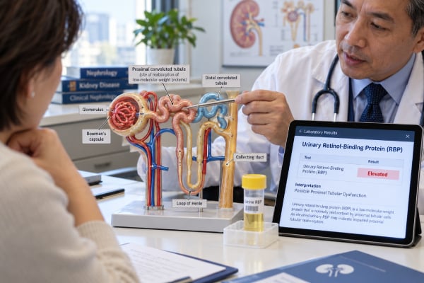
A urinary retinol-binding protein test measures retinol-binding protein 4 (RBP4), a small protein that is normally filtered by the kidneys and then almost completely reclaimed by the proximal tubules. When those tubules are injured or their reabsorptive machinery does not work properly, urinary RBP can rise sharply. This makes the marker useful for detecting proximal tubular dysfunction and low-molecular-weight proteinuria, including patterns seen in Fanconi syndrome, some inherited tubular disorders, medication or toxin exposure, and certain acute or chronic kidney diseases. Urinary RBP is not a routine screening test like urine albumin-to-creatinine ratio (uACR). It is usually ordered when the clinical question specifically involves tubular injury or when conventional urine protein findings do not fit a glomerular pattern. Interpretation depends heavily on the assay, the laboratory reference interval, urine creatinine, kidney function, and the rest of the tubular workup.
- High urinary RBP usually points to impaired proximal tubular reabsorption rather than classic glomerular albumin leakage.
- RBP is a low-molecular-weight protein and may be reported as a concentration or as an RBP-to-creatinine ratio.
- There is no single universal “normal range” across assays; use the reference interval printed by the testing laboratory.
- Causes include Fanconi syndrome, inherited proximal tubule disorders, some medications and toxins, acute tubular injury, and tubulointerstitial disease.
- A high result is interpreted with urine glucose, phosphate, bicarbonate-related findings, amino acids, albumin, total protein, creatinine, and eGFR.
Table of Contents
- What Urinary RBP Measures
- Why RBP Rises in Tubular Injury
- How the Test Is Performed
- How to Interpret High RBP
- Conditions Associated With High RBP
- RBP Versus Other Urine Protein Markers
- Follow-Up After an Abnormal Result
What Urinary RBP Measures
Retinol-binding protein 4 is a small circulating protein with a molecular weight of about 21 kDa. In blood, it transports vitamin A in association with transthyretin. A fraction of RBP that is not bound in a larger complex can pass through the glomerular filter.
Under normal conditions, the proximal tubule retrieves almost all of that filtered RBP through receptor-mediated endocytosis. The megalin-cubilin system on proximal tubular cells is central to this process. The filtered protein is taken up, processed, and degraded rather than lost in large quantities in urine.
When proximal tubular reabsorption fails, low-molecular-weight proteins escape into urine. RBP is one of the proteins that can reveal this problem. Other tubular markers include alpha-1 microglobulin, beta-2 microglobulin, and urinary cystatin C.
This is the core difference from urine albumin testing. Albuminuria most often reflects increased passage of albumin across the glomerular filtration barrier, while urinary RBP more directly reflects defective proximal tubular handling of filtered small proteins.
A urinary RBP test can therefore help identify a tubular proteinuria pattern that a routine albumin-focused dipstick might miss or underestimate.
Why RBP Rises in Tubular Injury
The proximal tubule performs far more than simple water reabsorption. It retrieves filtered glucose, phosphate, bicarbonate, amino acids, uric acid, and many small proteins. Damage to this part of the nephron can therefore produce a recognizable cluster of urinary losses.
RBP rises when filtered RBP cannot be efficiently taken back up by proximal tubular cells. In severe dysfunction, urinary RBP can increase by orders of magnitude above normal. The magnitude can be striking because normal proximal tubular reabsorption is extremely efficient.
This is why urinary RBP is useful in conditions such as Fanconi syndrome, where the proximal tubule loses multiple normally reclaimed substances. A patient may have high urinary RBP together with glucose in the urine despite a normal blood glucose level, phosphate wasting, aminoaciduria, bicarbonate loss, or uric acid wasting.
The biological meaning of an elevated RBP is therefore different from the meaning of a high total urine protein alone. Total protein gives a quantity; RBP helps describe which nephron segment is malfunctioning.
However, RBP is not perfectly specific for one disease. Proximal tubular dysfunction can result from inherited disorders, medications, heavy metals, monoclonal light chains, ischemia, inflammation, or other forms of kidney injury. The test localizes the pattern more than it identifies the final diagnosis.
How the Test Is Performed
Urinary RBP is usually measured in a spot urine specimen. Some laboratories report the absolute concentration, while others normalize RBP to urine creatinine and report an RBP-to-creatinine ratio. Creatinine normalization helps reduce the effect of urine concentration.
No universal preparation is required, but the ordering clinician or laboratory may specify a first-morning sample or other collection conditions. Following the same collection method over time is helpful when the test is being used for monitoring.
Unlike common protein dipsticks, RBP generally requires a targeted laboratory assay. Immunoassays are widely used, but methods, calibrators, units, and reference limits are not fully standardized across laboratories. This is an important practical limitation.
For that reason, an RBP value should not be interpreted using a cutoff copied from another laboratory or research paper. The correct comparison is the reference interval attached to the test result. If serial monitoring is important, using the same laboratory and assay can reduce method-related variation.
The urine sample may be tested alongside total protein, albumin, creatinine, glucose, phosphate, or other markers. A spot urine protein test shows overall protein excretion, while RBP adds information about tubular handling.
The clinician will also usually check serum creatinine and eGFR. Advanced reduction in filtration can alter the relationship between plasma RBP, filtered load, and urinary excretion, so kidney function remains part of the interpretation.
How to Interpret High RBP
A high urinary RBP result supports proximal tubular dysfunction when the clinical setting is appropriate. The strength of that conclusion depends on how elevated the marker is and whether other tubular abnormalities are present.
| Finding | What it suggests | What to check next |
|---|---|---|
| High RBP with low albumin fraction | Tubular low-molecular-weight proteinuria | Other proximal tubule losses, medications, toxins, inherited causes |
| High RBP plus glycosuria with normal blood glucose | Proximal tubular dysfunction | Phosphate, bicarbonate, uric acid, amino acids |
| High RBP plus substantial albuminuria | Mixed glomerular and tubular injury may be present | uACR, urine sediment, eGFR, underlying disease |
| Mild isolated RBP elevation | Possible early or limited tubular abnormality | Repeat test and clinical correlation |
The absence of a universal reference range is important enough to repeat. Published studies may report RBP in mg/L, µg/L, mg/g creatinine, µg/mmol creatinine, or other units. Assays may also recognize different molecular forms. A number that is abnormal in one method cannot safely be transferred to another.
A very high urinary RBP result is generally more convincing for proximal tubular dysfunction than a borderline elevation. Still, even a marked value is not a diagnosis by itself. The history may reveal a medication known to injure proximal tubules, occupational or environmental exposure, a plasma cell disorder, or a hereditary syndrome.
RBP can also be elevated in some acute kidney injury settings before or alongside changes in serum creatinine. This has driven interest in RBP as a biomarker of tubular injury, but routine clinical use varies by institution and indication.
Conditions Associated With High RBP
Several groups of disorders can produce high urinary RBP.
Fanconi syndrome and inherited proximal tubule disorders
Fanconi syndrome describes generalized proximal tubular dysfunction. Causes include inherited disorders such as cystinosis, Dent disease, and Lowe syndrome, as well as acquired conditions. RBP can be especially useful because low-molecular-weight proteinuria is a core feature.
If an inherited disorder is suspected, the age of onset, family history, growth, bone health, kidney stones, nephrocalcinosis, hearing or eye findings, and other systemic features can help narrow the diagnosis.
Medication and toxin-related tubular injury
Some medications can damage the proximal tubule. The specific risk depends on drug, dose, duration, comorbidities, and concurrent therapies. Certain antiviral agents, chemotherapies, and other nephrotoxic drugs have been linked to proximal tubular dysfunction.
Heavy metals such as cadmium can also cause proximal tubular injury. A high RBP result in a person with relevant occupational or environmental exposure may support further toxicologic evaluation.
Monoclonal protein disorders
Immunoglobulin light chains can injure proximal tubular cells and can cause Fanconi syndrome or other forms of tubulopathy. If the protein pattern is unusual, testing such as urine immunofixation or serum free light chains may be considered.
Tubulointerstitial and acute kidney injury
Inflammatory tubulointerstitial diseases, ischemic injury, and other causes of proximal tubular damage can increase low-molecular-weight proteins in urine. RBP is therefore sometimes used as a functional marker of tubular reabsorptive capacity rather than as a disease-specific marker.
RBP Versus Other Urine Protein Markers
Different urine markers answer different questions.
RBP versus albumin: Albumin is the main marker used for CKD detection, staging, and cardiovascular risk assessment. RBP is more specialized and is used when proximal tubular dysfunction is suspected.
RBP versus total protein: Total urine protein includes albumin, immunoglobulins, low-molecular-weight proteins, and other proteins. It shows how much protein is present but not which type predominates.
RBP versus beta-2 microglobulin: Both can indicate proximal tubular dysfunction. RBP has historically been considered more stable in acidic urine than beta-2 microglobulin, which can degrade at low pH. This can make RBP more practical in some settings.
RBP versus alpha-1 microglobulin: Alpha-1 microglobulin is also a useful low-molecular-weight tubular marker and is relatively stable in urine. Choice between markers often depends on local laboratory availability, assay performance, and the clinical question.
RBP versus dipstick protein: Standard dipsticks mainly detect albumin. A patient with significant tubular proteinuria can therefore have a weak dipstick response despite abnormal low-molecular-weight protein loss. When this mismatch occurs, a targeted test such as RBP, alpha-1 microglobulin, or beta-2 microglobulin can be informative.
For a broad evaluation, urine total protein, albumin, and one or more tubular markers can be combined rather than treated as competing tests.
Follow-Up After an Abnormal Result
The next step after high urinary RBP is to determine whether the abnormality is persistent, whether proximal tubular losses extend beyond protein, and what is causing the dysfunction.
Common follow-up may include:
- Repeat urinary RBP using the same assay when possible
- Serum creatinine and eGFR
- Urine total protein and albumin
- Serum and urine glucose
- Serum phosphate and urine phosphate handling when indicated
- Serum bicarbonate and acid-base assessment
- Uric acid testing
- Amino acid testing in selected cases
- Medication and supplement review
- Testing for monoclonal proteins when clinically appropriate
- Exposure history for heavy metals or other nephrotoxins
A clinician may also evaluate potassium, calcium, vitamin D status, and bone health if there is evidence of chronic phosphate wasting or Fanconi syndrome.
Treatment is directed at the cause rather than at RBP itself. A medication-related tubulopathy may improve after the responsible drug is changed or stopped when medically appropriate. An inherited disorder may need disease-specific treatment and long-term nephrology follow-up. A monoclonal protein disorder requires hematologic evaluation. Electrolyte and bicarbonate losses may need replacement.
Urgent assessment is appropriate if tubular dysfunction is accompanied by rapidly rising creatinine, severe electrolyte abnormalities, profound weakness, dehydration, reduced urine output, or other signs of acute kidney injury. A high RBP result without acute symptoms is usually evaluated in a planned clinical workup, but it should not be ignored when it is clearly abnormal or supported by other tubular findings.
Why urinary RBP is useful when routine tests look incomplete
A common reason to order urinary RBP is a mismatch between the clinical picture and routine protein testing. For example, a patient may have phosphate wasting, glycosuria with normal blood glucose, and metabolic acidosis but only a small amount of albumin on dipstick. In that setting, a low-molecular-weight protein assay can reveal substantial proximal tubular dysfunction that an albumin-focused test does not capture.
Urinary RBP can also help distinguish a predominantly tubular process from a purely glomerular one. This distinction is especially useful in disorders where both compartments can be involved. A person with chronic glomerular disease may develop secondary tubular injury, while someone with a monoclonal gammopathy may have both overflow of light chains and direct proximal tubular toxicity.
Because RBP reflects tubular reabsorptive function, it can change even when serum creatinine changes little. Serum creatinine is influenced by overall filtration, while RBP is more localized to proximal tubular handling. The markers therefore provide complementary rather than redundant information.
Pre-analytical and assay issues
Specialized biomarkers are only as useful as the method used to measure them. Urinary RBP is generally more stable in acidic urine than beta-2 microglobulin, which is one reason it has been favored in some settings. Even so, sample handling, storage time, assay calibration, and the form of RBP recognized by the method can affect results.
Creatinine-normalized ratios also require context. A person with very low muscle mass may excrete less creatinine, which can make a marker-to-creatinine ratio appear relatively high. Children, frail adults, and people with major changes in muscle mass may therefore need age- or population-appropriate interpretation.
Reference intervals can also differ by age and laboratory. The safest approach is to use the reporting laboratory’s range and, for serial measurements, repeat testing with the same method when possible. Research cutoffs should not be substituted casually for clinical assay limits.
How RBP fits with Fanconi syndrome testing
When Fanconi syndrome is suspected, RBP is one part of a pattern rather than a stand-alone confirmation. Clinicians look for inappropriate urinary losses of substances that the proximal tubule normally reabsorbs. The pattern may include:
- Glycosuria despite normal or only mildly elevated plasma glucose
- Phosphate wasting and low serum phosphate
- Bicarbonate loss causing a normal-anion-gap metabolic acidosis
- Aminoaciduria
- Uric acid wasting
- Potassium abnormalities
- Low-molecular-weight proteinuria
The combination helps distinguish generalized proximal tubular dysfunction from an isolated abnormal biomarker.
Long-term consequences depend on the cause. Chronic phosphate wasting can contribute to osteomalacia, bone pain, fractures, or impaired growth in children. Chronic bicarbonate loss can affect bone and muscle function. For these reasons, the significance of a high urinary RBP is not limited to kidney filtration—it can identify a transport defect with systemic consequences that need targeted replacement and treatment.
Urinary RBP is also best understood as a functional marker rather than a direct measurement of cell death. A proximal tubule can lose reabsorptive capacity because of reversible transport dysfunction, toxic injury, inflammation, or inherited defects. Therefore, a high RBP does not automatically mean permanent structural damage. Serial results, recovery of electrolyte handling, and changes in eGFR help show whether the process is resolving or progressing.
The test is not appropriate as a general population kidney screen. For routine CKD detection, uACR and eGFR have stronger evidence, standardized risk categories, and broader availability. RBP becomes valuable when a clinician has a specific reason to suspect tubular disease or when conventional albumin-based testing leaves an important question unanswered.
When RBP is ordered after exposure to a potentially nephrotoxic medication or toxin, baseline information can improve interpretation. A rise from a prior normal level may be more informative than a single mildly abnormal value in a person whose baseline is unknown. The decision to change therapy still depends on the overall risk-benefit balance, other laboratory abnormalities, and whether safer alternatives exist.
References
- Toward Assessment of Tubular Function in Kidney Disease 2025 (Review)
- Protein handling in kidney tubules 2025 (Review)
- The Clinical Significance of Urinary Retinol-Binding Protein 4: A Review 2022 (Review)
- Proficient Novel Biomarkers Guide Early Detection of Acute Kidney Injury: A Review 2022 (Review)
- Urine MMP7 as a kidney injury biomarker. 2024 (Review)
- Urine retinol-binding protein 4: a functional biomarker of the proximal renal tubule 2014 (Review)
Disclaimer
Urinary retinol-binding protein is a specialized kidney marker whose reference range and clinical meaning depend on the assay and the reason for testing. An abnormal result should be interpreted with kidney function, urine albumin and total protein, other proximal tubular markers, medications, and medical history. This article is educational and does not replace evaluation by a clinician or nephrologist.