A spot urine protein test measures protein in a single urine sample and is commonly used to screen for or monitor proteinuria. The most useful quantitative version is usually the urine protein-to-creatinine ratio (UPCR), which compares total urine protein with urine creatinine to reduce the effect of how concentrated or dilute the sample is. A spot test is much easier than collecting urine for 24 hours and is often accurate enough for routine clinical decisions. High protein can reflect glomerular kidney disease, tubular injury, overflow proteins, urinary tract inflammation, or a temporary response to exercise, fever, dehydration, or acute illness. The result is most meaningful when it is repeated if necessary and interpreted with urine albumin, kidney function, blood pressure, urine sediment, and symptoms. A very high UPCR, rising creatinine, blood in the urine, or edema generally needs more urgent evaluation than a small isolated increase.
- A spot UPCR below about 150 mg/g is generally normal to mildly increased in adults, while values above 500 mg/g are severely increased.
- UPCR is usually more useful than protein concentration alone because it corrects partly for urine dilution.
- A first-morning sample is often best for confirmation, especially when a random result is mildly abnormal.
- High total protein can come from albumin, low-molecular-weight proteins, or monoclonal light chains, so the protein type may matter.
- Heavy or persistent proteinuria, especially with edema, hematuria, high blood pressure, or falling eGFR, should be medically evaluated.
Table of Contents
- What a Spot Urine Protein Test Measures
- Spot UPCR Versus Dipstick and 24-Hour Testing
- Normal and High Spot Protein Results
- Causes of High Spot Urine Protein
- Collection Timing and Result Variability
- What Follow-Up Testing Can Show
- When High Protein Needs Faster Evaluation
What a Spot Urine Protein Test Measures
A spot urine protein test measures protein in one urine specimen rather than over a fixed period. The result may be reported as protein concentration, such as mg/dL, but concentration alone changes substantially with hydration. For that reason, clinicians often request a urine protein-to-creatinine ratio.
UPCR includes all measurable urine protein, not only albumin. That is useful when the clinician wants a broad estimate of protein loss or when non-albumin protein could be important.
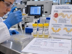
Healthy kidneys limit protein loss in two main ways. The glomerular filtration barrier prevents most larger proteins from entering the filtrate, and the proximal tubules reclaim many small proteins that do pass through. Proteinuria can therefore arise from glomerular leakage, tubular reabsorption failure, excess circulating small proteins, or inflammation and bleeding lower in the urinary tract.
A spot test does not identify the mechanism by itself. A high number tells the clinician how much protein is present relative to creatinine, while other tests help determine what kind of protein is present and why.
Spot UPCR Versus Dipstick and 24-Hour Testing
Each method has strengths and limitations.
Urine dipstick: A protein dipstick is fast and useful for screening, but it is semiquantitative and mainly detects albumin. It can underestimate tubular proteins and immunoglobulin light chains. The reading is also affected by urine concentration and pH.
Spot UPCR: UPCR measures total protein and creatinine quantitatively. It is convenient, requires one sample, and avoids most 24-hour collection errors. For many outpatient situations, it is the preferred way to quantify total proteinuria.
24-hour urine protein: A 24-hour urine protein test measures the actual amount of protein excreted over a full day. It can be useful when a spot result is discordant with the clinical picture, in selected pregnancy or glomerular disease settings, or when a timed value is specifically required. Its major weakness is inaccurate collection.
UPCR is often interpreted as roughly corresponding to grams of protein excreted per day when expressed as g/g in an average adult, but this is only an approximation. Someone with unusually low muscle mass may excrete less creatinine, making the ratio appear higher for a given amount of protein. A muscular person may have the opposite effect.
Normal and High Spot Protein Results
Adult reference ranges vary somewhat by laboratory, but common categories are:
| UPCR | Approximate category | Clinical interpretation |
|---|---|---|
| <150 mg/g | Normal to mildly increased | Usually not significant proteinuria |
| 150–500 mg/g | Moderately increased | Repeat and assess cause in context |
| >500 mg/g | Severely increased | More substantial kidney or systemic abnormality is possible |
| Approximately 3,000–3,500 mg/g or higher | Nephrotic range | Heavy protein loss requiring focused evaluation |
These are broad clinical categories rather than exact universal cutoffs. Laboratories may report mg/mg, mg/mmol, or g/g instead of mg/g. Always compare like units.
The albumin-to-creatinine ratio uses different thresholds because it measures albumin only. A uACR below 30 mg/g is A1, 30–300 mg/g is A2, and above 300 mg/g is A3. A UPCR and uACR from the same person can therefore look numerically different without being contradictory.
A single abnormal UPCR does not prove chronic kidney disease. Persistence over time matters, and temporary influences should be considered before a mild elevation is labeled chronic.
Causes of High Spot Urine Protein
Glomerular disease is a common cause of substantial proteinuria. Damage to the filtration barrier allows albumin and other plasma proteins into urine. Diabetic kidney disease, focal segmental glomerulosclerosis, membranous nephropathy, lupus nephritis, and other glomerulopathies can produce this pattern.
Tubular injury causes loss of low-molecular-weight proteins that should have been reabsorbed in the proximal tubule. A tubular proteinuria test may be considered when total protein is elevated but albumin does not account for most of it.
Overflow proteinuria occurs when blood contains very high levels of a small protein that is filtered faster than the tubules can reclaim it. Monoclonal immunoglobulin light chains in plasma cell disorders are the classic example. Myoglobin from severe muscle injury and hemoglobin from intravascular hemolysis are other possibilities.
Postrenal protein can come from inflammation, infection, or bleeding in the urinary tract. A urinary tract infection with many white blood cells or blood may therefore raise measured protein without a primary glomerular disorder.
Temporary increases are also common. Strenuous exercise, fever, dehydration, acute illness, severe hyperglycemia, and marked hypertension can raise urine protein. In adolescents and young adults, orthostatic proteinuria causes higher daytime protein but a normal first-morning value.
Collection Timing and Result Variability
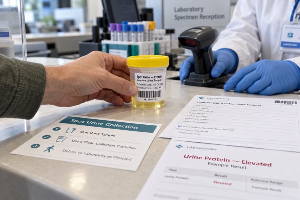
A random urine specimen is convenient for screening. If the result is unexpected or only mildly elevated, a first-morning specimen often gives a cleaner answer.
The first-morning sample reduces several sources of variation. The person has been recumbent, has not exercised, and has usually had a relatively stable hydration period. This makes it especially useful when distinguishing persistent proteinuria from orthostatic or activity-related protein excretion.
Before a confirmatory test, it is reasonable to avoid heavy exercise for about a day if medically appropriate. Testing during fever, acute infection, or severe uncontrolled blood sugar may be postponed when safe because these conditions can cause transient proteinuria.
A clean-catch sample helps reduce contamination. Menstrual blood, heavy vaginal discharge, or obvious urinary bleeding can alter a protein result. If contamination is likely, repeating the sample under better conditions is often more useful than overinterpreting the first number.
Serial testing should be as consistent as possible. A trend from 300 to 800 mg/g is more convincing if both samples were first-morning collections analyzed by the same laboratory than if one was collected after exercise and the other after overnight rest.
What Follow-Up Testing Can Show
The follow-up question is usually: Is the protein persistent, where is it coming from, and is kidney function affected?
Common next tests include:
- Repeat UPCR
- uACR to determine the albumin component
- Serum creatinine and eGFR
- Urinalysis and microscopy
- Blood pressure measurement
- Serum albumin when proteinuria is heavy
- Blood glucose or HbA1c if diabetes is relevant
- Additional serologic or hematologic studies when a systemic cause is suspected
A large difference between UPCR and uACR can be informative. If total protein is high but albumin is only a small fraction, the clinician may investigate tubular proteins or monoclonal light chains. This may include urine protein electrophoresis, immunofixation, or targeted low-molecular-weight protein assays.
Urine sediment also matters. Red blood cell casts or dysmorphic red cells suggest glomerular inflammation. White blood cell casts may suggest interstitial or infectious kidney disease. Fatty casts can occur in heavy nephrotic proteinuria.
The relationship with eGFR helps define severity but should not be oversimplified. Proteinuria can be substantial while eGFR remains normal, especially early in disease. Conversely, some people with advanced CKD have little proteinuria. Both markers are needed for a complete assessment.
When High Protein Needs Faster Evaluation
Mild proteinuria discovered during an acute illness may simply need repeat testing after recovery. The urgency changes when protein is heavy, persistent, or accompanied by signs of kidney dysfunction.
Faster evaluation is appropriate for:
- UPCR in the nephrotic range
- Rapidly increasing proteinuria
- New leg, facial, or abdominal swelling
- Low blood albumin
- Blood in the urine, especially with casts
- Rising serum creatinine or falling eGFR
- Markedly reduced urine output
- Severe or newly uncontrolled blood pressure
- Symptoms suggesting autoimmune disease, infection, plasma cell disease, or another systemic condition
A very high UPCR may be part of nephrotic-range proteinuria. If low serum albumin and edema are also present, the clinical picture may be nephrotic syndrome and requires evaluation of the underlying glomerular disease and complications.
Urgent medical care is appropriate for shortness of breath, chest pain, severe generalized swelling, one-sided painful leg swelling, confusion, or very low urine output. These symptoms can indicate complications such as fluid overload, thrombosis, severe kidney injury, or another acute condition.
For an asymptomatic person with a modestly elevated spot result, the most useful next step is usually a repeat quantitative sample and a review of kidney function rather than assuming the worst. Spot testing is valuable because it is easy to repeat, but the clinical meaning comes from persistence, magnitude, protein type, and the rest of the kidney evaluation.
Why the same protein concentration can mean different things
A laboratory may report urine protein concentration in mg/dL as well as UPCR. Concentration is highly dependent on urine water content. For example, 30 mg/dL in a very concentrated morning sample does not carry the same meaning as 30 mg/dL in a very dilute specimen. UPCR helps solve this problem by indexing the protein to creatinine.
This is also why comparing raw concentrations from different visits can be misleading. If one sample was collected after overnight fluid restriction and another after drinking several glasses of water, the protein concentration may change even if daily protein excretion is similar. Ratios are generally better for trend monitoring.
When a 24-hour collection still adds value
Spot testing has replaced many routine 24-hour collections, but timed urine is still useful in selected situations. It can help when the UPCR seems inconsistent with symptoms or serum albumin, when creatinine excretion is unusually low or high, or when a disease-specific protocol requires a timed measurement.
The quality of the 24-hour result depends on collection accuracy. Missing a void makes protein excretion look falsely low, while extending the collection beyond 24 hours can make it look high. A urine creatinine excretion result may help clinicians judge whether the collection is plausible, although this assessment is also affected by muscle mass and diet.
Interpreting changes during treatment
In established proteinuric kidney disease, clinicians often use serial UPCR to judge whether therapy is reducing protein loss. The goal depends on the diagnosis. A fall from nephrotic-range values to a much lower level can be clinically important even if the result remains abnormal.
When following a trend, it is useful to keep the test method consistent. A change from a dipstick result of “3+” to a UPCR of 1,200 mg/g cannot be interpreted as a precise percentage reduction because the two methods measure protein differently. Repeated quantitative UPCR values provide a cleaner comparison.
Blood pressure and eGFR should be reviewed at the same time. Some kidney-protective treatments lower intraglomerular pressure, which may reduce proteinuria and cause a small early change in filtration. A clinician distinguishes expected treatment effects from true acute kidney injury using the size, timing, and persistence of the changes.
Situations where non-albumin protein deserves special attention
A spot total protein result is particularly useful when albumin is not expected to tell the whole story. Examples include suspected plasma cell disorders, Fanconi syndrome, tubulointerstitial disease, and severe muscle injury. In these settings, relying only on uACR can underestimate the total protein burden.
The practical clue is often discordance: UPCR is high, but uACR is much lower than expected. That pattern should not be converted into a diagnosis by arithmetic alone, but it tells the clinician to consider electrophoresis, immunofixation, serum free light chains, or targeted tubular markers rather than repeating an albumin-only test.
A spot protein result can also be useful when monitoring known glomerular disease. In that setting, the question is often not whether protein is present but whether it is falling enough to suggest remission or rising enough to suggest relapse. Disease-specific definitions differ, so the same UPCR may carry a different implication in minimal change disease, IgA nephropathy, lupus nephritis, or diabetic kidney disease.
Children and adolescents need age-specific reference ranges. Young children can have a higher normal protein-to-creatinine ratio because creatinine excretion is lower, while adolescents commonly show orthostatic proteinuria. A first-morning specimen and pediatric thresholds reduce the risk of labeling a benign pattern as chronic disease.
Pregnancy likewise uses a specific clinical framework. Spot protein-to-creatinine ratio is often used to assess significant proteinuria in suspected preeclampsia, but obstetric thresholds and blood pressure findings govern interpretation. A routine adult CKD category should not be substituted for pregnancy-specific guidance.
Finally, urine appearance is unreliable. Foamy urine can occur with concentrated urine, a strong urinary stream, or cleaning products in the toilet, while substantial proteinuria may produce no obvious visual change. Quantitative testing is the only dependable way to measure the amount of protein.
Because creatinine appears in the denominator, body composition can influence UPCR. A frail person with low muscle mass may produce less urine creatinine, making the ratio appear higher for a given protein excretion, while a very muscular person can show the opposite effect. This limitation rarely makes spot testing unusable, but it explains why clinicians sometimes confirm surprising results with repeat testing or a timed collection.
For routine follow-up, a quantitative ratio is generally more useful than repeatedly checking only dipstick grades, because it provides a number that can be compared over time and related to treatment response.
This consistency is especially helpful when decisions depend on whether proteinuria is improving, stable, or clearly worsening. When possible, compare follow-up samples collected under similar conditions and interpreted by the same laboratory method, because this reduces avoidable variation and makes a real change in protein excretion easier to recognize.
References
- KDIGO 2024 CKD Guideline 2024 (Guideline)
- Methods for Diagnosing Proteinuria-When to Use Which Test and Why: A Review 2025 (Review)
- Measuring albuminuria or proteinuria: does one answer fit all? 2023 (Review)
- Recommendations for pre-analytical urine sample handling to allow optimal assessment of albuminuria and proteinuria. 2026 (Review)
- Diagnosis of Chronic Kidney Disease and Assessing Glomerular Filtration Rate. 2023 (Review)
- Protein handling in kidney tubules 2025 (Review)
Disclaimer
A spot urine protein result cannot by itself diagnose the cause of proteinuria or determine how serious a kidney condition is. Abnormal results should be interpreted with repeat testing, eGFR, blood pressure, urine microscopy, albumin measurements, and medical history. Seek urgent care for severe swelling, breathing difficulty, chest pain, very low urine output, or other acute symptoms.