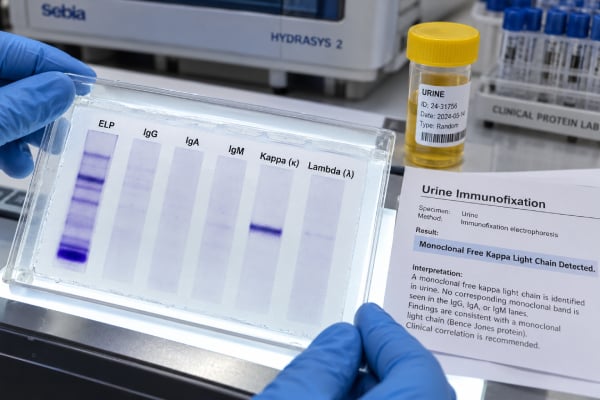
A urine immunofixation test is a sensitive laboratory method used to determine whether urine contains a monoclonal immunoglobulin protein, especially free kappa or lambda light chains. A positive result can support the diagnosis or monitoring of plasma cell and B-cell disorders such as multiple myeloma, AL amyloidosis, or monoclonal gammopathy of renal significance (MGRS), but it does not diagnose any of these conditions by itself. Immunofixation is qualitative or semiquantitative: its main job is to identify the type of monoclonal protein rather than measure total daily protein loss. It is often used with urine protein electrophoresis (UPEP), serum protein electrophoresis, serum immunofixation, and serum free light-chain testing. Modern serum testing is highly sensitive for monoclonal gammopathy screening, yet urine studies remain clinically useful in light-chain disease, renal presentations, amyloidosis evaluation, and selected monitoring situations. Interpretation depends on whether the band is monoclonal, which light chain is involved, how much protein is being excreted, kidney function, and whether there is evidence of organ damage.
- Urine immunofixation identifies the immunoglobulin type of a monoclonal urine protein, most often free kappa or free lambda light chains.
- A positive monoclonal band is abnormal but does not by itself prove multiple myeloma.
- UPEP helps separate and quantify protein fractions, while immunofixation is more sensitive for typing a monoclonal protein.
- Serum electrophoresis, serum immunofixation, and serum free light chains are usually interpreted alongside urine results.
- Monoclonal protein plus kidney injury can require urgent evaluation for myeloma-related kidney disease or MGRS, sometimes including kidney biopsy.
Table of Contents
- What urine immunofixation detects
- How the test works
- Positive, negative, and equivocal results
- Conditions linked to monoclonal urine proteins
- Kidney disease, light chains, and MGRS
- Collection and related blood and urine tests
- What happens after a positive test
What urine immunofixation detects
Urine immunofixation electrophoresis, often abbreviated uIFE, looks for a discrete population of immunoglobulin protein made by one clone of plasma cells or B cells. The most clinically important urinary findings are monoclonal free light chains—either kappa or lambda.
Normal immune systems contain many plasma cell clones making many different antibodies. That creates a broad, polyclonal mixture of immunoglobulins. A monoclonal gammopathy arises when one clone expands and produces identical immunoglobulin molecules or components. In urine, the clonal product may appear as a restricted light-chain band.
Historically, monoclonal light chains in urine were called Bence Jones proteins. Modern immunofixation is far more specific than old heat-precipitation methods because it identifies the immunoglobulin class or light-chain type using antisera.
uIFE is not primarily a “how much protein?” test. A UPEP test can quantify a monoclonal component and show the overall protein pattern, while immunofixation answers the typing question: is the restricted protein kappa, lambda, or associated with a heavy chain?
How the test works
The laboratory first separates urinary proteins by electrophoresis. The separated proteins are then exposed to antisera directed against immunoglobulin heavy chains and light chains. Where a specific antibody binds its matching immunoglobulin component, a visible band or pattern forms.
A narrow, discrete band in one light-chain lane can indicate a monoclonal protein. A broad diffuse pattern suggests polyclonal proteins rather than one clone. Laboratory specialists interpret the migration pattern, intensity, and corresponding lanes.
Urine often contains lower protein concentrations than serum, so the specimen may be concentrated before testing. This improves sensitivity but also means pre-analytical handling matters. Very dilute urine can make a small monoclonal protein harder to detect.
Modern laboratories may use agarose gel or capillary-based methods for electrophoresis and immunotyping. Some centers are adopting mass spectrometry for monoclonal protein detection, but electrophoresis and immunofixation remain widely established clinical tools.
Because immunofixation is highly sensitive, it can remain positive when the monoclonal protein concentration is too small to form a clearly quantifiable M-spike on routine electrophoresis. That sensitivity is useful for confirmation, but it also means the result must be interpreted with the clinical picture rather than equated with malignancy.
Positive, negative, and equivocal results
A positive urine immunofixation means the laboratory detected a restricted immunoglobulin pattern consistent with a monoclonal protein. The report may state “monoclonal free kappa light chain,” “monoclonal free lambda light chain,” or another typed pattern.
A positive result is abnormal, but it does not specify the diagnosis. The same kind of monoclonal protein can occur in multiple myeloma, light-chain MGUS, AL amyloidosis, MGRS, some lymphomas, or other clonal disorders. Diagnosis depends on the amount of clonal disease, symptoms, organ involvement, bone marrow findings, imaging, serum studies, and sometimes tissue biopsy.
A negative result means no monoclonal protein was detected by that assay in that specimen. It lowers the likelihood of urinary monoclonal protein but does not completely exclude a plasma cell disorder. Some disorders produce mainly serum protein, produce very low quantities, or fluctuate below the detection limit. Serum free light chains and serum immunofixation may still be abnormal.
An equivocal or faint band can occur when the amount is very small, the specimen is dilute, or the pattern is technically difficult to classify. The laboratory may recommend repeat testing, correlation with serum immunofixation, or another method.
Polyclonal light chains are different. They can increase with inflammation, infection, autoimmune disease, or reduced kidney function without showing the sharp restricted pattern of one clone. The distinction between monoclonal and polyclonal is the central interpretive purpose of immunofixation.
Conditions linked to monoclonal urine proteins
Multiple myeloma is the condition most people associate with a monoclonal urine protein, but the laboratory finding spans a broader disease spectrum. Myeloma is a plasma cell malignancy that can cause anemia, bone lesions, hypercalcemia, kidney injury, and other organ damage. Some myelomas produce large quantities of free light chains that enter urine.
MGUS is a clonal plasma cell or B-cell condition with a lower tumor burden and no attributable myeloma-defining organ damage. It is common with increasing age. A monoclonal protein in a person with MGUS still requires appropriate risk assessment and follow-up.
AL amyloidosis occurs when monoclonal light chains misfold and form amyloid deposits in organs such as the kidneys, heart, nerves, liver, or gastrointestinal tract. Kidney involvement often presents with albuminuria or nephrotic-range protein loss. Testing usually combines serum and urine immunofixation with serum free light chains because no single method is sufficiently sensitive on its own.
MGRS describes kidney diseases caused by a nephrotoxic monoclonal immunoglobulin from a clone that may be too small to meet conventional criteria for myeloma or lymphoma. The concept is important because a small clone can still cause progressive kidney damage.
Other B-cell or plasma cell disorders can also produce monoclonal proteins. The laboratory result identifies the protein; the clinical workup identifies the disease.
A urine free light-chain test may provide quantitative kappa and lambda values, but immunofixation gives stronger evidence about clonality when a distinct band is present.
Kidney disease, light chains, and MGRS
Kidney involvement changes the importance of a monoclonal protein. Some monoclonal light chains are directly toxic to kidney structures. Others form deposits or crystals, activate complement, or create obstructing casts.
In myeloma cast nephropathy, a very high filtered light-chain load combines with proteins in the distal nephron to form casts that obstruct and injure tubules. This can cause rapidly worsening kidney function and is a medical urgency.
In AL amyloidosis and monoclonal immunoglobulin deposition disease, abnormal proteins deposit in glomeruli or other kidney compartments. These diseases can produce heavy albuminuria and nephrotic syndrome rather than the same pattern seen in cast nephropathy.
MGRS covers several additional lesions, including light-chain proximal tubulopathy, proliferative glomerulonephritis with monoclonal immunoglobulin deposits, and certain complement-mediated processes linked to monoclonal proteins. Because the lesion cannot be determined from immunofixation alone, kidney biopsy is often essential when MGRS is suspected.
A striking difference between total urine protein and urine albumin can raise suspicion for light-chain proteinuria. For example, a high UPCR with only modest uACR suggests a large non-albumin component. That pattern can prompt uIFE and serum free light-chain testing, though it is not specific for a monoclonal disorder.
Collection and related blood and urine tests
Urine immunofixation may be performed on a random sample or a 24-hour collection, depending on the laboratory and clinical purpose. A 24-hour specimen can be useful when daily monoclonal protein excretion also needs to be quantified. The sample must be collected completely because missed voids can underestimate the protein burden.
For a 24-hour collection, the usual process is to discard the first void at the starting time, collect every subsequent urine sample for the next 24 hours, and include the final void at the ending time. Storage instructions vary by laboratory.
A monoclonal gammopathy workup commonly combines several tests:
- Serum protein electrophoresis (SPEP) to look for and quantify an M-protein in blood.
- Serum immunofixation to identify the immunoglobulin type.
- Serum free light chains to measure free kappa, free lambda, and the serum ratio.
- UPEP to separate and quantify urine protein fractions and urinary M-protein.
- Urine immunofixation to sensitively type urinary monoclonal protein.
A complete blood count, creatinine/eGFR, calcium, albumin, and sometimes imaging and bone marrow biopsy are added when myeloma or another plasma cell disorder is being evaluated.
For initial screening, modern guidance often emphasizes serum SPEP, serum immunofixation, and serum free light chains because the combination is highly sensitive and avoids dependence on a 24-hour urine collection. Urine immunofixation remains particularly relevant when AL amyloidosis is suspected, when renal disease suggests light-chain injury, or when a known urinary monoclonal protein is being monitored.
What happens after a positive test
The next step after a positive uIFE is not to assume cancer; it is to define the clone, quantify disease burden, and look for organ effects.
If serum studies have not already been done, they are usually obtained. The clinician reviews complete blood count, calcium, creatinine, eGFR, serum albumin, and total protein. Bone imaging may be needed when myeloma is suspected. A bone marrow biopsy can determine the percentage and characteristics of clonal plasma cells or B cells.
Kidney findings often determine urgency. Rapidly rising creatinine, heavy proteinuria, or a suspected nephrotoxic monoclonal protein may prompt immediate hematology-nephrology coordination. A kidney biopsy can establish whether the monoclonal protein is actually causing the kidney lesion, which is crucial for diagnosing MGRS.
A small monoclonal band without organ damage may ultimately be classified as MGUS or another precursor condition and monitored rather than treated. By contrast, a small clone that is causing kidney injury may require therapy even if it does not meet classic malignancy criteria.
Monitoring strategy depends on the disease. Serum free light chains are often convenient for light-chain myeloma response assessment, while serum and urine electrophoresis or immunofixation may be used for selected patients. Laboratory methods and clinical guidelines continue to evolve, so the exact panel is individualized.
The key point is that uIFE is a protein-typing test. Its result becomes clinically meaningful only after it is linked to the amount of monoclonal protein, serum findings, kidney function, symptoms, and tissue or marrow evidence when needed.
Common reporting phrases and how to read them
Urine immunofixation reports can be terse, so understanding the wording helps. “No monoclonal protein detected” means the assay did not identify a restricted immunoglobulin band in that specimen. It is reassuring but does not exclude every plasma cell disorder, especially if serum studies or clinical findings remain suspicious.
“Monoclonal free kappa light chain detected” or “monoclonal free lambda light chain detected” is a clearer abnormal finding. It indicates clonality of the urinary light chain but still does not specify whether the underlying condition is MGUS, myeloma, AL amyloidosis, MGRS, or another B-cell or plasma cell disorder.
A report may say “faint restricted band” or “suspicious band.” Small bands can be difficult to classify, particularly in dilute urine or after treatment when disease burden is low. The laboratory may recommend repeat testing or correlation with serum immunofixation and serum free light chains.
“Polyclonal increase” means multiple immunoglobulin populations are present rather than one dominant clone. This can occur with inflammation, infection, autoimmune disease, or reduced renal clearance. Polyclonal findings can be abnormal without representing a monoclonal gammopathy.
Another important phrase is “M-protein too small to quantify.” Electrophoresis may be unable to measure a reliable peak area while immunofixation still detects and types the band. This is one reason immunofixation is considered a confirmation and sensitivity-enhancing test rather than merely a duplicate of UPEP.
When monitoring treated disease, a previously positive immunofixation can become negative as the monoclonal protein falls. That is generally a favorable laboratory change, but response classification may require serum testing, marrow assessment, imaging, and disease-specific criteria. A negative urine immunofixation by itself does not prove complete eradication of the clone.
Patients should also know that a positive result is not caused by ordinary dietary protein intake. Immunofixation is detecting an immunoglobulin pattern, not simply the amount of protein eaten. Hydration can affect concentration and sensitivity, but it does not create a true monoclonal band.
Timing can matter after treatment. Therapeutic monoclonal antibodies and changes in immunoglobulin production can complicate some serum electrophoretic patterns, while urine findings may evolve differently. Specialist laboratories interpret results in the context of treatment history and prior band location rather than reading each study in isolation.
Kidney function can also affect how much monoclonal light chain reaches urine. Reduced GFR can increase serum free light chains while altering urinary excretion, and tubular injury can increase urinary loss. That is why a faint urine band does not necessarily mean a small total body burden, and a negative urine study does not guarantee absence of a circulating clone.
When a positive uIFE is discovered incidentally in someone without symptoms, the appropriate next step is structured evaluation rather than emergency assumptions. The clinician typically confirms serum studies, reviews blood counts, calcium and kidney function, and decides whether hematology referral or additional imaging is needed based on the whole profile.
For follow-up, comparing the new report with the original typed band can be useful. A different faint band after treatment may not represent the same clone and sometimes reflects immune recovery or assay complexity. Hematology laboratories are best placed to judge whether a new pattern represents persistent disease, relapse, or a separate benign finding.
References
- Multiple myeloma: 2024 update on diagnosis, risk-stratification, and management 2024 (Review)
- Diagnosis and Management of Multiple Myeloma: A Review 2022 (Review)
- A review of clinical guidelines, laboratory recommendations and external quality assurance programs for monoclonal gammopathy testing 2023 (Review)
- Recommendations for the study of monoclonal gammopathies in the clinical laboratory. A consensus of the Spanish Society of Laboratory Medicine and the Spanish Society of Hematology and Hemotherapy. Part I: Update on laboratory tests for the study of monoclonal gammopathies 2023 (Position Statement)
- Urine Protein Immunofixation Electrophoresis: Free Light Chain Urine Immunofixation Electrophoresis Is More Sensitive than Conventional Assays for Detecting Monoclonal Light Chains and Could Serve as a Marker of Minimal Residual Disease. 2023
- Monoclonal gammopathies of renal significance 2023 (Review)
Disclaimer
This article is for general education and cannot diagnose multiple myeloma, MGUS, AL amyloidosis, or MGRS. Urine immunofixation must be interpreted with serum monoclonal protein studies, kidney function, symptoms, and sometimes bone marrow or kidney biopsy findings. Rapid kidney deterioration, severe weakness, confusion, dehydration, marked swelling, or other acute symptoms need prompt medical evaluation.