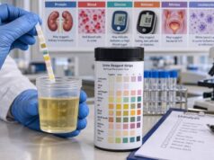
A urine myoglobin test looks for myoglobin, an oxygen-binding protein that normally stays inside skeletal and heart muscle cells. When muscle is damaged, myoglobin can enter the bloodstream, pass through the kidneys, and appear in urine. A positive result can support a diagnosis of significant muscle injury or rhabdomyolysis, but urine myoglobin is not the main test used to diagnose or monitor rhabdomyolysis. Blood creatine kinase (CK) is generally more reliable because myoglobin rises and clears quickly, so a urine sample can be negative even after major muscle damage. Myoglobin can also turn urine red-brown or tea-colored and can contribute to acute kidney injury, especially when muscle breakdown is severe, dehydration is present, or other kidney stressors coexist. Because rhabdomyolysis can also cause dangerous potassium and other electrolyte abnormalities, a positive urine myoglobin result should be interpreted with symptoms, CK, kidney function, electrolytes, and the clinical situation rather than by itself.
- Urine myoglobin usually indicates muscle-cell breakdown and is most often considered when rhabdomyolysis is suspected.
- A negative urine myoglobin result does not rule out rhabdomyolysis because myoglobin can disappear from blood and urine within hours.
- A urine dipstick may read “blood” with few or no red blood cells on microscopy when myoglobin is present.
- CK is usually the key laboratory marker for rhabdomyolysis; many clinicians use CK above 1,000 U/L or more than 5 times the upper limit of normal as a diagnostic threshold in the right setting.
- Dark urine with severe muscle pain, weakness, swelling, reduced urine output, or recent major exertion, heat illness, trauma, seizures, or drug exposure needs prompt medical evaluation.
Table of Contents
- What the Urine Myoglobin Test Measures
- What a Positive Urine Myoglobin Result Means
- Myoglobin, Rhabdomyolysis, and CK
- Kidney Risk and Other Complications
- Common Causes of Myoglobinuria
- How the Test Is Performed and Interpreted
- When to Seek Medical Care
What the Urine Myoglobin Test Measures
Myoglobin is a small heme-containing protein found mainly in muscle. Its job inside muscle cells is to bind and store oxygen. Under normal conditions, only very small amounts circulate in blood or appear in urine. When skeletal muscle cells are injured, however, they can release large amounts of myoglobin into the bloodstream.
Because myoglobin is small, the kidneys filter it readily. Once the amount in blood rises enough, it can appear in urine, a finding called myoglobinuria. Depending on the amount present, urine may look normal or may become pink, red, brown, or tea-colored.
A laboratory urine myoglobin assay measures myoglobin directly. This is different from the routine urine dipstick “blood” pad. The dipstick reacts to heme pigment, so it can become positive from intact red blood cells, free hemoglobin, or myoglobin. If the dipstick is strongly positive for blood but urine microscopy shows few or no red blood cells, myoglobinuria or hemoglobinuria becomes more likely. The urine blood dipstick test therefore provides a useful clue, but it cannot distinguish the pigments on its own.
Reference ranges for direct urine myoglobin vary by laboratory and assay. Many laboratories simply report the result as negative or positive, while others provide a concentration. Because methods differ and myoglobin is unstable in urine, the laboratory’s own reference interval should be used rather than a universal cutoff.
What a Positive Urine Myoglobin Result Means
A positive urine myoglobin result means enough myoglobin has entered the urine to be detected. The main implication is recent or ongoing muscle injury. The result does not identify the cause or tell how much kidney damage has occurred.
Rhabdomyolysis is the most important condition associated with substantial myoglobin release. It occurs when skeletal muscle breaks down and releases intracellular contents, including CK, potassium, phosphate, and myoglobin. The syndrome can range from a mild CK elevation with few symptoms to a medical emergency with electrolyte abnormalities, acute kidney injury, compartment syndrome, shock, or cardiac rhythm problems.
A positive urine myoglobin result is most meaningful when it fits the clinical picture. Examples include dark urine after extreme exercise, muscle crushing after trauma, prolonged immobilization, seizures, heat stroke, or exposure to a drug known to injure muscle.
The result is less useful as an isolated screening test. Myoglobin appears early and clears rapidly. Blood myoglobin may rise within a few hours of muscle injury and then fall much sooner than CK. Urine myoglobin can therefore be absent by the time a person is tested, even when CK remains markedly elevated.
A positive result also does not automatically mean the kidneys are failing. Some people with substantial myoglobinuria maintain normal kidney function, while others develop acute kidney injury. Risk depends on the overall severity of muscle breakdown, circulating volume, acid-base status, comorbid illness, and other kidney stressors.
The amount of myoglobin in urine also does not map neatly to the amount of muscle damage. A very high early concentration can fall rapidly as the kidneys clear the protein, while CK remains elevated. Urine output and hydration status can change the measured concentration as well. For that reason, clinicians generally do not use a single urine myoglobin value to grade rhabdomyolysis severity or decide whether a patient is safe for discharge.
Timing can be especially important after exertion. A person who develops severe muscle pain and cola-colored urine soon after a demanding workout may have detectable myoglobin early, but someone tested the next day may already have a negative urine assay while CK is still climbing. The same principle applies after seizures, trauma, or prolonged immobilization. The test is best understood as a time-sensitive clue rather than a durable record of muscle injury. This is also why serial CK measurements are usually more useful than repeating urine myoglobin over and over.
Myoglobin, Rhabdomyolysis, and CK
For suspected rhabdomyolysis, serum CK is usually the central laboratory test, not urine myoglobin. CK is an enzyme released from damaged muscle and remains elevated longer than myoglobin, making it more dependable for diagnosis and serial monitoring.
Many clinical references define rhabdomyolysis using a CK concentration above about 1,000 U/L or more than 5 times the laboratory upper limit of normal in the appropriate clinical setting. There is no single CK value that perfectly predicts kidney injury, and a high CK must be interpreted with symptoms, timing, cause, and other laboratory results.
A clinician evaluating possible rhabdomyolysis may order:
- CK, often repeated to identify the peak and confirm that levels are falling;
- serum creatinine and blood urea nitrogen to assess kidney function;
- potassium, phosphate, calcium, bicarbonate, and other electrolytes;
- urinalysis and urine microscopy;
- liver-associated enzymes such as AST and ALT, which can rise because muscle also contains these enzymes;
- an electrocardiogram when potassium abnormalities or arrhythmia risk are a concern.
An acute kidney injury blood test panel can help show whether muscle breakdown is affecting filtration or electrolyte balance.
Why urine myoglobin can be negative in true rhabdomyolysis
Myoglobin has a short circulating half-life. It can be filtered and cleared before a patient reaches care, whereas CK often peaks later, commonly over the first one to three days. Urine concentration, sample timing, assay design, and myoglobin instability can further reduce sensitivity.
That timing difference explains an important rule: a negative urine myoglobin test cannot safely exclude rhabdomyolysis when symptoms and CK suggest it. Conversely, a positive result can support the diagnosis but should not replace CK or kidney-function testing.
Myoglobin versus hemoglobin in urine
Both myoglobin and free hemoglobin can make a urine dipstick positive for “blood” without intact red cells on microscopy. Hemoglobinuria points toward red blood cell destruction in the circulation, while myoglobinuria points toward muscle injury. A urine hemoglobin evaluation, blood counts, bilirubin, haptoglobin, CK, and the clinical history can help distinguish these possibilities.
Kidney Risk and Other Complications
Myoglobin can contribute to acute kidney injury through several mechanisms. It can promote oxidative injury inside kidney tubules, combine with other material to form obstructing casts, and worsen damage when kidney blood flow is reduced. Dehydration and acidic urine may increase the kidney’s exposure to these harmful effects.
The risk of acute kidney injury is not determined by urine color or urine myoglobin alone. Clinicians assess the full picture, including creatinine, urine output, CK, potassium, bicarbonate, phosphate, calcium, vital signs, and the underlying cause. Tools such as the McMahon score may be used in hospitalized patients to estimate the risk of severe complications.
Warning signs of kidney involvement include:
- rising serum creatinine;
- falling urine output;
- fluid retention or swelling;
- worsening potassium or acid-base abnormalities;
- nausea, confusion, or other symptoms in severe kidney dysfunction.
Creatinine is especially important because it reflects filtration more directly than urine myoglobin. A creatinine blood test is commonly repeated during significant rhabdomyolysis to detect acute changes.
Rhabdomyolysis also threatens more than the kidneys. Damaged muscle releases potassium, and severe hyperkalemia can trigger dangerous heart rhythms. Phosphate may rise, calcium can shift, and extensive tissue injury can cause compartment syndrome. In severe illness, disseminated intravascular coagulation and shock can occur.
This is why treatment decisions are based on the whole syndrome rather than on the urine myoglobin value. Intravenous fluids are commonly used when clinically appropriate to support kidney perfusion and urine flow. Fluid type and rate are adjusted to the patient’s condition, urine output, heart function, and kidney status. Routine bicarbonate or diuretic use solely to prevent kidney injury is not supported by strong evidence in all patients and should not be self-directed.
Clinicians also avoid using CK or myoglobin as the sole trigger for dialysis. Kidney replacement therapy is considered when acute kidney injury causes problems such as severe hyperkalemia, refractory acidosis, fluid overload, or uremic complications. Some specialized filters can remove myoglobin from blood, but current practice still centers on standard indications for kidney replacement therapy rather than treating a laboratory pigment level by itself.
The best prevention strategy is early recognition and correction of reversible contributors. That may include stopping the exertion or heat exposure, treating seizures, relieving a compartment syndrome, reviewing medications and drug interactions, correcting severe electrolyte disorders, and restoring circulating volume when appropriate. Monitoring continues until CK is clearly trending down and kidney function and electrolytes are stable.
Common Causes of Myoglobinuria
Any process that causes substantial skeletal muscle injury can release myoglobin. Common or important causes include:
| Cause category | Examples | Useful clue |
|---|---|---|
| Exertion | Unaccustomed intense exercise, military training, endurance events | Severe muscle pain or weakness after heavy activity |
| Heat | Heat stroke, severe hyperthermia | High body temperature, confusion, collapse |
| Trauma | Crush injury, major accidents, prolonged compression | Swollen or injured muscle groups |
| Prolonged muscle activity | Seizures, severe agitation | Recent prolonged convulsions or repetitive contractions |
| Immobilization | Long periods unconscious or unable to move | Pressure injury and muscle tenderness after awakening |
| Drugs and toxins | Some statins, illicit drugs, alcohol, toxic exposures | Timing with medication change, overdose, or exposure |
| Medical disorders | Infections, electrolyte disorders, inflammatory or inherited muscle disease | Systemic illness or recurrent episodes |
Statin-associated muscle symptoms are common, but severe rhabdomyolysis from statins is uncommon. Risk can increase with certain drug interactions, high doses, severe illness, kidney impairment, or other predisposing factors. People should not stop a prescribed statin solely because urine looks dark; they should seek clinical assessment so CK, kidney function, and alternative causes can be checked.
Recurrent rhabdomyolysis after relatively modest exercise, fasting, illness, or other repeated triggers may prompt evaluation for an inherited metabolic or muscle disorder.
How the Test Is Performed and Interpreted
A urine myoglobin test usually requires a freshly collected urine sample. No special diet or fasting is typically needed unless other tests are ordered at the same time. Because myoglobin can degrade and the result is highly time-sensitive, prompt handling is important.
Direct laboratory methods can use immunoassays or other techniques to detect myoglobin. Some institutions no longer rely heavily on direct urine myoglobin testing because CK, routine urinalysis, microscopy, and clinical assessment often provide more actionable information.
When reading a result, consider four questions:
- Was the sample collected soon after the suspected muscle injury? A late sample can be negative after myoglobin has cleared.
- Is the urine dipstick positive for blood, and are red blood cells actually present on microscopy? A mismatch supports pigment rather than true hematuria.
- What is the CK level and trend? CK is more reliable for confirming ongoing muscle injury.
- Are kidney function and electrolytes normal? These results determine urgency and complication risk more directly than urine myoglobin alone.
A red or brown urine color is not specific for myoglobin. Blood, hemoglobin, some foods, medications, porphyria, and other pigments can change urine color. Dark urine from dehydration alone is usually deep yellow or amber rather than cola-colored, but appearance cannot reliably determine the cause.
Microscopy provides a practical cross-check. Numerous red blood cells support true hematuria, whereas a strongly heme-positive dipstick with few or no red cells points toward free pigment. That pattern still cannot tell myoglobin from hemoglobin, so CK, blood counts, hemolysis tests, symptoms, and recent events are used to separate muscle breakdown from red-cell destruction.
If rhabdomyolysis is suspected, do not try to “flush it out” by drinking extreme amounts of water without medical guidance. Severe cases need monitored fluid management, and excessive water can be unsafe in people with impaired kidney function or electrolyte disorders.
When to Seek Medical Care
Possible rhabdomyolysis deserves prompt evaluation because serious complications can develop even when symptoms initially seem limited.
Seek urgent medical care for dark red-brown urine together with any of the following:
- severe or rapidly worsening muscle pain, tenderness, weakness, or swelling;
- major crush injury or prolonged compression;
- heat illness, fainting, confusion, or very high body temperature;
- recent seizure or prolonged immobilization;
- markedly reduced urine output;
- palpitations, chest symptoms, severe weakness, or other signs that could reflect an electrolyte problem;
- recent drug overdose, toxic exposure, or a medication-associated muscle reaction.
The classic combination of muscle pain, weakness, and dark urine is not present in every case. Some people have only one of these features, so a normal-looking urine sample does not exclude rhabdomyolysis.
If a test already shows urine myoglobin, the appropriate follow-up generally includes CK, creatinine, electrolytes, and clinical evaluation of the cause and severity. A potassium blood test is particularly important when muscle breakdown is substantial because potassium can rise quickly and affect the heart.
For a mild exercise-related CK elevation without symptoms or kidney abnormalities, clinicians may recommend rest, hydration, medication review, and repeat testing. More severe cases often require emergency or hospital care, intravenous fluids, serial laboratory monitoring, and treatment of the trigger. Dialysis is reserved for standard indications of severe acute kidney injury, such as refractory electrolyte or acid-base problems or fluid overload; it is not used simply because urine myoglobin is positive.
References
- Rhabdomyolysis: an American Association for the Surgery of Trauma Critical Care Committee Clinical Consensus Document 2022 (Position Statement)
- Molecular Mechanisms of Rhabdomyolysis-Induced Kidney Injury: From Bench to Bedside 2022 (Review)
- Exertional Rhabdomyolysis in Athletes: Systematic Review and Current Perspectives 2023 (Systematic Review)
- Rhabdomyolysis 2025 (Review)
- Advances in rhabdomyolysis: A review of pathogenesis, diagnosis, and treatment 2026 (Review)
- Updates on Rhabdomyolysis: A Clinically Oriented Narrative Review 2026 (Review)
Disclaimer
This article is for general educational use and is not a diagnosis or treatment plan. Suspected rhabdomyolysis can become a medical emergency because of kidney injury and electrolyte abnormalities, even if urine myoglobin is negative. Seek prompt medical care for dark urine with severe muscle symptoms, reduced urine output, heat illness, major trauma, seizures, or significant drug or toxin exposure.