
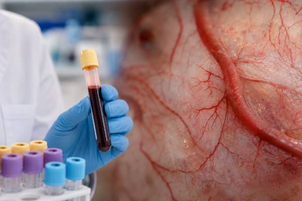
A vascular endothelial growth factor test measures a protein signal involved in blood-vessel growth, vessel permeability, tissue repair, and responses to low oxygen. Most clinical blood assays measure a form of VEGF-A in plasma, although methods differ in which isoform they detect. The test has a recognized role in evaluating and monitoring POEMS syndrome, a rare plasma-cell disorder with neuropathy and multisystem findings. Outside that setting, an elevated VEGF result is usually nonspecific. Hypoxia, inflammation, healing, platelet release, vascular disease, and cancer-related signaling can all raise the concentration. Serum and plasma results are especially difficult to compare because platelets store and release VEGF during clotting. A normal result does not rule out local angiogenesis or POEMS syndrome, and a high value is not a general cancer test. Interpretation must match the specimen, assay, clinical question, medications, and disease-specific evaluation.
- VEGF promotes endothelial-cell survival, migration, permeability, and new-vessel formation.
- Many assays measure VEGF-A, but isoform coverage and reference intervals vary by laboratory.
- Platelet release makes serum values much higher and biologically different from carefully processed plasma values.
- POEMS syndrome is the main established clinical setting for a circulating VEGF measurement.
- VEGF blood testing does not diagnose cancer, retinal disease, inflammation, or vascular injury by itself.
Table of Contents
- The VEGF Family and Angiogenesis
- What the Blood Test Measures
- Serum, Plasma, and Platelet Effects
- VEGF Testing in POEMS Syndrome
- Other Causes of High VEGF
- Normal, Low, and Treatment-Affected Results
- What Should Be Tested Next
- Practical Interpretation and Follow-Up
The VEGF Family and Angiogenesis
VEGF is not one molecule. The human VEGF family includes VEGF-A, VEGF-B, VEGF-C, VEGF-D, and placental growth factor. These proteins bind related receptors but have different roles. VEGF-A is the best-known driver of blood-vessel angiogenesis. VEGF-C and VEGF-D are more closely associated with lymphatic-vessel growth, although their signaling overlaps.
VEGF-A itself has several splice variants. VEGF-A165 is a major form, but shorter and longer isoforms differ in tissue binding, diffusion, and biological effect. Some assays detect one variant more strongly than others. A report labeled simply “VEGF” therefore needs a method description before its value can be compared with research studies or another laboratory.
VEGF is produced by many cell types, including macrophages, platelets and megakaryocytes, fibroblasts, kidney cells, tumor cells, retinal cells, and cells in healing tissue. Low oxygen is a major trigger. When oxygen falls, hypoxia-inducible factors increase VEGF gene expression so nearby endothelial cells can form or remodel vessels.
VEGF-A binds mainly to VEGF receptor 1 and VEGF receptor 2 on endothelial cells. VEGFR2 drives much of the classic response: endothelial survival, migration, proliferation, nitric-oxide production, and increased permeability. Supporting cells, extracellular matrix, integrins, inflammatory mediators, and mechanical forces determine whether the new vessel becomes stable and useful.
Angiogenesis is normal and necessary in embryonic development, menstrual cycling, placental growth, wound healing, and adaptation to exercise or reduced blood flow. It is not automatically a sign of disease. The body balances pro-angiogenic signals such as VEGF with inhibitory signals and vessel-maturation pathways.
Pathological angiogenesis occurs when this balance is persistent or poorly organized. Tumors may exploit VEGF to recruit abnormal vessels. Diabetic retinopathy, retinal vein occlusion, and age-related macular degeneration may involve harmful vessel leakage or growth in the eye. Rheumatoid synovium and inflamed tissues can produce VEGF as immune cells, fibroblasts, and endothelial cells interact.
High VEGF does not always produce effective circulation. In systemic sclerosis, for example, VEGF may be elevated while microvascular loss and malformed vessels continue. Chronic or excessive signaling can produce leaky, disorganized vessels rather than normal repair. The concentration alone cannot show whether angiogenesis is successful, harmful, or biologically blocked downstream.
VEGF also increases vascular permeability. This allows proteins and fluid to leave vessels, which can support inflammation and repair but can contribute to edema, effusions, retinal swelling, or tissue pressure. This permeability effect helps explain several manifestations of POEMS syndrome.
What the Blood Test Measures
Most available clinical tests use an immunoassay to quantify circulating VEGF in EDTA plasma. Results are commonly reported in picograms per milliliter. The laboratory’s reference interval is method specific and should not be replaced with a cutoff found on another report or website.
The exact analyte matters. One laboratory may use an assay specific for VEGF-A165, while another may detect a broader group of VEGF-A isoforms. A method that measures VEGF-A does not measure VEGF-C, VEGF-D, placental growth factor, receptor activation, or all angiogenic activity.
The result represents immunoreactive protein present in the submitted specimen at one time. It does not directly measure how much VEGF is being produced throughout the body. The protein may be concentrated in tissue, attached to extracellular matrix, bound to receptors or medicines, stored inside platelets, degraded, or cleared before collection.
A blood concentration is also not a vessel count. It does not measure capillary density, blood flow, leakage, oxygen delivery, or the quality of new vessels. Imaging, pathology, retinal examination, capillaroscopy, and organ-function studies answer different questions.
Some laboratories use VEGF testing specifically for suspected POEMS syndrome and therapeutic follow-up. Others offer a broader laboratory-developed test without a disease-specific ordering recommendation. The same number may therefore be interpreted differently depending on assay validation and clinical use.
Reference intervals vary substantially. Differences reflect antibody pairs, calibration standards, detected isoforms, specimen type, processing, and the population used to establish the interval. A value should be classified only against the interval printed by the performing laboratory.
An isolated elevation supports increased measurable VEGF in that sample; it does not identify the source. An isolated normal value means the assay did not find an above-range concentration under those collection conditions. Neither statement establishes whether VEGF signaling is active within a particular nerve, retina, tumor, joint, lung, or wound.
Serial testing can be useful only when the same question, specimen type, laboratory, and method are maintained. A numerical change after switching platforms may be analytical rather than biological. The report should be reviewed for assay changes over time.
Serum, Plasma, and Platelet Effects
Platelets are central to VEGF interpretation. They store VEGF and release it when activated. Serum is produced by allowing blood to clot, so clotting activates platelets and releases their contents. Serum VEGF is therefore usually much higher than plasma VEGF and includes a large ex vivo platelet contribution.
That difference is not a minor unit conversion. Serum and plasma answer different biological questions. Serum may reflect the VEGF released during clot formation and the number and activation state of platelets, while promptly processed plasma more closely approaches the freely circulating concentration.
A serum value should not be compared with a plasma reference interval. A treatment trend should not switch between the two. Even two plasma samples may differ if one contains more residual platelets than the other.
For plasma testing, the laboratory may require EDTA blood, separation from cells within a defined time, transfer to a clean tube, immediate refrigeration or freezing, and frozen transport. Some protocols use additional centrifugation to reduce platelets, while others validate a standard spin. The performing laboratory’s exact instructions control.
Delayed separation can change the result because blood cells remain active after collection. Rough handling, difficult venipuncture, prolonged tourniquet use, temperature changes, hemolysis, agitation, and repeated freeze-thaw cycles may alter release or recovery. A sample can look visually normal while still containing activated platelets.
Platelet count also matters. Thrombocytosis can increase the amount of stored VEGF available for release, especially in serum. Thrombocytopenia may reduce it. Platelet activation from inflammation, surgery, vascular injury, exercise, or collection trauma can influence measurement independently of tissue angiogenesis.
A 2022 systematic review found substantial effects from specimen system and preanalytic methodology across circulating VEGF studies. This helps explain why apparently precise thresholds often fail to transfer between laboratories or published cohorts.
VEGF has limited stability under some validated conditions. One major reference laboratory requires plasma to be refrigerated for no more than 24 hours or frozen, with no room-temperature storage after centrifugation. Another specifies critical frozen transport. Collection staff should consult the test directory before drawing the specimen.
When a result is unexpected, the clinician should ask whether serum or plasma was used, how quickly cells were removed, whether the sample was frozen as required, whether hemolysis occurred, and whether platelet count was unusually high. Repeating the test with the same flawed process will not resolve a preanalytic problem.
VEGF Testing in POEMS Syndrome
POEMS syndrome is a rare multisystem disorder caused by a clonal plasma-cell process. The name represents polyneuropathy, organomegaly, endocrinopathy, monoclonal plasma-cell disorder, and skin changes, but patients do not need every feature represented by the acronym.
VEGF is one of the most useful laboratory biomarkers in this condition. Markedly elevated concentrations can support the diagnosis in a patient with the correct neuropathy and plasma-cell findings, and falling values after treatment can support a response. This is the major setting in which a blood VEGF result has established clinical meaning beyond general angiogenesis research.
The neuropathy is typically progressive, symmetrical, and sensorimotor, often beginning distally with weakness, numbness, pain, or gait difficulty. Nerve-conduction studies may resemble chronic inflammatory demyelinating polyneuropathy, which is a common reason for delayed diagnosis.
Additional clues include a lambda-restricted monoclonal protein or plasma-cell disorder, sclerotic bone lesions, enlarged liver, spleen, or lymph nodes, edema, ascites, pleural effusions, pulmonary hypertension, papilledema, thrombocytosis or erythrocytosis, endocrinopathies, hyperpigmentation, hemangiomas, white nails, or increased body hair.
VEGF may contribute to vessel leakage, edema, skin findings, papilledema, and nerve injury, although it does not explain every feature. Its level generally reflects disease activity better than the small plasma-cell burden does.
A high VEGF result does not diagnose POEMS alone. Diagnostic frameworks require a demyelinating polyneuropathy and a monoclonal plasma-cell disorder, plus additional major and minor criteria. Castleman disease, chronic inflammatory neuropathies, myeloma-spectrum disorders, infection, cancer, and other causes of elevated VEGF must be considered.
A normal concentration does not fully exclude POEMS. Timing, prior corticosteroids or treatment, assay method, specimen handling, and biological variation can lower the value. Clinical suspicion should remain high when the neuropathy and systemic features fit, especially if testing conditions were uncertain.
The diagnostic threshold is method and specimen dependent. Published serum cutoffs should not be applied to a plasma assay, and a Mayo plasma reference limit should not be transferred to a different platform. Specialist interpretation is important because the separation between POEMS and other disorders depends on validated local data.
For monitoring, use the same laboratory and specimen type whenever possible. A decrease after radiation, systemic therapy, or stem-cell transplantation can support response, but clinical improvement in neuropathy may lag behind the biochemical change. A rising value can prompt reassessment, yet relapse is confirmed with the entire clinical and hematologic picture.
POEMS evaluation commonly includes neurologic examination, electromyography and nerve-conduction studies, serum and urine protein studies, immunofixation, serum free light chains, bone-marrow examination, whole-body imaging for sclerotic lesions, blood counts, endocrine testing, eye examination, cardiopulmonary assessment, and evaluation for fluid overload.
Other Causes of High VEGF
Outside POEMS syndrome, high VEGF is nonspecific. It may indicate a response to low oxygen, endothelial injury, inflammation, tissue repair, platelet release, or a disease that uses angiogenic signaling. The value usually cannot distinguish among these possibilities.
Cancer is an important biological context. Many tumors and surrounding stromal or immune cells increase VEGF in response to hypoxia and oncogenic signaling. VEGF can support vessel growth, permeability, and tumor progression. However, circulating concentrations overlap widely among people with and without cancer. A VEGF blood test is not recommended as a general cancer screen and cannot locate, classify, or stage a tumor.
When cancer is suspected, the appropriate pathway is based on symptoms and organ: examination, imaging, endoscopy, standard tumor markers only where validated, and tissue pathology. A high VEGF result should not lead directly to anti-angiogenic treatment.
Inflammatory diseases can increase VEGF because activated macrophages, fibroblasts, synovial cells, and endothelial cells participate in tissue remodeling. Rheumatoid arthritis, systemic sclerosis, vasculitis, inflammatory bowel disease, psoriasis, and other conditions have been associated with altered VEGF in research. The overlap is too broad for one concentration to diagnose or grade these disorders.
In systemic sclerosis, pooled studies show higher serum or plasma VEGF on average, particularly in some advanced vascular patterns. Yet patients can have ineffective angiogenesis despite high VEGF, and evidence for routine individual management remains limited. Nailfold capillaroscopy, pulmonary testing, echocardiography, autoantibodies, skin examination, and organ surveillance are more established.
Low oxygen can raise VEGF in lung disease, ischemia, anemia, sleep-disordered breathing, high-altitude exposure, or poorly perfused tissue. The degree and timing vary, and a blood value cannot identify the hypoxic source. Oxygen saturation, blood counts, cardiopulmonary testing, vascular imaging, and symptom-directed evaluation are more direct.
Wound healing, surgery, trauma, burns, and tissue regeneration may temporarily increase VEGF. Exercise can stimulate local angiogenic adaptation. These physiological or repair responses should be considered before interpreting a modest elevation as pathology.
Eye diseases such as neovascular age-related macular degeneration, diabetic macular edema, and retinal vein occlusion involve local VEGF activity. Diagnosis and treatment are based on retinal examination and imaging, especially optical coherence tomography and angiography when indicated. A routine systemic VEGF level does not replace ophthalmic assessment.
Kidney disease can affect endothelial biology and protein clearance, while preeclampsia involves an anti-angiogenic imbalance that includes soluble VEGF receptor 1 rather than a simple high circulating free-VEGF state. Standard obstetric testing, blood pressure, urine protein, blood counts, liver tests, and fetal assessment guide care.
Thrombocytosis, platelet activation, and specimen handling may be the main explanation for an elevated result, particularly in serum. The first interpretation step should therefore be analytical and hematologic before a rare angiogenic disorder is assumed.
Normal, Low, and Treatment-Affected Results
A normal VEGF result does not mean angiogenesis is absent. VEGF may be produced and consumed locally, bound to tissue or receptors, present as an isoform the assay detects poorly, or released intermittently. A retinal lesion, healing wound, inflamed joint, or tumor can have strong local signaling without a high plasma value.
A low result is rarely interpreted as a primary deficiency. VEGF is essential in development and vascular maintenance, but routine blood testing is not validated to diagnose “low angiogenesis,” poor circulation, fatigue, immune weakness, or impaired healing. Treatment should not aim to raise the laboratory number.
Medicines can lower production or alter detection. Corticosteroids, chemotherapy, anti-inflammatory treatment, and therapy directed at the underlying plasma-cell clone may reduce VEGF. In POEMS syndrome, this can be a desired treatment response when it matches clinical improvement.
Anti-VEGF therapies require special caution. Bevacizumab binds VEGF-A systemically and is used in selected cancers and other contexts. Aflibercept acts as a decoy receptor, and multiple anti-VEGF agents are injected into the eye for retinal disease. Drug-bound VEGF may be measured differently depending on the assay.
One reference laboratory specifically warns that bevacizumab interferes with VEGF detection. A low or altered result during treatment may reflect drug binding and assay design rather than true absence of pathway activity. The laboratory should be contacted before ordering or interpreting the test in a treated patient.
Circulating VEGF is generally not the routine biomarker used to choose or dose anti-VEGF cancer therapy. Tumor type, molecular features where relevant, imaging response, symptoms, blood pressure, urine protein, kidney function, wound-healing risk, bleeding or thrombosis, and drug-specific safety monitoring are more important.
Likewise, ophthalmologists monitor visual acuity, optical coherence tomography, retinal fluid, hemorrhage, and examination findings—not a serum VEGF target. Systemic levels may change after intravitreal treatment, but their clinical meaning is not sufficiently standardized for routine dosing decisions.
A normal result after therapy should never be interpreted as proof that cancer is gone, retinal disease is inactive, or POEMS is cured. The correct endpoint depends on the disease. Laboratory trends support, rather than replace, clinical response assessment.
What Should Be Tested Next
The next step depends on why VEGF was ordered. For progressive demyelinating neuropathy with systemic findings, investigate POEMS syndrome directly. This includes monoclonal-protein testing, serum free light chains, blood counts, bone imaging, marrow assessment, endocrine studies, eye examination, and cardiopulmonary evaluation in addition to VEGF.
A flow cytometry immune panel does not diagnose POEMS, although it may be useful for a separate immune question. Plasma-cell clonality usually requires protein studies, marrow pathology, immunophenotyping designed for plasma cells, and sometimes molecular testing.
For suspected inflammation, C-reactive protein, erythrocyte sedimentation rate, complete blood count, metabolic panel, urinalysis, autoantibodies, imaging, and organ-specific studies usually provide more actionable information. A broad cytokine panel may characterize immune activation in specialized settings but will not convert VEGF into a disease-specific marker.
For suspected vascular or hypoxic disease, assess oxygen saturation, hemoglobin, lung function, sleep breathing, cardiac status, pulses, vascular imaging, and tissue perfusion as indicated. A VEGF level cannot show whether an artery is blocked or whether an organ is receiving enough oxygen.
For an eye complaint, urgent ophthalmic examination is more important than blood testing. Sudden visual loss, a curtain-like shadow, new flashes and floaters, severe eye pain, or rapidly distorted central vision requires prompt assessment.
For possible cancer, use age- and risk-appropriate screening and symptom-directed diagnostics. Imaging or biopsy should not be ordered solely because of a small VEGF elevation without a supporting clinical reason.
When platelet contribution is possible, review the complete blood count and specimen type. A repeat in correctly processed plasma may be more informative than extensive imaging after an isolated serum elevation. The performing laboratory can clarify whether platelet-poor preparation is part of its validated protocol.
If results from two laboratories disagree, compare the detected isoform, antibody method, specimen matrix, reference interval, processing instructions, and treatment status. Converting values mathematically is not reliable when the assays measure different molecular pools.
Practical Interpretation and Follow-Up
Begin with the report rather than the word “VEGF.” Confirm whether the analyte is VEGF-A or a particular isoform, whether the specimen is plasma or serum, the units, method, reference interval, and collection requirements. Note any comment about bevacizumab or other interference.
Then define the clinical question. A value ordered for suspected POEMS syndrome has a different evidentiary role from one ordered in an inflammation panel, oncology study, fertility investigation, or wellness evaluation. The same elevation should not be given the same meaning in all settings.
For a high result, first check specimen integrity and platelet effects. Review platelet count, collection difficulty, processing delay, temperature, freeze-thaw history, and whether serum and plasma were confused. Next look for the phenotype: progressive neuropathy and monoclonal protein, active inflammation, hypoxia, healing injury, retinal disease, cancer, or recent treatment.
For a normal result, do not close the evaluation when clinical suspicion remains strong. In possible POEMS syndrome, verify the method and collection, assess whether treatment or corticosteroids preceded testing, and complete the required neurologic and plasma-cell workup. In other diseases, use the established diagnostic pathway rather than repeating VEGF indefinitely.
Serial measurements should use the same laboratory, specimen, method, and timing relative to treatment. Document the intervention between tests. A substantial decline in confirmed POEMS syndrome may support response; outside that disease, an isolated rise or fall often has uncertain meaning.
Do not use a VEGF value to start supplements, “anti-angiogenic” diets, or unproven therapies. Angiogenesis is essential for normal vessels and healing, while excessive or abnormal angiogenesis is disease specific. Manipulating the pathway without a diagnosis can cause harm or delay effective care.
Urgent care is based on symptoms. Seek immediate assessment for sudden vision loss, severe shortness of breath, chest pain, signs of stroke, rapidly worsening weakness, confusion, severe edema with breathing difficulty, or other evidence of organ failure. Progressive weakness, numbness, gait change, unexplained fluid retention, or multisystem symptoms warrant timely specialist evaluation even when VEGF is normal.
A clinically useful conclusion is narrow and transparent: the measured VEGF concentration is above, within, or below the laboratory’s interval for that specimen and method. It may support POEMS syndrome when the required clinical criteria are present, but otherwise it is a nonspecific marker influenced by platelets, hypoxia, inflammation, repair, disease, and treatment. The next decision should be based on the suspected organ or syndrome, not on angiogenesis in the abstract.
References
- Vascular Endothelial Growth Factor, Plasma 2026 (Laboratory Test Directory)
- Vascular Endothelial Growth Factor 2026 (Laboratory Test Directory)
- Comprehensive Diagnosis and Management of POEMS Syndrome 2022 (Review)
- A systematic review and meta-analysis of preanalytical factors and methodological differences influencing the measurement of circulating vascular endothelial growth factor 2022 (Systematic Review and Meta-Analysis)
- Vascular endothelial growth factor signaling in health and disease 2025 (Review)
- Vascular endothelial growth factor as a potential biomarker in systemic sclerosis: a systematic review and meta-analysis 2024 (Systematic Review and Meta-Analysis)
Disclaimer
This article is for general education and does not diagnose disease or replace evaluation by a qualified clinician. VEGF results require interpretation with the exact assay, specimen, handling, medications, symptoms, and disease-specific testing. Seek urgent care for sudden vision loss, severe breathing difficulty, neurologic symptoms, or rapid clinical deterioration.





