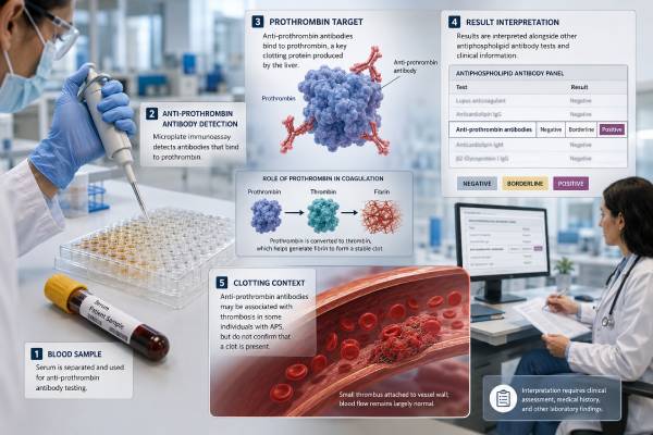
The anti-prothrombin antibody test looks for autoantibodies that recognize prothrombin, also called coagulation factor II. Prothrombin is a normal blood protein that the body converts to thrombin during clot formation. Anti-prothrombin antibodies belong to the broader antiphospholipid antibody family and have been studied in people with unexplained thrombosis, pregnancy complications, lupus, and possible antiphospholipid syndrome.
This is a non-criteria test. A positive anti-prothrombin result does not diagnose APS, and the evidence is less consistent than for lupus anticoagulant, anticardiolipin IgG/IgM, and anti-beta-2 glycoprotein I IgG/IgM. It is also essential to distinguish antibodies against prothrombin alone from antibodies against a phosphatidylserine-prothrombin complex, known as aPS/PT. The two assays can identify different antibody populations and should not be interpreted as equivalent. The result becomes useful only when the exact assay, antibody class, titer, persistence, standard APS profile, and confirmed clinical events are considered together.
- The test detects IgG and/or IgM autoantibodies that bind prothrombin, the precursor of thrombin.
- A positive result is a non-criteria finding and cannot establish antiphospholipid syndrome by itself.
- Anti-prothrombin and aPS/PT tests measure related but distinct antibody targets.
- There is no universal cutoff; result units and positive ranges depend on the laboratory method.
- Clotting risk is more clearly defined by the full APS profile, especially lupus anticoagulant and persistent multi-marker positivity.
- Symptoms of a possible stroke, pulmonary embolism, or deep vein thrombosis require emergency evaluation regardless of antibody results.
Table of Contents
- Prothrombin and the Immune Target
- Anti-Prothrombin Versus aPS/PT Antibodies
- Who May Be Tested and Why
- Reading the Laboratory Result
- What the Result Says About Clotting Risk
- Pregnancy and Obstetric Risk
- Role in APS Diagnosis
- Preparation, Repeat Testing, and Follow-Up
Prothrombin and the Immune Target
Prothrombin is made in the liver and circulates in an inactive form. When the coagulation system is activated, factor Xa and factor Va convert prothrombin to thrombin on a phospholipid surface in the presence of calcium. Thrombin then converts fibrinogen to fibrin, activates platelets, and amplifies several other clotting reactions.
Anti-prothrombin antibodies recognize parts of this protein. Their binding behavior depends on how prothrombin is presented in the assay. Some antibodies bind prothrombin coated directly onto a laboratory plate. Others bind more strongly when prothrombin attaches to a negatively charged phospholipid such as phosphatidylserine and changes shape. That distinction explains why a patient may have a positive anti-prothrombin test and a negative aPS/PT test, or the reverse.
Most commercial tests use a solid-phase immunoassay and report separate IgG and IgM results. Some laboratories also offer IgA, although its clinical role is less established. The report may use arbitrary units, units per milliliter, an index, or a qualitative category. Unlike a prothrombin time test, this antibody assay does not measure how many seconds blood takes to clot.
Anti-prothrombin antibodies can be detected in APS, systemic lupus erythematosus, other autoimmune conditions, infections, and occasionally in people without a clear related disease. Detection alone does not prove that the antibody interferes with coagulation in the body.
The name can also cause confusion with antithrombin, a separate natural anticoagulant protein. An anti-prothrombin antibody test does not measure antithrombin activity or diagnose antithrombin deficiency. It also differs from a prothrombin gene mutation test, which looks for an inherited F2 gene variant associated with venous thrombosis.
How antibody binding could affect coagulation
Prothrombin-related antibodies may influence coagulation in several ways. They can alter how prothrombin attaches to phospholipid surfaces, change the rate at which thrombin is generated, interfere with natural anticoagulant pathways, or form immune complexes that activate endothelial cells and complement. Different antibodies may have opposite laboratory effects depending on the epitope they recognize. Some interfere with clotting reactions in a test tube, while others appear more closely linked to thrombosis.
This diversity helps explain why “anti-prothrombin positive” is not a single biological state. Two patients with similar numerical results may carry antibodies that bind different regions of prothrombin and behave differently. Current clinical assays measure binding, not the full functional effect of each antibody. That limitation is another reason treatment cannot be selected from the antibody value alone.
Anti-Prothrombin Versus aPS/PT Antibodies
The clinical literature often discusses two prothrombin-related antibody groups:
- aPT antibodies: antibodies measured against prothrombin alone;
- aPS/PT antibodies: antibodies measured against prothrombin bound to phosphatidylserine.
| Feature | Anti-prothrombin | Anti-phosphatidylserine/prothrombin |
|---|---|---|
| Laboratory target | Prothrombin presented alone or on a manufacturer-specific surface | Prothrombin bound to phosphatidylserine |
| Common abbreviation | aPT | aPS/PT |
| Association with lupus anticoagulant | Variable | Often stronger |
| Evidence for thrombosis risk | Inconsistent across studies | More consistent, especially with higher titers and persistent positivity |
| APS criteria status | Non-criteria | Non-criteria |
A systematic review of thousands of participants found that both groups had associations with APS manifestations, but the strength and reproducibility varied. Later work has generally given aPS/PT more attention because of its relationship with lupus anticoagulant and thrombotic outcomes.
The difference is not merely wording. Antibody binding can depend on a conformational change that occurs when prothrombin contacts phosphatidylserine. Direct aPT assays may expose different epitopes and identify antibodies that do not behave the same way in coagulation reactions.
When a laboratory panel reports “prothrombin antibody,” verify whether it means direct aPT or aPS/PT antibody testing. The reference interval, manufacturer, and antigen description usually resolve the issue. If the report does not specify the target, the clinician can contact the laboratory.
Who May Be Tested and Why
Anti-prothrombin testing is not a routine screening test. It is most often used as part of an extended evaluation after standard testing and clinical review.
Possible reasons include:
- objectively confirmed venous or arterial thrombosis without a clear provoking factor;
- recurrent thrombosis despite an incomplete or conflicting APS laboratory profile;
- stroke, transient ischemic attack, or unusual-site thrombosis at a younger age;
- recurrent pregnancy loss, fetal death, placental insufficiency, or severe early preeclampsia;
- lupus with thrombotic, hematologic, or obstetric features;
- a strong APS-like history with repeatedly negative criteria antibodies;
- research or specialist risk-stratification protocols.
The test generally adds little when the pretest probability is low. Testing a healthy person without thrombosis, pregnancy morbidity, lupus, or another relevant clinical feature increases the chance of finding an isolated result with uncertain meaning.
A clinician should first confirm that a suspected clot truly occurred. Deep vein thrombosis and pulmonary embolism require imaging. Stroke requires appropriate neurologic assessment and imaging. Pregnancy morbidity should be documented by gestational age, ultrasound, placental findings, and obstetric records when available.
An extended result is most informative when it answers a specific question. Examples include whether a non-criteria antibody supports specialist concern in a patient with APS-like manifestations, whether an apparently isolated lupus anticoagulant has a related solid-phase antibody, or whether a broader autoantibody pattern is present in lupus.
It should not be ordered as a shortcut around a complete antiphospholipid syndrome blood test panel. Criteria assays remain the foundation of laboratory assessment.
Reading the Laboratory Result
A result needs four identifiers: antibody class, numerical value, unit, and laboratory cutoff. The words “positive” or “negative” without those details are not enough for careful interpretation.
Negative
A negative result means the assay did not detect anti-prothrombin antibody above its threshold. It does not rule out APS, because direct aPT antibodies are not required for classification or diagnosis. It also does not exclude aPS/PT antibodies or any of the criteria markers.
Borderline or low positive
A value just above the cutoff may result from weak biological reactivity, normal assay variation, recent infection, inflammation, or a transient immune response. Borderline results are especially difficult to compare between laboratories. One low-positive result rarely carries enough weight to change long-term treatment.
Moderate or high positive
A clearly elevated result may be more likely to represent a real antibody response, but no universal aPT titer defines high thrombotic risk. The meaning depends on the assay, persistence, clinical events, and other antibodies. A high direct aPT value should not be automatically assigned the risk estimates reported for aPS/PT.
IgG versus IgM
Persistent IgG autoantibodies are often considered more clinically persuasive, while isolated low-level IgM may be more likely to be transient. This is a general interpretive tendency, not an absolute rule. Some studies have found clinical associations for IgM, and some IgG results are incidental.
| Question | Why it matters |
|---|---|
| Was the target prothrombin alone or a phosphatidylserine-prothrombin complex? | The assays detect different antibody populations |
| Was the value far above the cutoff? | Weak results are more vulnerable to analytical and transient variation |
| Did it remain positive later? | Persistence is more convincing than a one-time result |
| Were criteria APS tests positive? | Criteria markers carry greater diagnostic and prognostic weight |
| Was there a confirmed APS-related event? | Antibody positivity without a clinical event is not APS |
The laboratory reference range cannot be replaced with a value found online. Assays use different coating methods, calibrators, control populations, and reporting scales. A result should be trended only with the same method whenever possible.
Examples of result patterns
An isolated low IgM aPT result during a respiratory infection may disappear and carry little long-term significance. Persistent high IgG aPT in a patient with lupus and an imaging-confirmed unprovoked clot is more concerning, although criteria antibodies still determine formal laboratory classification. A positive aPS/PT result with lupus anticoagulant is a different pattern again and may have stronger published links to thrombosis than direct aPT alone.
The absence of criteria antibodies changes the discussion but does not end it. A specialist may review whether testing occurred during anticoagulation, whether lupus anticoagulant results were technically valid, and whether an extended marker was repeated. Even then, the conclusion may remain “possible non-criteria APS phenotype” rather than a definite diagnosis.
What the Result Says About Clotting Risk
Direct anti-prothrombin antibodies have been associated with venous and arterial thrombosis in some studies, but results have not been uniform. Differences in study design, assay method, antibody class, cutoff, and patient population make a single risk estimate unreliable.
The broader APS profile provides more dependable risk information. Lupus anticoagulant is strongly associated with thrombosis. Persistent positivity for all three criteria groups—lupus anticoagulant, anticardiolipin, and anti-beta-2 glycoprotein I—is generally considered a high-risk profile. Titers, persistence, prior thrombosis, and traditional cardiovascular or venous thromboembolism risks all contribute.
Prothrombin-related antibodies may help explain why some patients have lupus anticoagulant activity. Certain antibodies bind prothrombin on phospholipid surfaces and interfere with phospholipid-dependent clotting assays in the laboratory. Paradoxically, a prolonged clotting time in vitro can coexist with increased clotting in the body. The test result should therefore not be interpreted as evidence of a bleeding tendency.
A positive aPT result does not predict when or where a clot will occur. It cannot calculate an individual annual risk. Important non-antibody factors include:
- previous unprovoked thrombosis;
- age and immobility;
- surgery or trauma;
- pregnancy and the postpartum period;
- estrogen-containing contraception or hormone therapy;
- cancer;
- smoking and obesity;
- hypertension, diabetes, and high cholesterol;
- inherited thrombophilia;
- active lupus or systemic inflammation.
The clinical response should be proportional to total risk, not the presence of one laboratory marker. A person with no events and isolated aPT positivity may need observation and risk-factor counseling rather than anticoagulation. A person with recurrent unprovoked thrombosis and persistent criteria antibodies may need long-term treatment regardless of the aPT result.
Pregnancy and Obstetric Risk
Anti-prothrombin and aPS/PT antibodies have been examined in recurrent miscarriage, fetal death, preeclampsia, fetal growth restriction, placental insufficiency, and preterm delivery. The evidence is more developed for aPS/PT than for direct aPT assays.
Possible mechanisms include interference with trophoblast function, placental inflammation, complement activation, and thrombosis or abnormal signaling at the maternal-fetal interface. APS-related pregnancy problems are not caused solely by large placental clots; inflammatory pathways and impaired placentation also appear important.
A positive direct aPT result should not be used to assign causation after a miscarriage. Early loss is common, and chromosomal abnormalities are a frequent explanation. Other evaluations may include uterine anatomy, thyroid function, diabetes, parental chromosome testing, and pregnancy tissue testing, depending on the history.
The pattern of events is crucial. Three early losses differ clinically from a fetal death after 10 weeks or delivery before 34 weeks because of severe preeclampsia. Records should include gestational age, fetal cardiac activity, growth measurements, blood pressure, placental pathology, and all standard APS results.
Treatment evidence for established obstetric APS supports clinician-directed low-dose aspirin and heparin in appropriate patients. The benefit of treating isolated direct anti-prothrombin positivity is uncertain. Aspirin and anticoagulants can cause bleeding and other adverse effects, so treatment should not be started solely from an extended panel result.
Pregnancy planning may involve maternal-fetal medicine, hematology, and rheumatology. A specialist can decide whether the history supports obstetric APS, a non-criteria APS phenotype, or another cause. The article on recurrent pregnancy loss antiphospholipid testing describes the broader evaluation.
Role in APS Diagnosis
Current APS classification criteria do not include direct anti-prothrombin antibodies. The 2023 ACR/EULAR system uses weighted clinical domains and laboratory domains based on lupus anticoagulant, anticardiolipin IgG/IgM, and anti-beta-2 glycoprotein I IgG/IgM.
Classification criteria are designed for research consistency, not as an automatic rule for every clinical decision. Even so, their laboratory choices reflect which assays are sufficiently standardized and validated. Direct aPT testing has not reached that level.
A clinician may still regard a persistent, strong anti-prothrombin result as supportive in a patient with a convincing APS-like history, particularly when criteria tests are negative or incomplete. This falls within specialist judgment and should be documented as such. It does not convert the marker into a criteria antibody.
The phrase seronegative APS is sometimes used when clinical manifestations strongly resemble APS but criteria antibodies remain negative. Extended testing can include aPS/PT, anti-domain I beta-2 glycoprotein I, anti-phosphatidylethanolamine, anti-phosphatidylserine, and direct aPT. No single extended marker has universal acceptance for diagnosing this debated entity.
Three points prevent overdiagnosis:
- A positive antibody without a relevant clinical event is not APS.
- A thrombosis with only a transient, weak non-criteria antibody does not automatically establish APS.
- Other causes of thrombosis or pregnancy morbidity still require evaluation.
The result may be most useful as one piece of a specialist narrative: it can support, but rarely settle, the diagnosis.
When the result may not change care
Some patients already have a clear reason for anticoagulation, such as recurrent unprovoked venous thrombosis. In that setting, an aPT result may refine the suspected cause without changing the immediate treatment. In other patients, the result may affect counseling around estrogen exposure, pregnancy, or surgery but still not justify daily medication. Before ordering or repeating the test, clinicians can ask which concrete decision would differ if the result were positive, negative, or unchanged.
Avoiding unnecessary serial testing is important. Antibody titers do not reliably track APS “activity” the way glucose tracks diabetes, and a falling aPT value does not prove that clot risk has disappeared. Clinical prevention plans should not be relaxed solely because a non-criteria antibody becomes negative.
Preparation, Repeat Testing, and Follow-Up
The test uses a standard venous blood sample. Fasting is usually unnecessary. Patients should tell the ordering clinician about recent infection, vaccination, surgery, pregnancy, autoimmune flare, and all medicines.
Anticoagulants can interfere with lupus anticoagulant clotting assays, but solid-phase antibody tests are generally less directly affected. Never stop warfarin, heparin, or a direct oral anticoagulant merely to obtain a cleaner laboratory result unless the prescribing clinician creates a safe plan.
A specialty laboratory may need several days to report the result. Mild soreness or bruising at the blood-draw site is common. Persistent bleeding deserves attention, especially in a patient taking anticoagulants.
There is no universally required repeat schedule for aPT. Clinicians often use an interval of at least 12 weeks when they want to assess persistence, mirroring criteria antibody practice. The same laboratory and assay should be used because switching methods may create an apparent change that is purely analytical.
After a positive result:
- obtain the complete report, including antigen, class, number, unit, and cutoff;
- review all criteria APS tests and whether they were repeated appropriately;
- verify any clot with imaging and reconstruct pregnancy events precisely;
- assess lupus and other autoimmune disease when symptoms suggest them;
- review temporary and conventional clotting risks;
- decide whether the result changes pregnancy, surgery, contraception, or anticoagulation planning.
A positive result is not a reason to self-start aspirin. Aspirin can cause gastrointestinal bleeding and interact with anticoagulants or anti-inflammatory drugs. Long-term anticoagulation carries even greater bleeding risk and requires a defined clinical indication.
Seek urgent care for sudden shortness of breath, chest pain, coughing blood, one-sided leg swelling, fainting, new weakness, facial droop, speech difficulty, sudden vision loss, or a severe new headache. The urgency comes from the symptoms, not from whether an antibody report is positive.
At a follow-up visit, useful questions include whether the assay measured direct aPT or aPS/PT, whether the result was sufficiently above the cutoff to be reproducible, and whether repeat criteria testing is needed. Ask how the finding changes the plan for travel, surgery, pregnancy, contraception, or future anticoagulation. A clear explanation should separate what is known from standard APS evidence from what remains uncertain about the non-criteria marker.
References
- An update on laboratory detection and interpretation of antiphospholipid antibodies for diagnosis of antiphospholipid syndrome: guidance from the ISTH-SSC Subcommittee on Lupus Anticoagulant/Antiphospholipid Antibodies 2025 (Guidance)
- Lupus anticoagulant and antiprothrombin antibodies 2025 (Review)
- The value of non-criteria antiphospholipid antibodies 2024 (Review)
- Understanding the Difference between Type-I and Type-II Anti-Prothrombin Autoantibodies: A Structural Perspective 2024 (Review)
- Anti-phosphatidylserine/Prothrombin Antibodies (aPS/PT) 2023 (Review)
- Anti-prothrombin (aPT) and anti-phosphatidylserine/prothrombin (aPS/PT) antibodies and the risk of thrombosis in the antiphospholipid syndrome. A systematic review 2014 (Systematic Review)
Disclaimer
This article provides general education about anti-prothrombin antibody testing and does not diagnose APS or determine treatment. Results must be interpreted with the exact assay, criteria antiphospholipid tests, documented clotting or pregnancy events, and individual bleeding risk. Do not start or stop aspirin or anticoagulation without medical supervision.