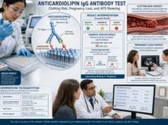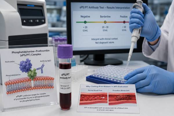
The phosphatidylserine-prothrombin antibody test looks for immune proteins that bind to a complex formed by phosphatidylserine and prothrombin. These antibodies, usually reported as aPS/PT IgG and IgM, are not part of the three standard laboratory criteria used to classify antiphospholipid syndrome (APS). Even so, they can provide useful evidence when a person has unexplained blood clots, pregnancy complications, a lupus anticoagulant pattern, or APS-like symptoms despite negative standard antibody tests. A positive result does not prove APS, predict exactly who will develop a clot, or automatically require treatment. Its meaning depends on the antibody type, strength, persistence, clinical history, other antiphospholipid antibodies, and factors such as medications or autoimmune disease. The test is best understood as an additional piece of a larger diagnostic and risk-assessment process rather than as a stand-alone answer.
- aPS/PT antibodies are non-criteria antiphospholipid antibodies directed against a phosphatidylserine-prothrombin complex.
- A positive result can support APS evaluation, especially when standard tests are negative or lupus anticoagulant testing is difficult to interpret.
- IgG positivity often has a stronger association with thrombosis than isolated low-level IgM, although results are assay- and population-dependent.
- Persistent, moderate-to-strong positivity is generally more meaningful than one borderline result during an acute illness.
- The test does not diagnose APS by itself and should be interpreted with clotting history, pregnancy history, lupus anticoagulant, anticardiolipin, and beta-2 glycoprotein I antibodies.
Table of Contents
- What the aPS/PT test detects
- Why the test is ordered
- How to read IgG, IgM, and negative results
- What the result may mean for clotting and pregnancy risk
- Relationship to lupus anticoagulant and standard APS tests
- Limitations, preparation, and repeat testing
- Next steps after an aPS/PT result
What the aPS/PT test detects
Phosphatidylserine is a negatively charged phospholipid normally concentrated on the inner surface of cell membranes. When cells are activated, injured, or undergoing programmed death, phosphatidylserine may become exposed on the outer membrane surface. Prothrombin, a clotting protein also called factor II, can bind to that phospholipid surface. The resulting phosphatidylserine-prothrombin complex creates a target that some immune systems recognize abnormally.
The aPS/PT blood test detects antibodies against this complex. Most laboratories use a solid-phase immunoassay and report two antibody classes:
- aPS/PT IgG, which may be more strongly associated with thrombosis and persistent autoimmune activity in several studies
- aPS/PT IgM, which can still be clinically relevant but is more likely to be difficult to interpret when isolated, weak, or temporary
A laboratory may report numerical units plus categories such as negative, borderline, weak positive, moderate positive, or strong positive. The units and cutoffs are not interchangeable between manufacturers. A value of 35 units on one platform cannot safely be compared with 35 units on another platform unless the laboratory confirms that the methods and reference ranges are equivalent.
This test is different from tests for antibodies against phosphatidylserine alone or prothrombin alone. The antigen used in an aPS/PT assay is the combined complex, and the antibodies detected may behave differently from antibodies measured by an anti-phosphatidylserine antibody test or an anti-prothrombin antibody test. These names are easy to confuse on laboratory menus, so the exact analyte should be checked before interpreting a result.
The test is called “non-criteria” because aPS/PT antibodies are not among the laboratory markers in the established APS classification systems. The standard laboratory markers remain lupus anticoagulant, anticardiolipin IgG or IgM, and anti-beta-2 glycoprotein I IgG or IgM. “Non-criteria” does not mean useless or disproven. It means that the marker has not been adopted as a required classification element, partly because assays, thresholds, and evidence are not standardized enough for universal use.
Classification criteria and clinical diagnosis are related but not identical. Criteria are designed mainly to create consistent research groups. A clinician may consider evidence outside those criteria when evaluating an individual patient, while still recognizing the uncertainty attached to that evidence.
Why the test is ordered
An aPS/PT test is usually ordered as an extension of an APS evaluation rather than as the first or only test. It may be considered when the clinical picture suggests an antiphospholipid-antibody disorder but the standard panel does not fully explain the findings.
Common reasons include:
- An unexplained deep vein thrombosis, pulmonary embolism, stroke, or clot in an unusual site
- Recurrent pregnancy loss, fetal death, severe placental insufficiency, early-onset preeclampsia, or fetal growth restriction
- A prolonged phospholipid-dependent clotting test or suspected lupus anticoagulant
- Systemic lupus erythematosus or another autoimmune condition with concerning vascular or obstetric features
- APS-like manifestations with negative or inconsistent standard antiphospholipid antibody results
- A need for additional risk information when standard antibodies are already present
The test can be particularly helpful in a suspected “seronegative” presentation. That term is sometimes used when a person has manifestations strongly suggestive of APS but repeatedly tests negative for the standard laboratory markers. An expanded seronegative APS test panel may include aPS/PT and other non-criteria antibodies. A positive non-criteria marker can make an immune-mediated explanation more plausible, but it does not remove the need to consider other causes of thrombosis or pregnancy loss.
Another reason to order aPS/PT is its close relationship with lupus anticoagulant. Many aPS/PT-positive patients also have a lupus anticoagulant pattern. Functional lupus anticoagulant assays can be distorted by direct oral anticoagulants, heparin, warfarin, factor deficiencies, inflammation, and preanalytical problems. Because aPS/PT is measured with an antibody-binding assay rather than a clotting-time assay, it is not affected in the same direct way by anticoagulant drugs. It may therefore offer supporting information when functional testing is unreliable. It cannot substitute for a properly performed lupus anticoagulant evaluation, however.
The test should not be ordered simply because a person bruises easily or has a slightly abnormal routine coagulation result. APS causes a tendency toward thrombosis, not a classic bleeding disorder, and many prolonged clotting times have explanations unrelated to antiphospholipid antibodies. A focused history helps determine whether this specialized test is likely to add value.
Before testing, the clinician usually considers whether the event was objectively confirmed, whether it was provoked by surgery or immobility, whether pregnancy complications meet recognized definitions, and whether inherited thrombophilia, malignancy, infection, anatomic problems, medication effects, or other autoimmune conditions are possible. The aPS/PT result is then interpreted within that broader differential diagnosis.
How to read IgG, IgM, and negative results
The laboratory’s own reference interval is the starting point. Because assays are not harmonized, there is no single universal aPS/PT cutoff that applies to all reports. Interpretation should use the exact method, units, and category printed by the performing laboratory.
| Report pattern | General interpretation | Important caution |
|---|---|---|
| IgG and IgM negative | No aPS/PT antibodies detected above that assay’s cutoff | Does not rule out APS or another cause of thrombosis |
| Borderline or weak positive | Low antibody signal near the cutoff | May be temporary, nonspecific, or method-dependent |
| Isolated IgM positive | Possible non-criteria antibody finding | Clinical association may be weaker when low-level and isolated |
| IgG positive | Often considered more concerning, especially if moderate-to-strong and persistent | Still not diagnostic without compatible clinical evidence |
| IgG and IgM positive | Broader immune response that may add weight to the finding | Risk depends on titer, persistence, standard antibodies, and history |
| Positive with lupus anticoagulant or other APS antibodies | May indicate a more substantial antiphospholipid antibody profile | Treatment decisions remain individualized |
A negative result means the assay did not detect aPS/PT antibodies above its threshold at that time. It does not exclude APS because aPS/PT is not required for diagnosis, and a person may instead have lupus anticoagulant, anticardiolipin, or anti-beta-2 glycoprotein I antibodies. It also does not exclude non-APS causes of a clot or pregnancy complication.
A borderline result deserves restraint. Values clustered around an assay cutoff may change category because of normal analytical variation. Transient antibodies may appear during infection, inflammation, or other immune activation. A borderline result without a relevant clinical event or other antibody abnormalities usually carries less weight than a clearly elevated result that remains present over time.
An isolated IgM result can be meaningful in the right context, but its positive predictive value is often lower than that of persistent IgG or combined positivity. The distinction is not absolute. Some patients with IgM antibodies have clinically important disease, and no result should be dismissed solely because of antibody class. The key question is whether the result fits the total pattern.
A moderate or strong IgG result may provide more persuasive evidence of an APS-related immune response, particularly when accompanied by lupus anticoagulant, standard solid-phase antibodies, or a well-documented unprovoked clot. However, even a strong result cannot establish how soon a clot will occur or prove that a past event was caused by the antibody.
Results should not be converted into a simple “low risk” or “high risk” label from the number alone. Risk is shaped by age, smoking, estrogen exposure, pregnancy, surgery, immobility, hypertension, diabetes, cholesterol, active lupus, prior thrombosis, and the complete antibody profile. A patient with a weak isolated result and no clinical events is very different from a patient with persistent IgG positivity, lupus anticoagulant, and recurrent unprovoked thrombosis.
What the result may mean for clotting and pregnancy risk
Research has repeatedly found associations between aPS/PT antibodies and thrombosis, particularly when the antibody is persistent, IgG, or present with lupus anticoagulant. Studies have also linked aPS/PT to arterial events, venous events, and severe APS phenotypes. These findings support the test’s potential value, but association is not the same as a precise prediction for an individual.
The strength of the evidence varies because studies use different assays, cutoffs, patient groups, and outcome definitions. Some enroll people with established APS or lupus, which increases the likelihood of positive findings. Others examine unselected patients with thrombosis. A result that performs well in a high-risk referral center may have a lower predictive value in someone tested without a strong clinical indication.
For a person who has never had a clot, a positive aPS/PT result does not automatically mean preventive anticoagulation is needed. Clinicians usually first assess whether standard high-risk features are present, such as lupus anticoagulant or multiple persistent criteria antibodies. They also address reversible risks, including smoking, dehydration, prolonged immobility, estrogen-containing medication, and uncontrolled cardiovascular risk factors. The harms of anticoagulation, especially bleeding, must be weighed against uncertain benefit.
For a person with a confirmed prior thrombosis, aPS/PT positivity may add support to an APS-related mechanism, but it is not the sole basis for choosing the duration or intensity of anticoagulation. The type of event, whether it was provoked, recurrence history, standard APS test results, bleeding risk, and other medical conditions remain central. A positive non-criteria antibody may be most influential when several pieces of evidence point in the same direction.
Pregnancy interpretation is similarly nuanced. aPS/PT antibodies have been studied in recurrent pregnancy loss and placenta-mediated complications. Some evidence suggests that they can identify risk not captured by standard antibodies, including in women with otherwise unexplained obstetric morbidity. Yet thresholds and treatment strategies are not standardized, and a positive result does not prove that every loss was immune-mediated.
A pregnancy loss evaluation should consider fetal chromosomal abnormalities, uterine anatomy, parental genetics, endocrine conditions, age-related factors, infection when clinically indicated, and other maternal disease. A recurrent pregnancy loss antiphospholipid panel is one component of that assessment. Treatment during a future pregnancy should be directed by a maternal-fetal medicine specialist, hematologist, rheumatologist, or reproductive specialist familiar with the complete history.
Symptoms that may signal an acute clot require urgent care regardless of antibody status. These include sudden shortness of breath, chest pain, coughing blood, one-sided leg swelling, new facial droop, weakness, speech difficulty, severe unusual headache, sudden vision loss, or a painful cold limb. A laboratory result should never delay emergency evaluation.
Relationship to lupus anticoagulant and standard APS tests
The most useful interpretation comes from viewing aPS/PT alongside the established APS laboratory tests. A standard antiphospholipid syndrome blood test panel generally includes:
- Lupus anticoagulant testing, using phospholipid-dependent clotting assays such as dRVVT and an aPTT-based method
- Anticardiolipin IgG and IgM antibodies
- Anti-beta-2 glycoprotein I IgG and IgM antibodies
Lupus anticoagulant is a functional laboratory phenomenon. The antibody interferes with phospholipid-dependent clotting reactions in the test tube, even though it is associated with clotting rather than bleeding in the body. aPS/PT testing is a solid-phase antibody assay. It detects antibody binding but does not directly measure clotting-time interference.
The two findings often overlap because antibodies against the phosphatidylserine-prothrombin complex may contribute to lupus anticoagulant activity in some patients. A positive aPS/PT result can therefore support the biological credibility of a positive lupus anticoagulant test. The overlap is incomplete: some people have one finding without the other.
Several result patterns are possible:
- Standard APS tests positive and aPS/PT positive: This may reinforce the presence of a broad antiphospholipid immune response. Clinical risk is often driven most strongly by the standard profile, especially lupus anticoagulant or multiple persistent criteria antibodies.
- Lupus anticoagulant positive and aPS/PT positive: The combination may strengthen concern for a meaningful prothrombotic antibody pattern, assuming anticoagulant interference has been addressed.
- Standard tests negative and aPS/PT positive: This may support a non-criteria or seronegative APS discussion but does not independently fulfill standard laboratory classification requirements.
- aPS/PT negative and standard tests positive: APS can still be present. The non-criteria test is not required.
- All antibody tests negative: APS becomes less likely, but timing, assay limitations, medication effects, and alternate diagnoses still need consideration.
The term “triple-positive APS” refers specifically to positivity in the three standard categories—lupus anticoagulant, anticardiolipin, and anti-beta-2 glycoprotein I—not to aPS/PT plus two other tests. Adding aPS/PT may refine research-based risk models, but it does not change the conventional definition of triple positivity.
A key advantage of aPS/PT testing is that anticoagulants do not create the same direct assay interference seen in clot-based lupus anticoagulant tests. This can be helpful when stopping anticoagulation is unsafe. Still, treatment should not be paused merely to obtain a cleaner test result without a clinician’s plan. Laboratories may use drug-removal methods, time blood collection near a medication trough, or interpret results with known limitations.
Limitations, preparation, and repeat testing
No special fasting is usually needed for an aPS/PT antibody test. The sample is drawn from a vein, and most routine medications do not need to be stopped for the antibody measurement itself. Patients should nevertheless give the ordering clinician and laboratory a complete list of anticoagulants, antiplatelet drugs, hormones, immune therapies, and recent illnesses because these details influence the surrounding APS evaluation.
The main limitations are not about sample collection; they concern standardization and clinical interpretation.
Assay variability: Different kits may use different antigen preparations, calibration systems, units, and positivity thresholds. Results from separate laboratories may not be directly comparable. Follow-up testing is easiest to interpret when performed with the same method.
No universally accepted clinical cutoff: Unlike established criteria antibodies, aPS/PT does not have a broadly accepted threshold tied to formal APS classification. Laboratories may define positivity from manufacturer data or local reference populations.
Transient positivity: Immune antibodies can appear temporarily. Acute infection, systemic inflammation, and other immune disturbances may reduce the specificity of a single result. Testing during an acute thrombosis may also complicate the overall evaluation because inflammatory markers and clotting assays can change.
Selection bias in research: Many studies involve patients already suspected of APS, lupus, or severe vascular disease. That can make the antibody appear more predictive than it would be in a low-risk population.
Uncertain treatment implications: There is not one evidence-based treatment pathway for every isolated aPS/PT-positive result. Management cannot be copied directly from studies of patients who meet standard APS criteria.
Persistence generally makes an antiphospholipid antibody result more credible. For standard APS laboratory criteria, positivity is confirmed on samples taken at least 12 weeks apart. Although aPS/PT is not itself a criteria test, clinicians often use a similar interval when deciding whether a non-criteria antibody is persistent. The purpose is to avoid treating a temporary immune response as a lasting risk marker. The broader principles of antiphospholipid antibody repeat testing are therefore relevant.
Repeat testing is not always necessary. A negative non-criteria test may not need to be repeated unless the clinical situation changes or the first sample was obtained under unusual circumstances. A weak positive in a person without compatible symptoms may warrant confirmation but not an extensive series of repeated panels. The ordering clinician should decide whether the result will change diagnosis, counseling, or treatment before ordering another test.
Testing relatives of an aPS/PT-positive patient is not routinely recommended. These antibodies are acquired immune markers, not a straightforward inherited trait. Family history may still affect the evaluation of thrombosis through other genetic or shared risk factors.
Next steps after an aPS/PT result
The next step is to connect the laboratory finding to a documented clinical question. A useful follow-up visit reviews the reason for testing, the exact result, the laboratory cutoff, and whether all standard APS markers were measured at an appropriate time.
After a negative result, the clinician may:
- Review standard lupus anticoagulant, anticardiolipin, and anti-beta-2 glycoprotein I results
- Look for other acquired or inherited causes of thrombosis when indicated
- Evaluate non-APS causes of pregnancy loss or placental disease
- Decide whether medication interference or timing makes any functional test unreliable
After a borderline or isolated low-positive result, the clinician may:
- Repeat the same assay after an appropriate interval, often at least 12 weeks
- Avoid assigning an APS diagnosis from the result alone
- Check whether infection or inflammation was present during the first draw
- Focus on modifiable vascular risks rather than beginning long-term anticoagulation automatically
After a persistent or clearly positive result, especially with compatible events, the clinician may:
- Confirm that standard APS testing is complete
- Review objective records of thrombosis or pregnancy complications
- Consider consultation with hematology, rheumatology, maternal-fetal medicine, or another relevant specialist
- Discuss the balance between recurrence prevention and bleeding risk
- Create a plan for pregnancy, surgery, hospitalization, long travel, or estrogen exposure
Patients should ask for a copy of the full report rather than relying on the word “positive.” Helpful questions include:
- Was the result IgG, IgM, or both?
- How far above the laboratory cutoff was it?
- Were lupus anticoagulant and standard APS antibodies also tested?
- Could an acute illness or medication have affected the evaluation?
- Should the test be repeated, and should it use the same laboratory method?
- Does the result change treatment now, or does it mainly change future risk planning?
A positive aPS/PT test can be meaningful, but its value is greatest when it resolves a specific uncertainty rather than creating a new label. The strongest interpretation usually comes from convergence: a persistent antibody, a convincing clinical event, and other laboratory or medical evidence pointing in the same direction. When those pieces do not align, caution is appropriate. Observation, repeat testing, and careful management of conventional risk factors may be safer than assuming that one non-criteria antibody defines lifelong disease.
References
- Integrating Anti-Phosphatidylserine/Prothrombin Antibodies Testing into Antiphospholipid Syndrome Diagnostics: A Multidomain, Expert Perception-Based Health Technology Assessment. 2026. Health technology assessment.
- Criteria and non-criteria anti-phospholipid antibodies in the different clinical forms of antiphospholipid syndrome. 2025. Original research.
- Noncriteria antiphospholipid antibodies in antiphospholipid syndrome. 2024. Review.
- Antibody profiles in the mosaic of “seronegative” APS syndrome. 2024. Original research.
- IgG and IgM Anti-Phosphatidylserine/Prothrombin Complex Antibody Detection May Improve Classification Accuracy of Systemic Lupus Erythematosus and Antiphospholipid Antibody Syndrome. 2025. Original research.
- An update on laboratory detection and interpretation of antiphospholipid antibodies for diagnosis of antiphospholipid syndrome: guidance from the ISTH-SSC Subcommittee on Lupus Anticoagulant/Antiphospholipid Antibodies. 2025. Guidance document.
Disclaimer
This article is for educational purposes and does not replace diagnosis or treatment from a qualified clinician. Antiphospholipid antibody results must be interpreted with medical history, examination, imaging, medication use, and other laboratory findings. Seek urgent medical care for symptoms of a possible blood clot, stroke, or pregnancy emergency.