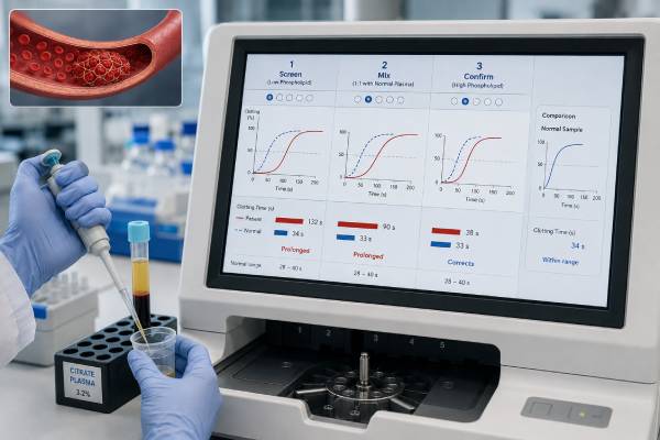
A lupus anticoagulant test looks for an antibody effect that slows phospholipid-dependent clotting reactions in the laboratory. The name is confusing: the antibody can occur without lupus, and in the body it is linked more strongly to blood clots than to bleeding. There is no single direct “lupus anticoagulant level.” Laboratories use a set of functional assays, usually dilute Russell viper venom time plus an aPTT- or silica-based method, followed by mixing and phospholipid confirmation steps when needed. A positive result can support antiphospholipid syndrome, but only when the clinical history is compatible and the antibody remains present on a second test at least 12 weeks later. A negative result lowers the likelihood that lupus anticoagulant is detectable under the test conditions, but it does not exclude other antiphospholipid antibodies or every cause of thrombosis. Anticoagulant medication is the most common reason a result becomes misleading or indeterminate.
- A positive lupus anticoagulant test means a phospholipid-dependent inhibitor was detected, not that a person has systemic lupus.
- Persistent lupus anticoagulant is one of the strongest laboratory risk markers for APS-related thrombosis.
- A negative result does not exclude anticardiolipin or anti-beta-2 glycoprotein I antibodies.
- The test usually requires screen, mix, and confirm logic rather than one isolated clotting time.
- Direct oral anticoagulants, heparin, and warfarin can distort the result, so medication timing must be documented.
- New chest pain, shortness of breath, one-sided swelling, or stroke symptoms needs emergency care, regardless of the laboratory result.
Table of Contents
- What Lupus Anticoagulant Is
- How Testing Detects It
- What a Positive Result Means
- What Negative and Indeterminate Results Mean
- Clotting and Pregnancy Risk
- Medication, Illness, and Test Errors
- Repeat Testing and Next Actions
What Lupus Anticoagulant Is
Lupus anticoagulant is a laboratory phenomenon caused by a varied group of antiphospholipid antibodies. These antibodies interact with phospholipid-binding proteins and interfere with the phospholipid surfaces needed for clotting reactions in a test tube. The result is a prolonged clotting time in certain sensitive assays.
The name dates from early observations in people with systemic lupus erythematosus whose plasma took longer to clot in vitro. Two parts of the name are now known to be misleading:
- Many people with lupus anticoagulant do not have lupus.
- The antibody is associated mainly with thrombosis and pregnancy complications, not with an anticoagulated state in the body.
Lupus anticoagulant is one of three established antiphospholipid antibody groups used in APS assessment. The others are anticardiolipin IgG and IgM and anti-beta-2 glycoprotein I IgG and IgM. Unlike those solid-phase immunoassays, lupus anticoagulant is detected through its effect on clot formation.
The antibody does not always cause disease. Some people have temporary positivity after infection. Others remain positive for years without a clot or pregnancy complication. Clinical risk depends on persistence, strength across assay systems, coexistence of other antibodies, prior events, autoimmune disease, and ordinary thrombosis risk factors.
Rarely, lupus anticoagulant coexists with low prothrombin or severe thrombocytopenia and contributes to bleeding. More often, a positive person who bleeds is bleeding because of anticoagulant medication, surgery, another factor inhibitor, or a separate hemostatic problem. A markedly prolonged aPTT with spontaneous bruising should not automatically be attributed to benign lupus anticoagulant.
The test is usually considered when there is:
- Unexplained venous or arterial thrombosis
- Stroke or transient ischemic attack at an unusually young age
- Recurrent or characteristic pregnancy morbidity
- An unexplained prolonged aPTT
- Systemic lupus or another autoimmune condition with a relevant history
- Previous antiphospholipid antibody positivity
Routine testing in people without a clinical reason can reveal transient or low-significance findings that are difficult to interpret.
How Testing Detects It
No single assay detects all lupus anticoagulants. Laboratories therefore use at least two phospholipid-sensitive test principles. The most common pairing is dilute Russell viper venom time, or dRVVT, and an activated partial thromboplastin time-based assay such as PTT-LA or silica clotting time.
The evaluation follows three linked ideas.
Screening
A screening reagent contains a limited amount or selected composition of phospholipid. If an antibody blocks the reaction, the clotting time becomes prolonged. The result is often normalized to pooled normal plasma and reported as a ratio.
A ratio around 1.20 is a common upper limit in some dRVVT systems, but there is no universal normal range. Laboratories validate cutoffs for their own instruments and reagents.
Mixing
The laboratory mixes patient plasma with normal pooled plasma, often in a 1:1 ratio. If the abnormal time corrects, a clotting-factor deficiency becomes more likely because the normal plasma supplied the missing factor. If the time remains prolonged, an inhibitor becomes more likely.
Weak lupus anticoagulants can dilute out and appear corrected, so mixing is not infallible. Laboratories use mixing-specific cutoffs or indices and may continue to confirmation even when the mix seems corrected.
Confirmation
The confirmation reagent contains more phospholipid. If extra phospholipid shortens the clotting time significantly, the inhibitor is phospholipid dependent. This response supports lupus anticoagulant.
A lupus anticoagulant reflex panel may perform these steps automatically after an abnormal screen. Not every report contains every test because a normal screen can stop that branch.
The final laboratory conclusion integrates both assay systems, the screen-mix-confirm pattern, anticoagulant exposure, and sample quality. A routine aPTT by itself is not a lupus anticoagulant test. Some lupus anticoagulants leave routine aPTT normal, while heparin or a factor deficiency can prolong it without lupus anticoagulant.
Solid-phase antibody tests remain important because anticoagulants affect them less directly. A person can have negative functional testing but positive anticardiolipin or anti-beta-2 glycoprotein I antibodies, and the reverse can occur.
What a Positive Result Means
A positive result means the laboratory found a pattern consistent with a phospholipid-dependent inhibitor. It does not mean that a clot is currently present, that the person will definitely develop one, or that systemic lupus is present.
For APS evaluation, the result gains weight when it is:
- Reproduced at least 12 weeks later
- Obtained without unaccounted anticoagulant interference
- Positive in more than one assay system or clearly positive in one validated system
- Accompanied by anticardiolipin or anti-beta-2 glycoprotein I antibodies
- Matched to an objectively documented clinical event
The strongest laboratory profile is persistent triple positivity: lupus anticoagulant plus anticardiolipin and anti-beta-2 glycoprotein I antibodies. This pattern is associated with a higher risk of first and recurrent thrombosis than isolated low-risk antibody findings. A triple-positive antiphospholipid antibody profile can also influence the choice of long-term anticoagulant after thrombosis.
An isolated positive result needs careful context. Temporary lupus anticoagulant can occur with viral or bacterial infection, major inflammation, and some medications. A first positive sample during hospitalization may disappear after recovery.
A person with a positive result but no clinical event is often described as an asymptomatic antiphospholipid antibody carrier rather than having APS. Management may focus on smoking cessation, blood pressure control, avoiding unnecessary estrogen exposure, and preventive measures during surgery or prolonged immobility. Full-dose anticoagulation is not automatically indicated.
A person with documented thrombosis and persistent lupus anticoagulant may have thrombotic APS. Venous events include deep vein thrombosis and pulmonary embolism. Arterial events include ischemic stroke, transient ischemic attack, and less commonly myocardial or limb ischemia. Small-vessel disease can affect kidneys, skin, lungs, brain, or other organs.
Classification criteria are designed for research and emphasize specificity. They should not be used as a self-diagnosis checklist. A specialist may diagnose and treat a clinically convincing case even when a research score is not met, or may reject APS when a classification item is technically present but a better explanation exists.
Does a stronger test mean a higher risk?
A more prolonged ratio can indicate a stronger in-vitro effect, but it is not a calibrated risk score. Reagent sensitivity, medication, factor levels, and antibody type alter the magnitude. Persistent positivity and the overall antibody profile are more useful than treating one ratio as a direct probability of thrombosis.
Four clinical profiles that should not be treated as equivalent
A person with one positive result during influenza, no clot history, and negative anticardiolipin and beta-2 glycoprotein I antibodies may have a transient finding. The immediate need is usually repeat testing after recovery, not a lifelong label.
A second person may have persistent lupus anticoagulant but no prior thrombosis. This is an asymptomatic high-risk laboratory marker, especially if another antibody is present. The plan may include aggressive control of smoking, blood pressure, cholesterol, and temporary prevention during surgery or prolonged immobility rather than continuous full-dose anticoagulation.
A third person may have persistent lupus anticoagulant after an unprovoked deep vein thrombosis. That history is much more compatible with thrombotic APS and often changes the expected duration and choice of anticoagulant therapy.
A fourth person may have lupus anticoagulant plus recurrent fetal loss or severe early placental disease but no personal thrombosis. This pattern may fit obstetric APS, which has a pregnancy-specific management plan and a separate assessment of long-term thrombosis risk.
The same word—positive—appears in all four profiles, yet the diagnosis, treatment, and follow-up differ. The result must be joined to the event history before it can guide care.
What Negative and Indeterminate Results Mean
A negative result means lupus anticoagulant was not detected by the methods and conditions used. It lowers the likelihood of an active, assay-detectable inhibitor, but it does not eliminate APS or all thrombosis risk.
Reasons a negative result may not be definitive include:
- The antibody reacts with a different assay principle
- A weak antibody was diluted in a mixing study
- High factor VIII during acute inflammation shortened an aPTT-based screen
- Anticoagulant medication caused a false-negative confirm pattern
- Residual platelets supplied phospholipid and neutralized the antibody
- The antibody was temporary and had already disappeared
- Other antiphospholipid antibodies are present instead
A negative lupus anticoagulant test should be interpreted with anticardiolipin and anti-beta-2 glycoprotein I results. It also does not exclude inherited thrombophilia, cancer-associated thrombosis, atrial fibrillation, vascular disease, or other causes of clotting.
An indeterminate result means the laboratory cannot confidently classify the pattern. This is common when a direct oral anticoagulant is present or when screen, mix, and confirm results conflict. Indeterminate is not equivalent to weak positive.
| Conclusion | What it says | Usual next step |
|---|---|---|
| Not detected | No qualifying phospholipid-dependent inhibitor pattern | Review other APS antibodies and clinical causes |
| Detected | At least one valid assay system supports lupus anticoagulant | Assess clinical history and repeat after at least 12 weeks |
| Indeterminate | Interference or discordance prevents classification | Address the cause and retest under safer conditions |
| Drug effect suspected | Anticoagulant likely altered the clotting assays | Use drug-aware laboratory strategy; do not stop treatment independently |
A portal may display individual abnormal ratios even when the final conclusion is negative. The laboratory may have found a prolonged screen that corrected in the mix or failed to show phospholipid dependence. Read the final interpretation before focusing on flagged components.
Clotting and Pregnancy Risk
Persistent lupus anticoagulant is generally the antiphospholipid marker most closely associated with thrombosis. The antibody may promote clotting by activating endothelial cells, platelets, monocytes, complement, and tissue factor pathways. Many researchers describe APS as a thrombo-inflammatory condition because immune activation and coagulation reinforce each other.
Actual risk varies. A positive test may be one “hit,” while a second trigger such as surgery, infection, pregnancy, immobility, smoking, estrogen therapy, or active lupus helps precipitate an event. This two-hit concept explains why some carriers remain event free for years and then develop thrombosis during a high-risk period.
Risk factors that deserve active management include:
- Previous thrombosis
- Triple-positive antibody profile
- Smoking
- Hypertension, diabetes, or high cholesterol
- Estrogen-containing contraception or hormone therapy
- Pregnancy and the six-week postpartum period
- Major surgery or prolonged immobility
- Active systemic autoimmune disease
- Cancer or severe infection
Pregnancy complications associated with APS include recurrent early miscarriage, fetal death, severe preeclampsia, placental insufficiency, fetal growth restriction, and medically indicated preterm birth. Lupus anticoagulant often predicts adverse pregnancy outcomes more strongly than isolated anticardiolipin or anti-beta-2 glycoprotein I positivity.
A positive result does not prove that APS caused a particular miscarriage. Chromosomal abnormalities, uterine factors, endocrine disease, and other placental problems remain common. A recurrent pregnancy loss antiphospholipid panel is most informative when interpreted with gestational ages, pathology, ultrasound findings, and repeat antibody testing.
Treatment depends on the clinical situation. Confirmed thrombotic APS often requires long-term anticoagulation. Obstetric APS without prior thrombosis is commonly managed with low-dose aspirin and prophylactic low-molecular-weight heparin during pregnancy, while a previous thrombosis may require therapeutic anticoagulation. These choices must be individualized because bleeding risk and medication safety differ.
A positive test alone is not a reason to start aspirin or a blood thinner without medical guidance. Preventive medication may benefit selected high-risk carriers, but indiscriminate treatment can cause gastrointestinal, intracranial, surgical, or obstetric bleeding.
Medication, Illness, and Test Errors
Lupus anticoagulant testing is unusually vulnerable to interference because it relies on clotting reactions. The medication intended to prevent thrombosis can create the same prolonged times that the test is trying to interpret.
Direct oral anticoagulants
Rivaroxaban frequently produces false-positive dRVVT results. Apixaban, edoxaban, and dabigatran can also distort screens and confirms. Drawing at the expected trough may reduce exposure but does not guarantee a drug-free sample.
Validated charcoal-based products can remove direct oral anticoagulants from plasma in some laboratories. A drug-specific anti-Xa or dilute thrombin test may be used to check residual medication. These approaches require laboratory expertise.
Heparin
Some reagents contain heparin neutralizers, but only up to a stated concentration. High-dose unfractionated heparin can overwhelm them. Low-molecular-weight heparin can also interfere, especially near peak concentration.
Warfarin
Warfarin reduces vitamin K-dependent factors, including factors II and X needed for dRVVT. Mixing with normal plasma may help but does not remove all uncertainty. Stopping warfarin solely for a test can be dangerous and must be managed by the treating team.
Acute illness and infection
Infection can induce temporary lupus anticoagulant. Acute thrombosis and inflammation alter factor VIII, C-reactive protein, and other coagulation proteins. Testing during the acute event may be clinically necessary, but later confirmation is essential.
Collection and processing
The blue-top citrate tube must be filled to the marked volume. Too much citrate prolongs clotting. Very high hematocrit may require citrate adjustment. Laboratories prepare platelet-poor plasma because residual platelets release phospholipid and can mask the antibody.
A clotted, hemolyzed, or markedly lipemic sample may be rejected. Delayed processing and repeated freeze-thaw cycles can also alter results. Reliable testing depends as much on specimen handling as on the calculation printed in the report.
Why two laboratories may disagree
One laboratory may use dRVVT plus silica clotting time, while another uses dRVVT plus PTT-LA. Their phospholipid composition, activators, pooled normal plasma, and cutoffs can differ. A weak antibody may therefore test positive at one site and negative at another. This does not automatically mean either laboratory made an error.
When results conflict, compare the dates, medications, acute illness, assay names, and raw ratios. Repeating at the same laboratory under stable conditions is usually more informative than sending samples to several laboratories at once. A specialist coagulation laboratory can help when the result will affect long-term anticoagulation or pregnancy management.
Repeat Testing and Next Actions
For APS evaluation, a positive lupus anticoagulant result should generally be repeated at least 12 weeks later. The interval distinguishes persistent autoimmunity from a transient response. A repeat at four or six weeks may provide clinical information but does not meet the usual persistence requirement.
Use the same laboratory when practical. Different reagent systems can produce different sensitivity and cutoffs. Keep the complete report, including screen ratios, confirm ratios, medication comments, and assay names.
After a positive result, ask the clinician:
- Was the sample reliable despite my medications?
- Which assay system was positive?
- Were anticardiolipin and anti-beta-2 glycoprotein I antibodies checked?
- Does my clot or pregnancy history fit APS?
- When should the repeat sample be collected?
- Should I avoid estrogen or take special precautions for surgery, travel, or pregnancy?
- Do I need hematology, rheumatology, or maternal-fetal medicine follow-up?
After a negative result, confirm that two lupus anticoagulant principles were used and review the other APS antibodies. If the clinical event remains unexplained, the workup may shift to cardiac, vascular, malignant, genetic, or obstetric causes.
After an indeterminate result, identify the precise interference. The solution may be a medically approved trough sample, drug-level testing, validated drug removal, or retesting after acute illness. Repeating under identical interfering conditions is unlikely to clarify the result.
Do not wait for antibody testing if symptoms suggest an acute clot. Call emergency services for sudden shortness of breath, chest pain, coughing blood, facial droop, speech difficulty, one-sided weakness, sudden vision loss, or a new painful swollen leg. During pregnancy, severe headache, visual changes, upper abdominal pain, high blood pressure symptoms, heavy bleeding, or reduced fetal movement also need urgent assessment.
A lupus anticoagulant result becomes useful when four pieces align: a valid laboratory pattern, appropriate clinical context, persistence over time, and a treatment plan that balances clotting and bleeding. None of those pieces should be inferred from one flagged clotting time.
Keep a dated record of previous clots, imaging reports, pregnancy outcomes, antibody results, and medication exposure at each blood draw. This history can prevent unnecessary repeat imaging, clarify whether an event was provoked, and show whether positivity truly persisted. It is especially valuable before changing anticoagulants, planning pregnancy, or scheduling major surgery. A one-page timeline is often more informative than a list of isolated laboratory values.
References
- Lupus anticoagulant detection and interpretation in antiphospholipid syndrome 2024 (Review)
- Testing for the lupus anticoagulant: the good, the bad, and the ugly 2024 (Review)
- An update on laboratory detection and interpretation of antiphospholipid antibodies for diagnosis of antiphospholipid syndrome: guidance from the ISTH-SSC Subcommittee on Lupus Anticoagulant/Antiphospholipid Antibodies 2025 (Guidance)
- Guidelines on the investigation and management of antiphospholipid syndrome 2024 (Guideline)
- New definitions for antiphospholipid syndrome 2024 (Review)
- 2023 ACR/EULAR antiphospholipid syndrome classification criteria 2023 (Classification Criteria)
Disclaimer
This article offers general education about lupus anticoagulant testing and cannot determine an individual diagnosis or treatment. A qualified clinician must interpret the complete coagulation panel, medication exposure, symptoms, and clotting or pregnancy history. Never stop anticoagulants or begin aspirin based only on a laboratory result.