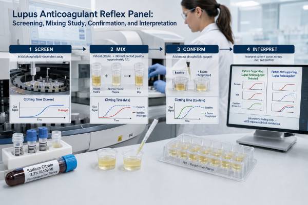
A lupus anticoagulant reflex panel is a stepwise group of clotting tests designed to determine whether a phospholipid-dependent inhibitor is present. The laboratory starts with sensitive screening assays, usually dilute Russell viper venom time and an aPTT- or silica-based method. When a screen is abnormal, predefined reflex rules add mixing and confirmation tests to the same specimen. The mixing step asks whether normal plasma corrects the prolonged time; the confirmation step asks whether extra phospholipid shortens it. The final result is an integrated interpretation, not a simple average of several numbers. Anticoagulant drugs, acute illness, factor deficiencies, and sample quality can make a panel falsely positive, falsely negative, or indeterminate. A positive panel supports lupus anticoagulant, but antiphospholipid syndrome also requires an appropriate clinical history and persistent antibody positivity on testing at least 12 weeks apart.
- “Reflex” means the laboratory automatically adds follow-up tests when an initial screen meets preset criteria.
- Most panels use two screening principles, commonly dRVVT plus an aPTT- or silica clotting time-based assay.
- A noncorrecting mix suggests an inhibitor, while phospholipid-dependent shortening on confirmation supports lupus anticoagulant.
- A normal screen may stop that test branch, so not every report contains every possible component.
- Anticoagulants are a major source of unreliable results, especially direct oral factor Xa inhibitors.
- A positive panel does not diagnose lupus, prove a current clot, or by itself establish APS.
Table of Contents
- What a Reflex Panel Does
- The Two Main Screening Branches
- How the Mixing Study Separates Deficiency From Inhibition
- How Confirmation Demonstrates Phospholipid Dependence
- Reading the Final Report
- Interference and Unreliable Panels
- Clinical Follow-Up and Repeat Testing
What a Reflex Panel Does
A reflex panel is an ordered laboratory algorithm. Instead of performing every test on every sample, the laboratory begins with one or more screens. An abnormal screen triggers additional tests according to rules validated by that laboratory. A normal screen may end that branch of the algorithm.
This approach has several advantages. It preserves sample, reduces unnecessary testing, shortens the time to an interpretation, and keeps related measurements on the same blood draw. It also allows the laboratory to apply consistent decision rules rather than asking the clinician to order separate confirmatory tests after each result returns.
A lupus anticoagulant reflex panel usually addresses three laboratory questions:
- Is a phospholipid-dependent screening time prolonged?
- Does the abnormality behave like an inhibitor when patient plasma is mixed with normal plasma?
- Does additional phospholipid reduce the inhibition?
The answers are integrated across at least two assay systems because lupus anticoagulants are heterogeneous. One antibody may affect a venom-based reaction, while another affects an intrinsic-pathway assay.
The word “panel” can be misleading because the components are not identical among laboratories. One panel may use dRVVT and silica clotting time. Another may use dRVVT and PTT-LA. Some include prothrombin time, thrombin time, anti-Xa testing, or anticoagulant-neutralization steps. Others add anticardiolipin and anti-beta-2 glycoprotein I antibodies only when a broader APS panel is ordered.
Before comparing two reports, check the test names, reagents, ratios, cutoffs, and interpretive comments. A panel labeled “lupus anticoagulant reflex” is primarily a functional coagulation evaluation. It may not include the solid-phase antibodies needed for a complete antiphospholipid syndrome blood test panel.
Reflex testing is appropriate when APS is clinically plausible, when an unexplained prolonged aPTT needs evaluation, or when a previous lupus anticoagulant result requires confirmation. Broad screening in people without thrombosis, characteristic pregnancy morbidity, or another relevant reason can identify transient antibodies that never cause disease.
The Two Main Screening Branches
Most modern panels begin with two phospholipid-sensitive assays that enter the coagulation cascade at different points. Using distinct principles increases the chance of detecting a heterogeneous lupus anticoagulant while limiting dependence on one reagent.
dRVVT branch
The dilute Russell viper venom time uses venom-derived factor X activation. The screening reagent contains limited phospholipid, making the reaction sensitive to phospholipid-dependent inhibition. The result is often normalized to pooled normal plasma and reported as a screen ratio.
If the screen ratio exceeds the local cutoff, the laboratory may reflex to a 1:1 mix with normal plasma and a high-phospholipid confirm reagent. The dRVVT test sequence is relatively specific for lupus anticoagulant but is strongly affected by some anticoagulant drugs.
aPTT or silica-based branch
The second screen activates the intrinsic pathway. Laboratories may use a lupus-sensitive aPTT reagent, PTT-LA, or silica clotting time. These reagents contain less phospholipid or a composition selected to increase sensitivity to lupus anticoagulant.
An abnormal screen can reflex to an aPTT mixing study and a phospholipid-rich confirmation method. Confirmation may use paired reagents with low and high phospholipid, a hexagonal-phase phospholipid assay, or another validated approach.
The two branches can disagree. A positive dRVVT with a negative PTT-LA may still represent lupus anticoagulant if the dRVVT screen, mix, and confirm pattern is valid. A positive intrinsic-pathway branch with a normal dRVVT may also be meaningful. The final conclusion should state which system detected the inhibitor.
Why a routine aPTT is not enough
A normal routine aPTT does not exclude lupus anticoagulant. Routine reagents are designed for broad coagulation screening and vary substantially in phospholipid concentration. Some are intentionally less sensitive to lupus anticoagulant so that common preoperative testing is not frequently prolonged by clinically incidental antibodies.
Likewise, an isolated prolonged routine aPTT does not prove lupus anticoagulant. Heparin, factor deficiencies, factor VIII inhibitors, and sample problems are common alternatives. The reflex panel supplies the inhibitor and phospholipid-dependence steps needed for a more specific conclusion.
Why screens are normalized
Clotting times in seconds vary with instrument, reagent lot, and normal plasma. Laboratories often divide the patient time by a pooled normal plasma time to create a ratio. A ratio near 1.20 is a common cutoff on some systems, but it is not universal. Each laboratory should establish or validate its own reference limits.
A screen just above the cutoff is not automatically a weak positive. The confirm response, mix behavior, anticoagulant exposure, and second assay determine whether the pattern supports lupus anticoagulant.
How the Mixing Study Separates Deficiency From Inhibition
The mixing study combines patient plasma with normal pooled plasma, commonly in equal volumes. Normal plasma supplies adequate levels of most clotting factors. The laboratory repeats the prolonged assay on the mixture and evaluates whether the time corrects.
If the abnormal time moves into the expected range, a factor deficiency becomes more likely. If it remains prolonged, an inhibitor becomes more likely because the inhibitory substance in the patient sample also acts on the added normal plasma.
This basic logic is useful, but the result is not a simple yes-or-no observation. Laboratories may use:
- A mixing-test-specific normalized ratio
- The index of circulating anticoagulant
- Percent correction
- A locally validated cutoff derived from normal and inhibitor samples
A screen cutoff should not automatically be applied to a 1:1 mix. Dilution changes the expected result distribution, so a dedicated mixing cutoff improves sensitivity.
Why a lupus anticoagulant mix may appear corrected
Mixing halves the concentration of the patient’s antibody. A weak lupus anticoagulant can be diluted below the assay’s detection threshold and appear to correct. This creates a false-negative mixing study. Some algorithms therefore perform confirmation even when the mix corrects, especially if the screen-confirm relationship strongly suggests phospholipid dependence.
Why a mix may fail to correct without lupus anticoagulant
Several other inhibitors prevent correction:
- Unfractionated heparin or a direct oral anticoagulant
- A specific factor inhibitor, especially factor VIII inhibitor
- A strong nonspecific inhibitor unrelated to antiphospholipid antibodies
- Very low levels of multiple clotting factors in severe illness
Clinical symptoms help distinguish these possibilities. Lupus anticoagulant is usually associated with thrombosis risk and often no bleeding. An acquired factor VIII inhibitor commonly presents with spontaneous bruising, soft-tissue bleeding, or internal bleeding.
Immediate versus incubated mixing
Some factor inhibitors become stronger after incubation at 37°C, while lupus anticoagulant is usually assessed with an immediate mix in dedicated LA algorithms. A separate prolonged-aPTT workup may include both immediate and incubated mixes. The patient may therefore see more than one mixing result on a comprehensive coagulation report.
The mixing step answers “inhibitor or deficiency?” It does not by itself prove that the inhibitor is phospholipid dependent. That requires the confirmation step.
How Confirmation Demonstrates Phospholipid Dependence
Confirmation uses an otherwise comparable clotting reaction with more phospholipid. If the added phospholipid shortens the clotting time substantially, the inhibitor behaves like lupus anticoagulant.
In dRVVT, the laboratory compares low-phospholipid screen and high-phospholipid confirm reagents. It may calculate a screen-to-confirm ratio after normalizing each time to pooled normal plasma. In an aPTT-based branch, confirmation may compare two phospholipid concentrations or use the hexagonal phospholipid neutralization test.
A valid lupus anticoagulant pattern generally contains:
- Prolongation in at least one sensitive screen
- Evidence of an inhibitor, with recognition that weak antibodies may be diluted in a mix
- Significant correction or shortening with excess phospholipid
- No better explanation from anticoagulant drugs or another coagulation abnormality
The order of mix and confirm is debated because each sequence has strengths and weaknesses. A screen-mix-confirm algorithm emphasizes proof of inhibition before confirmation but can miss weak antibodies that dilute out. A screen-confirm-mix sequence may capture phospholipid dependence earlier but can be less straightforward when factor deficiency or anticoagulant effect is present. Modern guidance allows laboratories to use validated algorithms and interpret all steps together.
Confirmation is not the same as repeating the test 12 weeks later. The confirm reagent verifies phospholipid dependence within the current specimen. Repeat testing verifies persistence over time. Both concepts are important but answer different questions.
A large confirm response does not quantify clinical severity. A person with a dramatic laboratory ratio does not necessarily have a larger clot or a more imminent event than someone with a smaller ratio. Risk depends on persistence, other antiphospholipid antibodies, prior events, and conventional thrombosis factors.
Reading the Final Report
The final interpretive line is usually more useful than any single component. Common conclusions include “lupus anticoagulant detected,” “not detected,” “indeterminate,” or “testing affected by anticoagulant therapy.”
| Panel pattern | Likely laboratory conclusion | Important qualification |
|---|---|---|
| Both screens normal | Lupus anticoagulant not detected | A very weak or assay-specific antibody may still be missed |
| One screen prolonged, mix noncorrecting, confirm positive | Lupus anticoagulant detected in that system | Exclude medication interference and confirm persistence |
| Screen prolonged, mix corrects, confirm negative | Factor deficiency more likely | Factor assays may be needed |
| Screen and confirm both prolonged without phospholipid response | Not a classic lupus anticoagulant pattern | Consider drug effect or specific inhibitor |
| Discordant or drug-affected branches | Indeterminate | Repeat under interpretable conditions |
A report may contain many abbreviations: SCT, dRVVT, DRVVS, DRVV confirmation, PTT-LA, ratio, mix, ICA, HEXLA, or neutralization. The most useful details to retain are the final conclusion, which branch was positive, the collection date, and whether an anticoagulant comment was present.
“Not detected” means no qualifying pattern appeared under the conditions tested. It does not rule out anticardiolipin or anti-beta-2 glycoprotein I antibodies because those are separate immune assays. It also does not exclude all causes of thrombosis or pregnancy loss.
“Detected” means the laboratory found a phospholipid-dependent inhibitor consistent with lupus anticoagulant. It does not diagnose systemic lupus erythematosus, despite the name. It does not prove that a current symptom is caused by a clot.
“Indeterminate” is not a weak diagnosis. It means the laboratory cannot reliably separate lupus anticoagulant from interference. The correct response is to address the interference, not to count the result as positive or negative.
A patient portal may flag several intermediate values even when the final panel is negative. Conversely, an individual screen ratio may look only slightly high while the integrated pattern is positive. Read the interpretive comment before drawing conclusions from red or bold numbers.
A sample report walkthrough
Suppose the dRVVT screen ratio is 1.34, above a laboratory cutoff of 1.20. The 1:1 mix remains elevated by the laboratory’s mixing criterion, and the high-phospholipid confirm reagent lowers the normalized result enough to produce a positive screen-to-confirm ratio. The PTT-LA branch is normal. If no interfering anticoagulant is present, the laboratory may conclude that lupus anticoagulant is detected by dRVVT. The normal second branch does not cancel the valid first branch.
Now consider a second report with a dRVVT screen ratio of 1.45 and an abnormal PTT-LA. Both mixing tests remain prolonged, but the confirm tests show little or no phospholipid-dependent correction. The patient took rivaroxaban that morning. In this setting, the laboratory may report anticoagulant interference rather than lupus anticoagulant because the screens, mixes, and confirms can all be altered by the drug.
A third report may show an isolated prolonged PTT-LA that corrects with normal plasma, while dRVVT is normal. That pattern is more compatible with an intrinsic factor deficiency. Factor VIII, IX, XI, or XII testing may follow, depending on bleeding history and the degree of prolongation.
These examples also show why the reflex panel should be interpreted as a decision tree. The first abnormal value determines which tests appear next, and each added result narrows the explanation. A long report is not evidence of a more severe disease; it often means only that the laboratory had to investigate an abnormal screen.
Interference and Unreliable Panels
Anticoagulant treatment affects the same clotting reactions used by the panel. Testing is easiest before treatment begins, but suspected thrombosis often requires immediate anticoagulation. Patient safety takes priority over obtaining an ideal laboratory specimen.
Direct oral anticoagulants
Rivaroxaban commonly creates false-positive dRVVT patterns. Apixaban, edoxaban, and dabigatran can also produce false-positive or false-negative effects depending on reagent and concentration. A trough sample may reduce interference but does not guarantee its absence.
Some reflex panels include an activated-charcoal drug-removal step when a screening pattern suggests a direct oral anticoagulant. Removal products can help but require local validation. A drug-specific anti-Xa or dilute thrombin assay may be used to assess residual medication.
Heparin
Many dRVVT reagents contain heparin neutralizers, and some panels add heparinase. Neutralization works only up to a stated heparin level. High-dose unfractionated heparin can still invalidate testing. Low-molecular-weight heparin effects depend on timing and reagent sensitivity.
Warfarin
Warfarin lowers factors II, VII, IX, and X. Low factor II and X can prolong dRVVT, while mixing may partly correct the deficiency. Specialized interpretation is needed, and stopping warfarin without a bridging plan can be dangerous.
Acute phase and sample factors
Acute infection may produce transient lupus anticoagulant. High factor VIII can shorten an aPTT-based screen and mask a weak antibody, while high C-reactive protein can prolong some aPTT reagents. Pregnancy, liver disease, disseminated intravascular coagulation, and severe illness can alter multiple factors.
Residual platelets release phospholipid and may neutralize lupus anticoagulant, causing false-negative results. Underfilled citrate tubes, high hematocrit, hemolysis, lipemia, or clotted specimens can also distort times. Proper preparation of platelet-poor plasma is therefore part of the diagnostic process.
Never skip or stop an anticoagulant dose solely for testing unless the prescribing clinician creates a safe plan. A report obtained under medication may still be useful when the laboratory knows the drug and applies validated mitigation, but limitations must remain visible in the interpretation.
Clinical Follow-Up and Repeat Testing
A positive lupus anticoagulant reflex panel is one laboratory component of APS evaluation. The other established antibodies are anticardiolipin IgG and IgM and anti-beta-2 glycoprotein I IgG and IgM. The combination of all three marker groups is called triple positivity and generally indicates a higher-risk profile.
APS also requires a relevant clinical manifestation, most commonly objectively confirmed thrombosis or characteristic pregnancy morbidity. Classification criteria used in research define specific domains, but clinicians consider the complete history, competing explanations, and treatment implications.
The first positive panel should usually be repeated at least 12 weeks later. A shorter interval can help investigate interference, but it does not establish persistent positivity. Use the same laboratory when practical, because reagents and cutoffs vary.
After a positive result, discuss:
- Whether the sample was interpretable despite medication
- Which assay branch was positive
- Whether anticardiolipin and beta-2 glycoprotein I antibodies were measured
- Whether the clinical event fits APS
- The exact date for repeat testing
- Risk reduction during surgery, pregnancy, immobility, or estrogen exposure
- Whether hematology, rheumatology, or maternal-fetal medicine review is appropriate
A positive result without a prior clot does not automatically require full-dose anticoagulation. Treatment decisions balance the antibody profile, personal history, cardiovascular risks, and bleeding risk. Aspirin and anticoagulants can cause serious harm when used without a clear indication.
After a negative result, ask whether both recommended assay principles were used. If suspicion remains high, review anticoagulant exposure and acute-phase conditions. Other thrombophilias, vascular disorders, or causes of pregnancy loss may need evaluation.
After an indeterminate result, the next test should solve the stated problem. That may mean collecting at a medically supervised drug trough, checking a drug level, using a validated neutralization method, or repeating after acute illness resolves.
Seek urgent medical care for sudden shortness of breath, chest pain, coughing blood, one-sided leg swelling, facial droop, speech difficulty, one-sided weakness, or sudden vision loss. The panel estimates an antibody phenomenon; it cannot rule in or rule out an emergency clot at the bedside.
A reflex panel is most valuable when its components are treated as a logical sequence. Screening identifies a sensitive abnormality, mixing tests its behavior, confirmation establishes phospholipid dependence, and repeat testing establishes persistence. The clinical history then determines whether the laboratory finding represents APS or an isolated antibody.
References
- Lupus Anticoagulant Reflex Panel 2025 (Official Laboratory Guidance)
- Comparison of different algorithms for lupus anticoagulant detection: a single-center experience 2024 (Study)
- An update on laboratory detection and interpretation of antiphospholipid antibodies for diagnosis of antiphospholipid syndrome: guidance from the ISTH-SSC Subcommittee on Lupus Anticoagulant/Antiphospholipid Antibodies 2025 (Guidance)
- Lupus Anticoagulant Testing for Diagnosis of Antiphospholipid Syndrome: A Perspective Informed by Local Practice 2025 (Review)
- Guidelines on the investigation and management of antiphospholipid syndrome 2024 (Guideline)
- 2023 ACR/EULAR antiphospholipid syndrome classification criteria 2023 (Classification Criteria)
Disclaimer
This article provides general information about lupus anticoagulant reflex testing and cannot interpret an individual panel or diagnose APS. Medication changes and decisions about aspirin or anticoagulation require a clinician who can review the complete report, symptoms, and medical history. Do not interrupt prescribed anticoagulant therapy for repeat testing without a supervised safety plan.