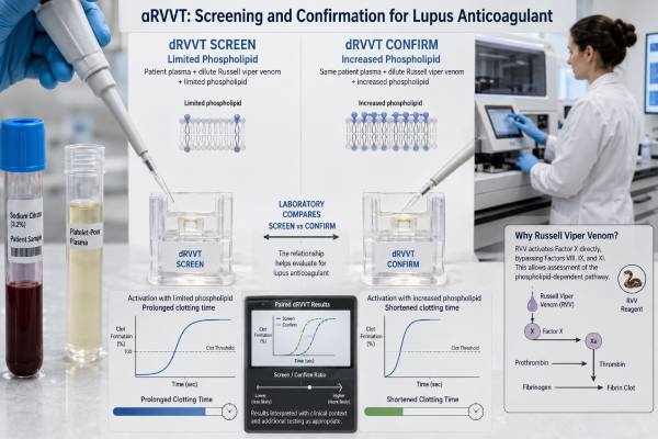
The dilute Russell viper venom time, usually shortened to dRVVT, is one of the main laboratory methods used to detect lupus anticoagulant. Despite its name, lupus anticoagulant is associated more often with abnormal clotting in the body than with bleeding, and many people who test positive do not have lupus. The dRVVT is not a single clotting-time number interpreted in isolation. It usually involves a low-phospholipid screening test, a phospholipid-rich confirmation test, and sometimes a mixing study. The pattern shows whether a phospholipid-dependent inhibitor is likely to be present. Results are especially sensitive to anticoagulant medicines, sample quality, acute illness, and laboratory-specific cutoffs. A positive pattern can support antiphospholipid syndrome when it persists for at least 12 weeks and accompanies a qualifying clot or pregnancy complication. A negative result does not exclude every lupus anticoagulant because no one assay detects all antibody types.
- dRVVT screens for lupus anticoagulant by measuring phospholipid-dependent clotting after direct activation of factor X.
- A prolonged screen is not enough for a positive result; phospholipid confirmation and the full test pattern are needed.
- Screen and confirm ratios are laboratory specific, although a normalized ratio near 1.2 is a common cutoff on some systems.
- Direct oral anticoagulants, heparin, and warfarin can cause misleading results, including false-positive or false-negative patterns.
- No fasting is usually required, but the laboratory must know every anticoagulant and the time of the last dose.
- A confirmed positive lupus anticoagulant test should usually be repeated after at least 12 weeks when APS is being evaluated.
Table of Contents
- How dRVVT Works
- The Screen, Mix, and Confirm Sequence
- Ratios and Common Result Patterns
- Medication and Sample Interference
- Clinical Meaning of a Positive Test
- Preparation and Repeat Testing
- Questions and Next Steps
How dRVVT Works
The dRVVT uses a laboratory reagent derived from Russell viper venom. The venom component directly activates clotting factor X. Activated factor X then works with factor V, calcium, phospholipid, and prothrombin to generate thrombin, which converts fibrinogen into a clot. Because the test enters the clotting pathway at factor X, it bypasses much of the intrinsic and extrinsic pathways measured by tests such as the activated partial thromboplastin time and prothrombin time.
The screening reagent contains a limited amount of phospholipid. This deliberate shortage makes the reaction sensitive to antibodies that interfere with phospholipid-dependent clotting. If lupus anticoagulant is present, it may bind to phospholipid-protein complexes and slow the reaction, prolonging the clotting time.
The confirmation reagent contains more phospholipid. The extra phospholipid can overwhelm or neutralize the antibody effect. When the clotting time becomes meaningfully shorter with the confirm reagent, the pattern supports phospholipid dependence, a defining laboratory feature of lupus anticoagulant.
The patient is never exposed to snake venom. The reagent is used only in the test tube with separated plasma. The amount and formulation are standardized by the assay manufacturer.
The dRVVT is useful because it is relatively specific for lupus anticoagulant and is less affected by deficiencies of factors VIII, IX, XI, and XII than an activated partial thromboplastin time-based method. It is not immune to other influences, however. Deficiencies or inhibitors involving factors II, V, or X, low fibrinogen, anticoagulant drugs, and substantial sample problems can alter the result.
Laboratories generally use at least two lupus anticoagulant methods based on different clotting principles. The second method is often a sensitive activated partial thromboplastin time assay such as the PTT-LA test. This paired approach matters because lupus anticoagulants are a heterogeneous group of antibodies. One antibody may strongly affect dRVVT while another is detectable only in an aPTT-based system.
The dRVVT does not measure the concentration of one specific antibody. It measures an antibody effect on a functional coagulation reaction. This distinction explains why a numerical dRVVT ratio does not correspond directly to an anticardiolipin or anti-beta-2 glycoprotein I antibody level.
The Screen, Mix, and Confirm Sequence
A complete dRVVT evaluation separates three questions: Is the screening clotting time prolonged? Does the abnormality behave like an inhibitor? Is the inhibitor dependent on phospholipid? Laboratories answer these questions with screen, mix, and confirm steps, although the exact sequence varies by guideline and local algorithm.
Screening test
The screen uses a low-phospholipid reagent. The patient’s clotting time is compared with a normal pooled plasma value or a locally established reference interval. A normal screen makes a dRVVT-detectable lupus anticoagulant less likely. An abnormal screen triggers further analysis rather than establishing positivity.
A prolonged screen can have several explanations:
- Lupus anticoagulant
- A therapeutic anticoagulant
- Deficiency or dysfunction of factor II, V, or X
- A specific clotting-factor inhibitor
- Low fibrinogen or another major coagulation abnormality
- Preanalytical or analytical error
Mixing study
For the mixing study, the laboratory usually combines patient plasma with normal pooled plasma in a 1:1 ratio and repeats the clotting assay. Normal plasma supplies clotting factors. If the prolonged time corrects substantially, a factor deficiency becomes more likely. If it remains prolonged, an inhibitor becomes more likely.
Mixing studies are useful but imperfect. Dilution can weaken a low-titer lupus anticoagulant enough to make the mixture appear corrected, producing a false-negative inhibitor pattern. Conversely, anticoagulant medicines can prevent correction. Laboratories may use a mixing-test-specific cutoff or an index calculation rather than applying the screen cutoff to the mixture.
Some algorithms perform screen, then confirm, then mix. Others use screen, mix, and confirm. A report may omit a mixing result when the screen and confirmation pattern is already clear or when local policy handles weak inhibitors differently. The absence of a reported mixing study does not necessarily mean that the laboratory performed an incomplete test.
Confirmation test
The confirm reagent contains a high phospholipid concentration. The laboratory compares the screen and confirm results, often after normalization to pooled normal plasma. A significant shortening in the confirm phase indicates that additional phospholipid reduced the inhibitory effect.
This phospholipid dependence distinguishes lupus anticoagulant from many factor deficiencies. It is the reason the test is called a confirmation step even though the final interpretation still depends on medication exposure, other coagulation results, and clinical context.
A lupus anticoagulant reflex panel may automatically add the mix and confirm tests when the initial screen is abnormal. Reflex testing reduces delays and ensures that the laboratory interprets the components together.
Ratios and Common Result Patterns
Raw clotting times in seconds vary with reagent lots, instruments, and local populations. Most laboratories therefore convert dRVVT times into normalized ratios. A common calculation divides the patient clotting time by the clotting time of normal pooled plasma tested with the same reagent.
The report may include:
- dRVVT screen time in seconds
- dRVVT screen ratio
- dRVVT mix ratio or mixing index
- dRVVT confirm time or ratio
- Screen-to-confirm ratio, sometimes called the normalized ratio
- A final interpretation such as “lupus anticoagulant detected,” “not detected,” or “indeterminate due to anticoagulant effect”
Many assay systems use a screen ratio around 1.20 as the upper limit, but this is not a universal normal range. A laboratory may establish a different cutoff from its own healthy reference population or validate the manufacturer’s value. The screen-to-confirm threshold also varies. The final interpretive comment is usually more useful than comparing a result with a cutoff found online.
| Screen | Mix | Confirm | Possible interpretation |
|---|---|---|---|
| Normal | Not needed | Not needed | No lupus anticoagulant detected by dRVVT; another method may still be positive |
| Prolonged | Does not correct | Shortens with excess phospholipid | Pattern supports lupus anticoagulant |
| Prolonged | Corrects | Little phospholipid effect | Factor deficiency is more likely |
| Prolonged | Does not correct | Little phospholipid effect | Consider anticoagulant drug, specific factor inhibitor, or another cause |
| Prolonged | Variable | Variable | Indeterminate; review drug level, sample timing, and other assays |
A positive dRVVT result means that the pattern is consistent with lupus anticoagulant under the laboratory’s method and conditions. It does not mean that a clot is present. It does not measure clot size or predict exactly when thrombosis will occur. It also does not diagnose systemic lupus erythematosus.
A negative dRVVT does not rule out lupus anticoagulant. The antibody may affect an aPTT-based assay instead, may be too weak for the current reagent, or may be masked by treatment or acute-phase changes. This is why the final lupus anticoagulant interpretation should include all methods used.
Some reports use “borderline,” “weak positive,” or “indeterminate.” These terms signal that one part of the pattern is close to a cutoff or that interference prevents a confident result. The appropriate response is usually not to assume positivity or negativity, but to review medications, repeat under better conditions, and compare with solid-phase antiphospholipid antibody tests.
Why the number of seconds can mislead
A dRVVT screen of 46 seconds may be abnormal in one laboratory and within range in another because the reagent concentration, analyzer, pooled plasma, and reference population differ. Even results produced by two versions of the same commercial kit may not be directly comparable after a reagent-lot change. Normalization reduces some of this variation by expressing the patient result relative to a normal plasma result tested under the same conditions.
The screen-to-confirm relationship is more important than the screen time alone. For example, two people can have the same prolonged screen ratio. In one, the confirm ratio falls substantially when phospholipid is added, supporting lupus anticoagulant. In the other, both screen and confirm remain prolonged to a similar degree, suggesting medication effect, a factor problem, or another inhibitor. A patient portal may flag both screens in red even though the laboratory conclusions differ.
A stronger ratio also should not be treated as a precise measure of future clot risk. Reagent sensitivity and antibody heterogeneity affect the size of the prolongation. Clinical risk assessment uses persistence, the presence of other antiphospholipid antibodies, prior thrombosis or pregnancy morbidity, and conventional risk factors. The ratio helps identify the laboratory phenomenon; it is not a stand-alone severity score.
Medication and Sample Interference
Anticoagulants are the most important source of misleading dRVVT results. They alter clotting reactions by design, so they can mimic or hide an inhibitor. The laboratory must know the exact drug, dose, last administration time, kidney function when relevant, and reason for treatment.
Direct oral anticoagulants
Factor Xa inhibitors such as rivaroxaban, apixaban, and edoxaban commonly prolong dRVVT. Rivaroxaban is particularly likely to create a screen-confirm pattern that resembles lupus anticoagulant. The thrombin inhibitor dabigatran can also interfere. Testing at a presumed trough level may reduce but does not eliminate the problem.
Some laboratories use activated-charcoal products or other adsorbents to remove direct oral anticoagulants from plasma. These methods can improve interpretation, but removal may be incomplete, may alter some samples, and should be validated locally. A drug-specific anti-Xa or dilute thrombin test may be needed to show whether clinically relevant medication remains.
Heparin
Many dRVVT reagents contain a heparin neutralizer that can handle unfractionated heparin up to a stated concentration. The neutralizer has limits. A high heparin level can still prolong the assay, and low-molecular-weight heparin may affect results depending on timing and reagent sensitivity. Drawing the sample just before the next dose may be considered when clinically safe and approved by the treating clinician.
Warfarin and other vitamin K antagonists
Warfarin lowers vitamin K-dependent factors, including factors II and X, which dRVVT needs. This can prolong the screen and complicate mixing studies. Interpreting dRVVT during warfarin treatment requires specialized algorithms and the accompanying prothrombin time or INR. Stopping warfarin solely to obtain a test can expose a patient to thrombosis and should never be done without a managed plan.
Acute illness and inflammation
Severe inflammation can complicate lupus anticoagulant testing. High C-reactive protein has a stronger known effect on some aPTT-based reagents, but studies also show that dRVVT times may change with substantial inflammation. Infection can also trigger temporary lupus anticoagulant. Testing during acute thrombosis or critical illness may be necessary, but a later sample often gives a clearer answer.
Sample handling
Residual platelets release phospholipid and may neutralize lupus anticoagulant, causing a false-negative result. Laboratories aim to prepare platelet-poor plasma, often with double centrifugation. Hemolysis, lipemia, an underfilled citrate tube, a clotted specimen, or a long delay before processing can also compromise testing.
The blood tube must fill to the required level because citrate anticoagulant is proportioned for a specific blood volume. People with a very high hematocrit may need an adjusted citrate volume. These preanalytical details are invisible to the patient but essential to reliable results.
Clinical Meaning of a Positive Test
Lupus anticoagulant is one of the three established laboratory marker groups used in antiphospholipid syndrome assessment. The other two are anticardiolipin antibodies and anti-beta-2 glycoprotein I antibodies. Among these markers, persistent lupus anticoagulant is often associated with the strongest clotting risk.
APS is considered when a person has a compatible clinical event and persistent antiphospholipid antibodies. Clinical events include objectively confirmed venous, arterial, or small-vessel thrombosis and defined obstetric complications. Research classification criteria are useful for consistent study enrollment, but clinicians diagnose and manage individual patients using the entire record.
A first positive dRVVT may be temporary. Infection, inflammation, medication interference, or analytical variation can produce a one-time result. For APS evaluation, the lupus anticoagulant should generally remain positive on a second occasion at least 12 weeks later.
Risk is higher when lupus anticoagulant occurs with anticardiolipin and anti-beta-2 glycoprotein I antibodies. This is called triple positivity. Persistent high-risk profiles carry more weight than an isolated weak result. The antibody pattern, prior event, age, smoking, estrogen exposure, surgery, pregnancy, cancer, immobility, and autoimmune disease all contribute to actual risk.
A positive test without a previous clot does not automatically require full-dose anticoagulation. Management may focus on controlling cardiovascular risks and using preventive measures during high-risk situations. A person with confirmed thrombotic APS often needs long-term anticoagulant treatment, but the choice depends on event type, recurrence, bleeding risk, and antibody profile.
The term “anticoagulant” causes understandable confusion. In the test tube, the antibody prolongs phospholipid-dependent clotting. In the body, it can promote clotting through cell activation, complement, tissue factor, and inflammatory pathways. Bleeding is uncommon unless another problem is present, such as low platelets, anticoagulant medication, a factor deficiency, or the rare lupus anticoagulant-hypoprothrombinemia syndrome.
A dRVVT result should not delay urgent treatment of suspected thrombosis. Sudden chest pain, shortness of breath, coughing blood, one-sided leg swelling, stroke symptoms, or sudden vision loss require immediate medical evaluation whether the most recent dRVVT was positive, negative, or never performed.
Preparation and Repeat Testing
No fasting or special diet is normally required. Hydration can make venipuncture easier, but it does not determine lupus anticoagulant status. The most important preparation is an accurate medication and clinical history.
Before the blood draw, provide:
- The name and dose of every anticoagulant
- The exact time of the most recent dose
- Recent changes in treatment or missed doses
- Current pregnancy status
- Recent infection, hospitalization, surgery, or thrombosis
- Any known factor deficiency or liver disease
- Prior lupus anticoagulant results and the testing laboratory
Do not skip an anticoagulant dose unless the prescribing clinician gives explicit instructions and has considered the clotting risk. A laboratory-perfect sample is not worth creating an unsafe gap in treatment.
When possible, initial testing may be performed before anticoagulation begins. In real life, a suspected clot often requires immediate treatment, so this timing is not always available. Clinicians and laboratories can select strategies based on the drug: testing at trough, measuring a drug level, using a validated removal product, relying more heavily on antibody immunoassays, or postponing the functional test.
Repeat testing for persistence must occur at least 12 weeks after the first positive result. Repeating sooner can help troubleshoot interference but does not satisfy the persistence interval used in APS frameworks. The later test should ideally use the same laboratory and include both dRVVT and a second principle-based assay.
A negative repeat result may indicate a transient antibody, but the interpretation depends on whether anticoagulant therapy or another factor could have masked it. A positive repeat is more persuasive when both samples were collected under interpretable conditions.
Solid-phase tests for anticardiolipin and beta-2 glycoprotein I antibodies are less affected by anticoagulants than dRVVT. They can be performed while treatment continues, although they do not replace lupus anticoagulant testing. The complete antiphospholipid antibody panel helps place a difficult dRVVT result in context.
Questions and Next Steps
The next step depends on whether the report is negative, positive, or uninterpretable and why the test was ordered.
For a negative dRVVT, check whether an aPTT-based lupus anticoagulant assay was also performed. If clinical suspicion remains strong, review timing, medication exposure, and whether the sample was collected during an acute event. Other causes of thrombosis or pregnancy complications may need investigation.
For a positive dRVVT, confirm that the report shows phospholipid dependence rather than only a prolonged screen. Review the mixing result, confirm ratio, anticoagulant exposure, and other antiphospholipid antibodies. Arrange repeat testing at least 12 weeks later if APS is being considered.
For an indeterminate result, ask what prevented interpretation. A direct oral anticoagulant level, repeat sample at a safer time, alternative venom assay, or specialist coagulation laboratory may resolve the uncertainty. Repeatedly ordering the same test under the same interfering conditions is unlikely to help.
Useful questions for the clinician or laboratory include:
- Was the dRVVT screen abnormal, and did the confirm phase show phospholipid dependence?
- Was a mixing study performed, and what cutoff was used?
- Could my anticoagulant explain the pattern?
- Was the plasma adequately platelet poor?
- Was a second lupus anticoagulant method performed?
- Were anticardiolipin and anti-beta-2 glycoprotein I antibodies measured?
- When should the test be repeated, and how can it be done safely?
- Does my history suggest APS or only an isolated laboratory finding?
A well-interpreted dRVVT is a multistep laboratory conclusion, not a single out-of-range number. The screen, mix, confirm response, medication context, second assay, repeat result, and clinical history must point in the same direction before the finding can guide major long-term decisions.
References
- Update on the Laboratory Diagnosis of Lupus Anticoagulant 2025 (Review)
- Testing for the lupus anticoagulant: the good, the bad, and the ugly 2024 (Review)
- Lupus Anticoagulant Testing for Diagnosis of Antiphospholipid Syndrome: A Perspective Informed by Local Practice 2025 (Review)
- Comparison of different algorithms for lupus anticoagulant detection: a single-center experience 2024 (Study)
- Lupus anticoagulant testing during anticoagulation, including direct oral anticoagulants 2022 (Review)
- 2023 ACR/EULAR antiphospholipid syndrome classification criteria 2023 (Classification Criteria)
Disclaimer
This article provides general information about dRVVT and lupus anticoagulant testing. Test interpretation and any decision about anticoagulant treatment require a clinician who can review the assay method, medications, symptoms, and clotting or pregnancy history. Never stop a blood thinner to improve test accuracy without a medically supervised plan.