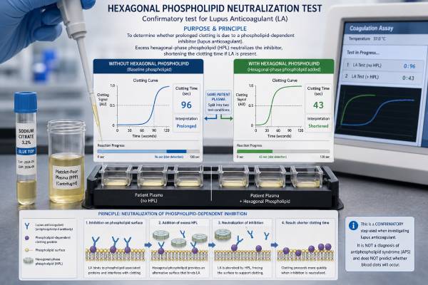
The hexagonal phospholipid neutralization test is an aPTT-based method used to confirm that a prolonged clotting time is caused by a phospholipid-dependent inhibitor, most often lupus anticoagulant. The laboratory tests plasma under two closely matched conditions: one without added hexagonal-phase phospholipid and one with it. If the added phospholipid shortens the clotting time by more than the assay’s cutoff, the result supports lupus anticoagulant activity. Laboratories may call the method Hex LA, HEXLA, hexagonal phase phospholipid, HPP, or STACLOT-LA. It is usually one component of a broader lupus anticoagulant evaluation, not a stand-alone diagnosis of antiphospholipid syndrome. Anticoagulant medicines, factor deficiencies, specific factor inhibitors, acute illness, and sample handling can distort the pattern. A confirmed positive result becomes clinically meaningful only after review of the full antibody profile, clotting or pregnancy history, and repeat testing at least 12 weeks later when APS is suspected.
- The test confirms phospholipid dependence by comparing an aPTT-type clotting time with and without hexagonal-phase phospholipid.
- A positive result usually means the clotting time shortened beyond a laboratory-defined number of seconds after phospholipid was added.
- Cutoffs are assay specific; some laboratories use a delta near 6–8 seconds, but the report’s own threshold controls interpretation.
- A positive hexagonal phospholipid test supports lupus anticoagulant, not systemic lupus and not a current blood clot.
- Direct oral anticoagulants, heparin, and warfarin can produce false or indeterminate results.
- Persistent positivity at least 12 weeks apart is required when the result is used to support APS classification.
Table of Contents
- What Hexagonal Phospholipid Means
- How the Neutralization Test Is Performed
- Interpreting the Delta Clotting Time
- How It Fits With Other Lupus Anticoagulant Tests
- False Results and Interference
- APS, Clotting, and Pregnancy Meaning
- Preparation, Repeat Testing, and Follow-Up
What Hexagonal Phospholipid Means
Phospholipids can arrange themselves in different physical structures. In many cell membranes they form a bilayer, with two organized layers facing each other. Under certain conditions, phospholipids can instead form a hexagonal phase. The confirmatory reagent used in this test commonly contains phosphatidylethanolamine arranged in a hexagonal phase, a form that can efficiently neutralize the clot-prolonging effect of many lupus anticoagulant antibodies.
Lupus anticoagulants are not one single antibody. They are a varied group of immunoglobulins that recognize phospholipid-binding proteins or related complexes. In laboratory clotting assays, these antibodies reduce the availability of phospholipid surfaces needed to assemble coagulation enzyme complexes. The reaction takes longer, so the activated partial thromboplastin time, or an aPTT-like assay, becomes prolonged.
Adding a concentrated, specially arranged phospholipid gives the antibodies more target surface. If the added material absorbs or neutralizes the antibody effect, clotting speeds up. This change demonstrates phospholipid dependence, one of the main requirements for identifying lupus anticoagulant in vitro.
The test does not measure the amount of phospholipid in the blood. It does not diagnose a phospholipid deficiency, and it does not expose the patient to phospholipid. All reactions occur after blood has been collected into a sodium citrate tube and processed into plasma.
Common names on reports include:
- Hexagonal phase phospholipid neutralization
- Hexagonal phospholipid neutralization test
- HPP or HPP neutralization
- Hex LA or HEXLA
- STACLOT-LA
- Lupus inhibitor with hexagonal phase confirmation
The exact reagents and calculations vary among manufacturers and laboratories. A positive result from one platform cannot be translated into an identical number on another. The final interpretive comment and the stated cutoff matter more than a general range found online.
The method is especially useful for lupus anticoagulants that affect the intrinsic-pathway clotting system measured by aPTT-sensitive reagents. It complements, rather than replaces, venom-based testing such as the dilute Russell viper venom time.
How the Neutralization Test Is Performed
The laboratory begins with platelet-poor citrated plasma. Many commercial versions mix the patient’s plasma with normal pooled plasma. The mixture helps supply clotting factors and focuses the assay on an inhibitor effect, although it also dilutes weak antibodies. Two reaction tubes are prepared under matched conditions.
In the first tube, the plasma mixture is incubated without the neutralizing hexagonal phospholipid. In the second, it is incubated with hexagonal-phase phosphatidylethanolamine. An LA-sensitive aPTT reagent and calcium are then added, and the instrument records the time until a clot forms in each tube.
The basic comparison is:
Clotting time without hexagonal phospholipid − clotting time with hexagonal phospholipid = delta clotting time
A large positive difference means the added phospholipid shortened the reaction. That pattern supports a phospholipid-dependent inhibitor. If the times remain similar, the prolonged aPTT may have another cause.
The method can be viewed as an integrated confirmation test because it creates a low-phospholipid and a phospholipid-neutralized condition within one assay. However, the laboratory still considers the preceding screen, mixing studies, dRVVT, medication exposure, and other coagulation results.
Why normal pooled plasma may be included
Normal pooled plasma contains adequate levels of most coagulation factors. Adding it can correct a mild factor deficiency and reveal whether an inhibitor remains active. If patient plasma has low factor VIII, IX, XI, or XII, the pooled plasma supplies the missing factor. A lupus anticoagulant, by contrast, can continue to interfere with phospholipid-dependent clotting after mixing.
This design has a tradeoff. A weak lupus anticoagulant may be diluted enough to escape detection. A negative hexagonal phospholipid assay therefore does not exclude every lupus anticoagulant.
The importance of incubation and reagent design
Commercial assays specify incubation times, temperatures, plasma proportions, and reagent volumes. These details affect sensitivity. Some methods include a heparin neutralizer up to a stated limit. Others are more vulnerable to unfractionated heparin or low-molecular-weight heparin. Laboratories should validate cutoffs and interference limits on their own instruments rather than relying only on a package insert.
The blood draw itself is brief, but processing is technically demanding. The tube must be filled correctly, mixed gently, and centrifuged to remove platelets. Residual platelets release phospholipid and can neutralize lupus anticoagulant before the formal confirmatory step, creating a falsely reassuring result.
Interpreting the Delta Clotting Time
The report may show two clotting times and a calculated delta, or it may report only positive, negative, or indeterminate. Some laboratories consider a delta of 6 seconds or more positive; others use approximately 8 seconds or a locally derived value. The cutoff depends on the assay and should never be imported from a different laboratory.
| Observed pattern | General meaning | What still needs review |
|---|---|---|
| Clotting time shortens beyond the cutoff | Phospholipid-dependent inhibitor supported | Anticoagulants, other LA assays, persistence, and clinical history |
| Little or no shortening | Test does not confirm lupus anticoagulant by this method | Factor deficiency, specific inhibitor, drug effect, or LA detectable by another assay |
| Result close to the cutoff | Weak or borderline phospholipid response | Analytical variation, medication level, and repeat testing |
| Both reactions markedly prolonged | Strong inhibitor or anticoagulant effect may be present | Drug-specific testing and a broader coagulation workup |
A positive delta shows that excess hexagonal phospholipid reduced the clotting inhibition. It is evidence for lupus anticoagulant activity, but it is not a direct antibody concentration. A delta of 20 seconds is not simply “twice as severe” as a delta of 10 seconds. Reagent sensitivity, baseline clotting time, anticoagulant exposure, and antibody characteristics influence the size of the difference.
A negative result has several possible meanings. There may be no lupus anticoagulant, the antibody may be too weak after mixing, or the antibody may react better in a dRVVT or another phospholipid-dependent system. A specific factor inhibitor can prolong aPTT without responding to phospholipid. Factor VIII inhibitors are particularly important because they can cause serious bleeding and require a different evaluation.
An indeterminate result often reflects competing effects. For example, a direct factor Xa inhibitor may prolong both test conditions unevenly. A laboratory may decline to call the result positive even when the calculated delta crosses the cutoff because the drug makes the pattern unreliable.
The result should be interpreted with the baseline aPTT. If a routine aPTT is normal, a sensitive lupus anticoagulant assay can still be positive because routine reagents differ in phospholipid content. If the aPTT is very prolonged, the clinician should consider bleeding history, factor levels, heparin exposure, and specific inhibitors instead of assuming that lupus anticoagulant explains everything.
Three examples of how the same flag can mean different things
Consider a person whose clotting time is 58 seconds without hexagonal phospholipid and 45 seconds with it. The 13-second shortening may exceed the laboratory cutoff and support lupus anticoagulant. If dRVVT is also positive, no interfering anticoagulant is present, and the same pattern recurs after 12 weeks, the laboratory evidence is strong. Whether the person has APS still depends on a documented clinical event.
In a second case, the times are 74 and 64 seconds, producing a 10-second delta that technically crosses the cutoff. The person took rivaroxaban six hours before the draw, and a drug-specific anti-Xa assay confirms substantial medication in the sample. The laboratory may report the hexagonal result as uninterpretable rather than positive because the drug can change both reactions unevenly.
In a third case, the times are 72 and 70 seconds, so phospholipid produces almost no correction. A mixing study also fails to correct, and the patient reports new bruising and muscle bleeding. This pattern should raise concern for a specific factor inhibitor, such as an acquired factor VIII inhibitor, rather than being dismissed as a negative lupus anticoagulant test. Factor assays and a Bethesda-type inhibitor measurement may be needed urgently.
These examples show why the delta must be read with medication timing, bleeding or clotting symptoms, mixing behavior, and the second lupus anticoagulant method. A portal may display one red number, while the laboratory conclusion depends on several linked observations.
How It Fits With Other Lupus Anticoagulant Tests
International laboratory guidance recommends at least two lupus anticoagulant test systems based on different principles. The most common pairing is an aPTT-based method and dRVVT. Hexagonal phospholipid neutralization usually serves as confirmation within the aPTT branch.
A typical pathway may include:
- An LA-sensitive aPTT or PTT-LA screen
- A mixing study to assess correction with normal plasma
- Hexagonal phospholipid neutralization to demonstrate phospholipid dependence
- dRVVT screen and confirm testing as a second assay principle
- A laboratory conclusion that integrates all results
Not every laboratory follows this exact order. Some perform confirmation before mixing to avoid losing weak antibodies through dilution. Some use silica clotting time rather than a PTT-LA reagent. Others report the hexagonal test only when a screening result meets reflex criteria.
The hexagonal method and dRVVT can disagree without either being technically wrong. Lupus anticoagulants differ in the phospholipid surfaces and cofactors they recognize. An antibody that strongly prolongs an aPTT-type test may leave dRVVT normal. The reverse also occurs.
The test should also be separated conceptually from solid-phase antibody assays. Anticardiolipin and anti-beta-2 glycoprotein I tests measure immunoglobulin binding, usually IgG and IgM. They do not depend on clot formation and are less directly affected by anticoagulant medicines. A full APS blood test panel combines these immune assays with functional lupus anticoagulant testing.
The platelet neutralization procedure is another older phospholipid confirmation approach. It uses platelet-derived phospholipid rather than hexagonal-phase phosphatidylethanolamine. Residual heparin, platelet preparation, and factor inhibitors can affect it. Many laboratories prefer modern integrated commercial assays, but local methods differ.
Because no single test detects every lupus anticoagulant, a negative hexagonal result should not overrule a clearly positive, well-controlled dRVVT. Likewise, one weak positive hexagonal result should not be interpreted without the rest of the panel.
False Results and Interference
Anticoagulant medication is the largest practical challenge. These drugs intentionally delay clotting and can create abnormal screen, mix, and confirm patterns.
Direct oral anticoagulants
Rivaroxaban, apixaban, edoxaban, and dabigatran can affect aPTT-based lupus anticoagulant assays to different degrees. A sample drawn at the lowest expected drug concentration may still contain enough medication to interfere. Activated-charcoal products can remove direct oral anticoagulants from plasma in some laboratories, but removal must be verified and the method validated.
Heparin
Unfractionated heparin strongly prolongs aPTT unless neutralized. Some hexagonal phospholipid reagents contain a neutralizer, but only up to a defined heparin concentration. High levels can overwhelm it. Low-molecular-weight heparin usually affects aPTT less, yet the effect depends on the drug, dose, timing, and reagent.
Warfarin
Warfarin lowers vitamin K-dependent clotting factors. Although the hexagonal test is aPTT based, sufficiently low factor levels and the mixed-plasma design can complicate results. A normal pooled plasma mix may partly compensate, but interpretation still requires the INR and broader coagulation pattern.
Factor deficiencies and inhibitors
Deficiencies of intrinsic-pathway factors can prolong the starting clotting time. Mixing with normal plasma often corrects them, but severe or combined deficiencies may remain confusing. A specific factor VIII inhibitor may fail to correct and can coexist with autoimmune disease. Unlike typical lupus anticoagulant, it is associated with bleeding rather than thrombosis.
Acute illness
Infection can cause temporary lupus anticoagulant. High inflammatory proteins may alter some aPTT reagents. Acute thrombosis also changes factor VIII and other coagulation proteins. Testing during hospitalization can be clinically necessary, but a later outpatient sample may provide a cleaner assessment.
Preanalytical problems
An underfilled blue-top tube leaves too much citrate relative to plasma and prolongs clotting. A very high hematocrit has a similar effect unless citrate volume is adjusted. Hemolysis, lipemia, clotting in the tube, delayed processing, and platelet contamination can all invalidate or distort the assay.
These problems explain why a patient portal result should not be self-diagnosed from the delta alone. The laboratory’s interpretive comment may identify drug interference or recommend a new specimen.
APS, Clotting, and Pregnancy Meaning
A confirmed lupus anticoagulant pattern is one of the established laboratory markers used in antiphospholipid syndrome evaluation. APS is a thrombo-inflammatory autoimmune condition associated with venous, arterial, or small-vessel thrombosis and certain pregnancy complications.
The antibody’s name is misleading. It was first recognized because it prolonged clotting in a laboratory tube, and it was observed in some people with lupus. In the body, lupus anticoagulant is more closely linked to clotting than bleeding, and many positive people do not have systemic lupus erythematosus.
Persistent lupus anticoagulant is often the strongest single laboratory risk marker in APS. Risk rises further when anticardiolipin and anti-beta-2 glycoprotein I antibodies are also positive. The combination of all three is called triple positivity and usually represents a high-risk profile.
A laboratory result alone does not diagnose APS. The clinical record must show a compatible event, and the antibody must generally remain positive at least 12 weeks later. Research classification criteria define specific thrombotic, obstetric, microvascular, cardiac valve, and hematologic domains, but clinical diagnosis remains individualized.
Pregnancy complications linked to APS include recurrent early loss, fetal death, severe preeclampsia, placental insufficiency, fetal growth restriction, and indicated preterm delivery. A positive hexagonal phospholipid test may help establish lupus anticoagulant within a recurrent pregnancy loss antiphospholipid panel, but it does not prove that APS caused a particular loss.
A positive result in a person without thrombosis or pregnancy morbidity may represent an asymptomatic antibody carrier state. Treatment is based on the whole risk profile, not the assay alone. Full-dose anticoagulation can cause major bleeding and is not automatically prescribed for an isolated laboratory finding.
Seek urgent care for sudden shortness of breath, chest pain, coughing blood, one-sided leg swelling, facial droop, speech difficulty, sudden weakness, or abrupt vision loss. These symptoms require immediate assessment regardless of the most recent lupus anticoagulant result.
Preparation, Repeat Testing, and Follow-Up
Fasting is not usually required. The most important preparation is to tell the ordering clinician and laboratory about every anticoagulant, the dose, and the exact time of the last dose. Do not withhold medication on your own.
Provide information about recent infection, surgery, hospitalization, pregnancy, thrombosis, liver disease, bleeding, and previous abnormal coagulation tests. Prior reports are useful because they show which methods were positive and whether the antibody persisted.
If the result is positive and APS is under consideration, repeat lupus anticoagulant testing at least 12 weeks later. The interval prevents a short-lived antibody response from being classified as persistent. Whenever possible, repeat testing should use the same laboratory and include both the aPTT-based and dRVVT branches.
After a positive result, ask:
- What were the clotting times with and without hexagonal phospholipid?
- What delta cutoff does this laboratory use?
- Did medication make the result uncertain?
- Were dRVVT and a mixing study also performed?
- Were anticardiolipin and anti-beta-2 glycoprotein I antibodies measured?
- Does my history contain a clinical feature of APS?
- When and under what medication conditions should testing be repeated?
After a negative result, determine whether another lupus anticoagulant method was performed. A negative hexagonal assay can coexist with a positive dRVVT. If all tests are negative but the clinical history remains strongly suggestive, a coagulation specialist can review assay timing, anticoagulants, and alternative diagnoses.
After an indeterminate result, the next test should address the reason for uncertainty. This may involve a drug-specific level, a sample collected at a medically approved trough, a validated anticoagulant-removal process, or testing after an acute illness resolves. Repeating the same assay without changing the interfering condition rarely solves the problem.
The hexagonal phospholipid neutralization test is most reliable when treated as a controlled comparison within a coordinated laboratory workup. Its main contribution is demonstrating that an aPTT-based inhibitor responds to added phospholipid, which helps distinguish lupus anticoagulant from other causes of a prolonged clotting time.
Keep copies of the complete reports rather than recording only “positive” or “negative.” The useful details include the assay name, clotting times, delta, cutoff, medication comment, collection date, and companion dRVVT result. These details help a specialist compare later samples and avoid treating two different platforms as if they were identical. They are also important before pregnancy, surgery, or a change in long-term anticoagulant therapy.
References
- Update on the Laboratory Diagnosis of Lupus Anticoagulant 2025 (Review)
- Testing for the lupus anticoagulant: the good, the bad, and the ugly 2024 (Review)
- Lupus Anticoagulant Testing for Diagnosis of Antiphospholipid Syndrome: A Perspective Informed by Local Practice 2025 (Review)
- Guidelines on the investigation and management of antiphospholipid syndrome 2024 (Guideline)
- Antiphospholipid Antibody Testing 2024 (Review)
- A hexagonal (II) phase phospholipid neutralization assay for lupus anticoagulant identification 1993 (Original Study)
Disclaimer
This article is educational and cannot determine whether an individual has lupus anticoagulant or antiphospholipid syndrome. A clinician and coagulation laboratory must interpret the result with medication exposure, other assays, symptoms, and event history. Never stop anticoagulant treatment for testing unless the prescribing clinician provides a safe plan.