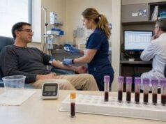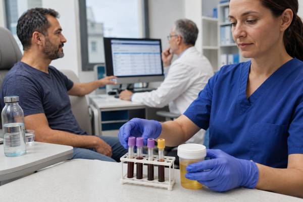
A diabetes insipidus hormone test panel evaluates whether excessive urine output comes from inadequate vasopressin release, kidney resistance to vasopressin, excessive water intake, or another cause of diuresis. The most useful panel does not rely on one hormone value. It links measured urine volume with serum sodium, plasma or serum osmolality, urine osmolality, glucose, kidney function, electrolytes, and—when needed—copeptin or antidiuretic hormone testing. Copeptin is often preferred to direct ADH measurement because it is more stable and easier to analyze. Some patients can be classified from paired blood and urine samples obtained during spontaneous hypernatremia. Others require supervised water deprivation, desmopressin response testing, or stimulated copeptin with hypertonic saline or arginine. The goal is to identify the physiologic pattern without causing dehydration, dangerous sodium change, or inappropriate desmopressin treatment.
- First confirm true high-volume, hypotonic urine rather than urinary frequency.
- Serum sodium and osmolality show whether the blood is dilute, normal, or concentrated.
- Urine osmolality shows whether the kidneys are conserving water appropriately.
- Copeptin is a stable surrogate for vasopressin release; direct ADH testing is less robust.
- Dynamic tests are supervised because water restriction and sodium shifts can be hazardous.
- Results distinguish AVP deficiency, AVP resistance, primary polydipsia, and solute diuresis.
Table of Contents
- What the Panel Includes
- Who Needs Testing
- Stepwise Testing Process
- Result Patterns
- Dynamic Tests and Cutoffs
- Causes, Confounders, and Follow-Up
- Urgent Safety and Questions
What the Panel Includes
Diabetes insipidus is a water-balance syndrome characterized by abnormally large volumes of dilute urine. Many specialists now use the terms arginine vasopressin deficiency, or AVP deficiency, for central diabetes insipidus and arginine vasopressin resistance, or AVP resistance, for nephrogenic diabetes insipidus. The older name remains common, but the disorder is unrelated to diabetes mellitus.
A useful diagnostic panel combines several measurements because each answers a different question.
| Test | Main question |
|---|---|
| 24-hour urine volume or timed output | Is true polyuria present? |
| Urine osmolality or specific gravity | Is the urine dilute, concentrated, or solute-rich? |
| Serum sodium | Is free-water loss or excess affecting blood concentration? |
| Plasma or serum osmolality | Is there an adequate osmotic stimulus for vasopressin release? |
| Copeptin | Is endogenous vasopressin release low, appropriate, or high for the stimulus? |
| Direct ADH or vasopressin | Can hormone release be estimated when copeptin is unavailable? |
| Glucose, urea, creatinine, calcium, potassium | Is another metabolic or kidney cause producing polyuria? |
Plasma osmolality can be measured directly or estimated from sodium, glucose, and urea. The directly measured value is preferred during formal dynamic testing. Laboratories commonly report milliosmoles per kilogram, abbreviated mOsm/kg. Serum and plasma osmolality are clinically similar for this purpose, although the specimen and method should be documented.
Urine osmolality reflects all dissolved particles in urine. A low value indicates water-rich urine; a high value indicates concentration or a large solute load. Specific gravity is a rapid surrogate, but glucose, protein, or contrast can increase specific gravity disproportionately. Osmolality is more informative when the diagnosis is uncertain.
Vasopressin is produced in the hypothalamus and released through the posterior pituitary. It binds V2 receptors in the kidney collecting ducts and increases aquaporin-2 water channels. AVP deficiency means the signal is inadequate. AVP resistance means the signal is present but the kidney response is impaired.
Copeptin is released with vasopressin from the same precursor. It is stable enough for routine handling and is now central to many modern diagnostic algorithms. A copeptin blood test is interpreted differently at baseline and after stimulation.
Direct ADH measurement is vulnerable to degradation, platelet binding, low circulating concentrations, and strict handling requirements. A result may be useful from an expert laboratory, but it should not be treated as interchangeable with copeptin or interpreted without paired osmolality.
Who Needs Testing
Testing begins when symptoms suggest pathologic water loss or intake. Common adult complaints include passing several liters of pale urine, waking repeatedly at night to urinate, intense thirst, preferring cold water, and needing constant access to fluids. Children may have heavy diapers, persistent bed-wetting, irritability, poor feeding, poor weight gain, slowed growth, fever without infection, constipation, or recurrent dehydration.
Urinary frequency alone is not polyuria. Bladder infection, overactive bladder, prostate disease, pelvic-floor dysfunction, and anxiety may cause frequent small voids. Measuring total output prevents an unnecessary endocrine workup.
In adults, more than about 3 liters in 24 hours often prompts evaluation, but more than 50 milliliters per kilogram per day is a more individualized threshold. Pediatric criteria vary by age and weight. A diary should include drink volume, urine volume, timing, nighttime symptoms, and medicines.
Testing is particularly important after pituitary or hypothalamic surgery, head trauma, neurosurgery, or treatment of a sellar mass. Sudden high urine output with rising sodium can indicate postoperative AVP deficiency. The pattern can be transient, permanent, or triphasic, with early polyuria followed by water retention and then recurrent deficiency.
Other indications include:
- unexplained hypernatremia with inappropriately dilute urine;
- recurrent dehydration despite access to water;
- long-term lithium exposure;
- high calcium or low potassium with polyuria;
- chronic kidney or tubulointerstitial disease;
- a family history of inherited AVP deficiency or resistance;
- pregnancy with new severe thirst and polyuria;
- hypothalamic disease affecting thirst regulation;
- possible primary polydipsia related to psychiatric disease, medication, or habit.
The initial panel also distinguishes water diuresis from solute diuresis. Uncontrolled diabetes mellitus causes glucose to pull water into urine. Sodium, urea, mannitol, high-protein feeding, radiographic contrast, and some medicines can also produce high-volume urine that is not maximally dilute.
Testing should not begin with unsupervised water restriction. A person with severe AVP deficiency can lose water rapidly and develop dangerous hypernatremia. Likewise, empiric desmopressin in primary polydipsia can cause water retention and severe hyponatremia.
Stepwise Testing Process
A stepwise process reduces unnecessary dynamic testing and identifies urgent cases early.
Step 1: Confirm Polyuria
A 24-hour collection is often the clearest starting point. Every void is measured from the first discarded morning sample through the first sample the next morning. Creatinine excretion may help assess completeness. In hospital, hourly output can be measured directly.
If total volume is below the polyuria threshold, the evaluation shifts toward urinary frequency, nocturia, sleep apnea, bladder disease, or other causes. If volume is high, urine osmolality identifies whether the diuresis is primarily water or solute.
Step 2: Determine Whether Urine Is Hypotonic
Urine osmolality below about 300 mOsm/kg is strongly hypotonic. Values between roughly 300 and 800 mOsm/kg may occur in partial AVP disorders, primary polydipsia, kidney disease, or mixed states. A value above 800 mOsm/kg generally demonstrates intact vasopressin release and kidney concentration and makes diabetes insipidus unlikely.
High urine volume with high osmolality suggests solute diuresis. Urinalysis, glucose, urea exposure, sodium intake or infusion, medicines, and kidney recovery are reviewed before hormone testing.
Step 3: Pair Blood and Urine
Serum sodium and plasma osmolality are collected near the urine sample. A high or high-normal sodium with dilute urine indicates that the kidneys are losing water despite a strong conservation signal. This pattern strongly raises AVP deficiency or resistance.
Low or low-normal sodium with dilute urine suggests that water intake may be driving the process, although partial disorders and medications can overlap. Normal sodium does not exclude AVP deficiency because intact thirst and free access to water may compensate for losses.
A typical adult plasma osmolality reference interval is approximately 275 to 295 mOsm/kg, but laboratories differ. A value above about 300 mOsm/kg is a strong osmotic stimulus. Interpretation relies on the relationship: concentrated plasma should be matched by concentrated urine in a healthy system.
Step 4: Exclude Metabolic and Renal Mimics
Blood glucose, hemoglobin A1c when appropriate, creatinine, urea, calcium, potassium, and medication history are reviewed. Cortisol deficiency can impair free-water excretion and mask AVP deficiency; after glucocorticoid replacement, polyuria may emerge. Thyroid disease and severe systemic illness can also change water handling.
Step 5: Select a Dynamic Test
Clear hypernatremia with dilute urine may permit diagnosis without deliberate dehydration, especially when copeptin is appropriately interpreted. Ambiguous cases proceed to stimulated copeptin or a supervised water-deprivation/desmopressin test. The choice depends on local expertise, age, comorbidities, assay access, and safety.
Preparation is protocol-specific. Patients must disclose desmopressin, diuretics, lithium, glucocorticoids, anticonvulsants, antidepressants, pregnancy, kidney disease, heart disease, seizure history, and recent vomiting or diarrhea. Medication changes should be made only by the responsible team.
Result Patterns
The panel is read as a physiologic pattern rather than as isolated “high” or “low” flags.
| Condition | Serum sodium or plasma osmolality | Urine osmolality | Copeptin or ADH pattern | Desmopressin response |
|---|---|---|---|---|
| Complete AVP deficiency | Often high if intake is inadequate; may be normal with free drinking | Very low | Inappropriately low | Large rise in urine osmolality |
| Partial AVP deficiency | Normal to high-normal | Low to intermediate | Low or borderline after stimulation | Moderate rise |
| AVP resistance | Normal-high or high | Low | High when adequately stimulated; baseline copeptin may be markedly high | Little or no rise |
| Primary polydipsia | Often low-normal | Low; may improve with controlled dehydration | Appropriate stimulated release | Variable, usually limited |
| Solute diuresis | Variable | Often above 300 mOsm/kg | Not the primary discriminator | Not diagnostic |
AVP Deficiency
In complete AVP deficiency, urine remains very dilute even while plasma osmolality and sodium rise. Copeptin is low relative to the osmotic stimulus. After desmopressin, the kidney can respond, so urine osmolality increases substantially and urine volume falls.
Partial deficiency is more difficult. Some endogenous hormone is released, and urine may concentrate into an intermediate range. Chronic high water intake can blur the distinction from primary polydipsia. Stimulated copeptin is particularly helpful.
AVP Resistance
In AVP resistance, the body releases vasopressin but the kidneys do not respond adequately. Copeptin is often high, especially after dehydration or at baseline in severe disease. Urine remains dilute and desmopressin causes little improvement in complete resistance. Partial resistance can show a modest response.
Acquired causes include lithium, hypercalcemia, hypokalemia, kidney disease, obstruction, and certain medicines. Inherited disease often presents in infancy or childhood, especially with recurrent hypernatremic dehydration and poor growth.
Primary Polydipsia
Excessive water intake suppresses vasopressin and lowers serum osmolality. With chronic intake, the kidney’s medullary gradient becomes less effective, so urine concentration during water deprivation may be delayed. Stimulated copeptin demonstrates that the hormone system can respond when given a standardized signal.
Primary polydipsia is not always psychiatric. Habit, dry mouth, medications, hypothalamic thirst disturbance, and attempts to follow excessive hydration advice can contribute. Treatment must avoid abrupt restriction when chronic hyponatremia is possible.
Mixed or Indeterminate Results
Kidney disease, partial AVP deficiency, partial resistance, prolonged polydipsia, recent desmopressin, and inadequate test stimulation can produce mixed results. A borderline panel should not be converted into a permanent diagnosis without reviewing protocol quality and repeating or changing the test when needed.
Dynamic Tests and Cutoffs
Dynamic testing deliberately changes osmolality or stimulates copeptin release. It should occur in a monitored setting with predefined stopping criteria.
Hypertonic Saline-Stimulated Copeptin
Hypertonic saline raises serum sodium under close observation. Sodium is checked repeatedly until the protocol target is reached, often at least 150 mmol/L, although exact targets vary. Copeptin is then measured, and sodium is lowered safely with oral water and, when needed, intravenous glucose solution.
A stimulated copeptin value at or below about 4.9 pmol/L has commonly supported AVP deficiency, whereas a higher response supports primary polydipsia. The assay and center’s validated cutoff take priority. The test has high diagnostic accuracy but requires experienced supervision because overly rapid sodium elevation is hazardous.
A baseline copeptin above about 21.4 pmol/L in a patient with confirmed hypotonic polyuria strongly supports AVP resistance. This threshold should not be used indiscriminately during acute stress, dehydration, or reduced kidney function.
Arginine-Stimulated Copeptin
Intravenous arginine stimulates copeptin without intentionally producing hypernatremia. Samples are collected at specified times, commonly including 60 minutes. It is easier to perform than hypertonic saline testing but was less accurate in a large head-to-head trial.
A value near the original cutoff around 3.8 pmol/L may fall into a diagnostic gray zone. Recent analyses support using lower and upper thresholds to identify high-probability AVP deficiency or primary polydipsia and sending intermediate results to a more definitive test. Cutoffs must match the assay and sampling schedule.
Water Deprivation and Desmopressin
During water deprivation, fluids are withheld while body weight, vital signs, urine volume, urine osmolality, serum sodium, and plasma osmolality are measured. Testing stops for excessive weight loss, rising sodium, orthostatic symptoms, or other safety limits. Desmopressin is then given to test kidney responsiveness.
A marked increase in urine osmolality after desmopressin supports AVP deficiency. Minimal change with persistently dilute urine supports AVP resistance. Partial states and chronic primary polydipsia overlap, which is why modern copeptin protocols may be preferred when available.
No patient should attempt a home water-deprivation test. The duration and stopping rules cannot be safely improvised, especially in children, pregnancy, impaired thirst, or complete AVP deficiency.
Direct ADH testing can be paired with osmolality, but poor stability and assay limitations reduce reliability. A low number is only meaningful if plasma is concentrated enough that hormone release should be strong. The ADH test result must therefore be interpreted as an osmolality-response relationship.
Causes, Confounders, and Follow-Up
Once AVP deficiency is identified, the next question is why. Pituitary MRI evaluates the hypothalamus, stalk, posterior pituitary, and nearby structures. Causes include surgery, trauma, craniopharyngioma, germinoma, metastasis, inflammatory and infiltrative disease, infection, autoimmune processes, and genetic disorders. Loss of the posterior pituitary bright spot is not diagnostic by itself.
Anterior pituitary testing may include morning cortisol, ACTH, free T4, TSH, prolactin, LH, FSH, sex hormones, IGF-1, and growth assessment. Multiple deficits suggest broader hypothalamic-pituitary disease. New AVP deficiency with headache or visual change requires urgent structural evaluation.
AVP resistance prompts review of lithium and other drugs, calcium, potassium, kidney function, obstruction, and family history. The 2025 international consensus on nephrogenic diabetes insipidus emphasizes early recognition, genetic assessment in appropriate patients, and structured prevention of dehydration.
Several factors confound the panel:
- Free access to water: can normalize sodium despite severe AVP deficiency.
- Impaired thirst: allows rapid hypernatremia and makes the condition more dangerous.
- Chronic polydipsia: reduces the kidney concentration gradient.
- Reduced kidney function: impairs concentration and may raise copeptin.
- Nausea, pain, stress, or infection: can raise copeptin independent of osmolality.
- Desmopressin: changes urine concentration for hours and may suppress endogenous measures.
- Glucocorticoid deficiency: can mask polyuria until cortisol is replaced.
- Pregnancy: placental vasopressinase increases vasopressin breakdown.
Treatment follows the subtype. AVP deficiency is generally treated with desmopressin and access to water. Education centers on avoiding both dehydration and hyponatremia. AVP resistance is treated by correcting the cause, reducing dietary solute when appropriate, and using selected kidney-directed therapies. Primary polydipsia requires a supervised behavioral and medical plan; desmopressin may be dangerous.
Follow-up uses symptoms, urine pattern, thirst, weight, sodium, and treatment safety. A fixed “normal urine volume” is less useful than a stable pattern that permits sleep and daily activity without sodium disturbance. Patients with structural disease also need interval pituitary and imaging follow-up.
Special Settings
Hospitalized and postoperative patients need a faster, more repetitive version of the panel. Hourly urine output is paired with serial sodium, fluid balance, and urine concentration. A single large urine volume after intravenous fluid does not prove AVP deficiency, but sustained dilute output with rising sodium is concerning. Desmopressin decisions should account for the possibility of a later antidiuretic phase, when retained water can abruptly lower sodium.
In pregnancy, new polyuria may be dismissed as normal urinary frequency. True high-volume dilute urine, especially with thirst and rising sodium, requires assessment for gestational AVP deficiency. Placental vasopressinase degrades native vasopressin more than desmopressin, but liver disease can increase risk by reducing enzyme clearance. Testing and treatment are coordinated with endocrinology and obstetric care.
For infants and young children, paired samples and genetic assessment may be more informative than prolonged dehydration. They cannot reliably communicate thirst, and fluid restriction can become dangerous quickly. Growth records, feeding history, recurrent fever, constipation, developmental progress, and family history add diagnostic value. In suspected inherited AVP resistance, early molecular testing can guide counseling and avoid repeated hazardous challenges.
People with adipsia or impaired thirst require a different safety plan. They may not respond to rising sodium by drinking. Scheduled fluid intake, daily weight targets, regular sodium checks, caregiver education, and carefully timed desmopressin may be necessary. Their apparently mild thirst history should never be used to dismiss a serious water-balance disorder.
Urgent Safety and Questions
Hypernatremic dehydration can develop quickly when a person with AVP deficiency cannot drink enough. Emergency symptoms include confusion, unusual drowsiness, severe weakness, fever, repeated vomiting, fainting, seizures, or reduced consciousness. Infants, older adults, hospitalized patients, and people with impaired thirst or mobility are at particular risk.
Hyponatremia is the major danger of excessive desmopressin effect combined with continued fluid intake. Worsening headache, nausea, vomiting, confusion, muscle cramps, marked fatigue, or seizures require prompt assessment. The patient’s sick-day and missed-dose plan should be written and readily available.
Questions to ask after a panel include:
- Was my 24-hour urine volume truly above the polyuria threshold?
- Was the urine hypotonic, or was this a glucose, urea, or sodium-driven diuresis?
- What were serum sodium, plasma osmolality, and urine osmolality at the same time?
- Was copeptin measured at baseline or after a validated stimulus?
- Did the dynamic test reach its sodium or timing target?
- Does the pattern favor AVP deficiency, AVP resistance, primary polydipsia, or remain indeterminate?
- Could lithium, calcium, potassium, kidney disease, cortisol deficiency, or another medicine affect the findings?
- Do I need pituitary MRI, genetic testing, or other pituitary hormone tests?
- What are my safe drinking and desmopressin instructions?
A well-designed panel tells a coherent story: how much urine is produced, whether it is dilute, whether the blood is concentrated enough to demand water conservation, whether the vasopressin system responds, and whether the kidneys obey that signal. That relationship is more reliable than any isolated “high” or “low” result.
References
- Diagnostic Tests for Diabetes Insipidus 2025 (Clinical Review)
- Central and nephrogenic diabetes insipidus 2025 (Review)
- Diagnosis and Management of Central Diabetes Insipidus in Adults 2022 (Review)
- International expert consensus statement on the diagnosis and management of congenital nephrogenic diabetes insipidus (arginine vasopressin resistance) 2025 (Consensus)
- Arginine or Hypertonic Saline–Stimulated Copeptin to Diagnose AVP Deficiency 2023 (Clinical Trial)
- Arginine Vasopressin Disorder (Diabetes Insipidus) 2024 (Clinical Review)
Disclaimer
This article is for general education and cannot diagnose diabetes insipidus or determine a desmopressin regimen. Water deprivation, hypertonic saline testing, medication changes, and desmopressin interruption require direct medical supervision. Seek urgent care for confusion, seizures, inability to drink, severe dehydration, or symptoms of dangerously high or low sodium.