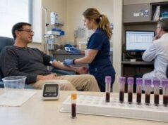
A follicle-stimulating hormone test measures FSH, a pituitary hormone that helps regulate ovarian follicle development, estradiol production, sperm production, puberty, and reproductive function. The result is commonly used in evaluations of irregular or absent periods, infertility, menopause, premature ovarian insufficiency, low testosterone, low sperm count, delayed or early puberty, and possible pituitary or hypothalamic disease. FSH is not interpreted from one universal normal range. Levels change with age, sex, menstrual-cycle phase, pregnancy, menopause, hormone treatment, and laboratory method. The most useful interpretation pairs FSH with symptoms and related tests such as LH, estradiol, testosterone, prolactin, thyroid studies, anti-Müllerian hormone, semen analysis, and sometimes pituitary imaging. High FSH often reflects reduced ovarian or testicular feedback, while low or inappropriately normal FSH with low sex hormones can point to hypothalamic or pituitary dysfunction.
- FSH is produced by anterior pituitary gonadotroph cells.
- Menstrual-cycle timing strongly affects results in people who ovulate.
- High FSH usually indicates reduced gonadal response, not pituitary failure.
- Low or normal FSH can be abnormal when estradiol or testosterone is low.
- FSH alone cannot predict natural pregnancy or egg quality.
- Hormonal medicines, illness, nutrition, and assay interference can alter results.
Table of Contents
- What FSH Does
- Why FSH Is Tested
- Preparation and Timing
- Normal Ranges and Patterns
- High FSH Results
- Low FSH and Pituitary Function
- Fertility, Next Steps, and Questions
What FSH Does
FSH is a glycoprotein hormone made by the anterior pituitary. The hypothalamus releases gonadotropin-releasing hormone, or GnRH, in pulses. GnRH stimulates the pituitary to release FSH and luteinizing hormone. The ovaries or testes then produce sex hormones and inhibins that feed back to the hypothalamus and pituitary.
In ovaries, FSH binds receptors on granulosa cells. It supports growth of follicles that contain immature eggs and promotes aromatase activity, which converts androgens into estradiol. As a follicle develops, estradiol and inhibin B generally reduce FSH. One dominant follicle continues growing, and a later LH surge triggers ovulation.
In testes, FSH acts mainly on Sertoli cells. Together with high local testosterone produced under LH stimulation, it supports spermatogenesis. Sertoli cells release inhibin B, which feeds back selectively on FSH secretion. A man can therefore have relatively preserved testosterone but high FSH when seminiferous-tubule function is impaired.
FSH changes substantially across life. It rises briefly in infancy during “mini-puberty,” remains low through much of childhood, increases during puberty, fluctuates during reproductive cycles, and rises after menopause when ovarian feedback declines. Values in children require age, sex, and pubertal-stage intervals.
FSH and LH should usually be interpreted together. Their absolute numbers and relationship to estradiol or testosterone reveal whether the gonads are failing despite strong pituitary stimulation or whether pituitary stimulation is insufficient. A luteinizing hormone test supplies the complementary signal.
The term “normal” can be misleading. A mid-range FSH may be appropriate when estradiol or testosterone is normal, yet inappropriately normal when the sex hormone is clearly low. Endocrine interpretation asks whether the pituitary response fits the physiologic situation.
Why FSH Is Tested
In people with ovaries, FSH is ordered for absent periods, irregular cycles, infertility, symptoms of low estrogen, suspected menopause, or ovarian dysfunction. It may help distinguish ovarian failure from hypothalamic or pituitary causes of amenorrhea.
Common indications include:
- no menstrual period by the expected age;
- periods that stop for several months when pregnancy is excluded;
- cycles that become irregular before age 40;
- hot flashes, vaginal dryness, sleep disturbance, or low bone density;
- infertility or an unexpectedly low response to ovarian stimulation;
- prior chemotherapy, radiation, ovarian surgery, or autoimmune disease;
- suspected Turner syndrome or another chromosome condition.
For premature ovarian insufficiency, current international guidance uses menstrual disturbance for at least four months plus an elevated FSH above 25 IU/L. A repeat measurement after four to six weeks is recommended when diagnostic uncertainty remains. The test does not have to be limited to a specific cycle day for this purpose, but hormonal contraception can obscure interpretation.
FSH is also used in fertility evaluation, often with estradiol on cycle day 2, 3, or 4. A higher early-follicular FSH can indicate reduced ovarian responsiveness. However, ovarian reserve is not the same as fertility, and one value does not measure egg quality or guarantee whether spontaneous pregnancy will occur. Age, antral follicle count, anti-Müllerian hormone, tubal status, ovulation, semen, and other factors matter.
In people with testes, FSH is ordered when low sperm count, infertility, small testes, delayed puberty, or low testosterone is being evaluated. High FSH supports primary testicular or seminiferous-tubule damage. Low or inappropriately normal FSH with low testosterone may indicate hypothalamic-pituitary dysfunction.
FSH may be part of a pituitary evaluation after surgery, radiation, head injury, a sellar mass, severe headache with visual symptoms, or multiple hormone abnormalities. It is commonly paired with LH, sex hormones, prolactin, TSH, free T4, morning cortisol, and IGF-1 in a pituitary hormone panel.
In children, FSH helps assess precocious or delayed puberty, ambiguous pubertal progression, and suspected gonadal dysfunction. A random FSH can be difficult to interpret early in puberty because hormone secretion is pulsatile and often first increases during sleep. GnRH stimulation or other tests may be needed.
FSH is not a general screening test for fatigue, weight change, low mood, or sexual symptoms without context. Those symptoms have many causes, and testing should be selected according to history and examination.
Preparation and Timing
FSH is measured from a blood sample. Fasting is not usually required unless other ordered tests require it. The collection time is less critical than for testosterone or cortisol, but menstrual-cycle day, hormone medication timing, and clinical state should be documented.
For an early-follicular fertility assessment, clinicians often request testing on cycle day 2, 3, or 4. Day 1 is the first day of full menstrual bleeding, not light spotting. FSH is then interpreted with estradiol. A seemingly reassuring FSH can be suppressed by an early estradiol rise, so both numbers are important.
For evaluation of premature ovarian insufficiency, testing may be performed regardless of cycle day when cycles are very irregular or absent. Pregnancy should be excluded first. If the result is borderline or inconsistent with symptoms, it may be repeated after four to six weeks.
Menopause in a person older than 45 is often diagnosed clinically from symptoms and menstrual history rather than a single FSH. During perimenopause, FSH can swing from high to premenopausal levels, so one normal result does not exclude the transition. Testing is more useful when age is younger, the history is atypical, or hormonal treatment complicates the picture.
Medicines and supplements to report include:
- combined hormonal contraception, progestins, estrogen, or testosterone;
- fertility medicines containing FSH, hCG, or GnRH analogues;
- selective estrogen-receptor modulators and aromatase inhibitors;
- antipsychotics or other drugs that raise prolactin;
- glucocorticoids, opioids, and anabolic steroids;
- high-dose biotin supplements.
Do not stop prescribed hormones solely to obtain a test. The clinician may interpret the result on treatment, schedule a washout when safe, or choose another marker.
Acute illness, undernutrition, significant weight loss, intense exercise, severe stress, and chronic systemic disease can suppress the hypothalamic-pituitary-gonadal axis. Testing during an unstable period may reflect adaptive suppression rather than a permanent disorder.
The laboratory should use age-, sex-, and phase-specific intervals. Results from different assays may not match exactly, so serial monitoring is clearest when performed with the same laboratory method.
When Repeat Testing Helps
Repeat testing is useful when the result conflicts with the history, was collected on the wrong cycle day, followed a recent hormone injection, or occurred during acute illness. The repeat should answer a defined question rather than simply checking whether the number changes. For example, an early-follicular panel can be repeated with estradiol when ovarian response is being assessed, while possible premature ovarian insufficiency is reconsidered after four to six weeks if the first result is uncertain.
A fluctuating result is not automatically a laboratory error. FSH secretion is pulsatile, and ovarian feedback changes across and between cycles. Perimenopause can produce particularly wide swings. In contrast, a persistently high value with low estradiol in a younger patient carries more weight than one isolated elevation.
When pituitary disease is suspected, repeating FSH alone is rarely sufficient. The repeat panel should usually include LH and the relevant sex hormone, plus prolactin, thyroid tests, and other pituitary axes according to symptoms. A low-normal FSH becomes more meaningful when testosterone or estradiol remains low on properly timed samples.
Laboratory interference is uncommon but should be considered when the result is physiologically impossible or changes dramatically between methods. High-dose biotin can interfere with some immunoassays. Heterophile antibodies and rare FSH variants may also produce misleading values. The laboratory can repeat the sample on another platform, perform dilution studies, or use blocking reagents when interference is suspected.
Normal Ranges and Patterns
FSH is usually reported in international units per liter, IU/L, equivalent numerically to mIU/mL. Reference intervals vary enough that the laboratory report should be used instead of a universal chart.
Illustrative adult ranges from commonly used immunoassays may look like this:
| Physiologic state | Illustrative FSH pattern |
|---|---|
| Early follicular phase | Approximately 3–10 IU/L |
| Midcycle | May rise to approximately 6–20 IU/L or higher |
| Luteal phase | Often approximately 1–9 IU/L |
| Postmenopause | Commonly above 20–30 IU/L, often much higher |
| Adult male | Often approximately 1–12 IU/L |
These are examples, not diagnostic cutoffs. Menstrual timing, age, assay, estradiol, and treatment can shift the interval.
A useful interpretation matrix pairs FSH with the target-gland hormone:
| FSH | Estradiol or testosterone | General pattern |
|---|---|---|
| High | Low | Primary ovarian or testicular dysfunction |
| Low or normal | Low | Hypothalamic or pituitary hypogonadism |
| Appropriate | Normal | Axis may be intact; symptoms need broader evaluation |
| High | Normal | Early or compensated gonadal dysfunction, cycle timing, or treatment effect |
| Low | High | Exogenous hormone, pregnancy, hormone-producing state, or negative feedback |
In menstruating adults, FSH varies from day to day. It may be transiently high as ovarian reserve declines, then lower in another cycle. A single value is therefore a snapshot rather than a fixed measure of reproductive capacity.
In men, FSH reflects seminiferous-tubule function more than testosterone production. High FSH with low sperm concentration suggests impaired spermatogenesis. Normal FSH does not guarantee a normal semen analysis, because focal testicular damage, obstruction, or genetic causes can occur without a large rise.
Children require pediatric ranges and pubertal staging. A value that is low for an adult can be normal before puberty. Conversely, pubertal-range FSH in a young child may support central puberty when interpreted with LH, sex hormones, growth acceleration, and bone age.
Urine FSH tests and home menopause kits provide only limited information. Hydration and hormone fluctuation affect urine concentration, and a positive or negative threshold cannot replace clinical evaluation. They are particularly unreliable for diagnosing perimenopause from one result.
High FSH Results
High FSH usually means the pituitary is increasing stimulation because ovarian or testicular feedback has weakened. This is called hypergonadotropic hypogonadism when sex hormones are low.
In people with ovaries, common explanations include natural menopause, premature ovarian insufficiency, Turner syndrome, prior ovarian surgery, chemotherapy, pelvic radiation, autoimmune ovarian injury, galactosemia, and certain genetic conditions. FSH may also rise temporarily after stopping hormonal suppression.
Natural menopause produces persistently reduced ovarian estradiol and inhibin, so FSH is often markedly elevated. During perimenopause, values fluctuate and should not be used alone to determine contraceptive need. Ovulation and pregnancy remain possible until menopause is established clinically.
Premature ovarian insufficiency occurs before age 40. It is not always complete or permanent; intermittent ovarian activity can occur. The current evidence-based guideline defines diagnosis using at least four months of disordered cycles and FSH above 25 IU/L, with repeat testing when uncertainty exists. Evaluation may include pregnancy testing, thyroid assessment, prolactin, chromosome analysis, FMR1 premutation testing, and selected autoimmune studies.
A high early-follicular FSH may suggest reduced ovarian response during fertility treatment. It does not specify the number of eggs remaining, egg quality, or the chance of natural conception. Age remains a stronger predictor of egg quality. Reproductive decisions should not be based on FSH alone.
In people with testes, high FSH may result from Klinefelter syndrome, testicular injury, torsion, infection, chemotherapy, radiation, genetic causes of impaired sperm production, or age-related testicular change. If testosterone is low and LH is also high, primary testicular failure is likely. If testosterone is normal but FSH is high, seminiferous-tubule damage may be more prominent than Leydig-cell dysfunction.
Rarely, a functioning pituitary gonadotroph adenoma produces biologically active FSH. These tumors can cause ovarian enlargement, menstrual disturbance, or testicular effects, but most pituitary tumors that stain for FSH are clinically nonfunctioning. An isolated modest elevation is far more likely to reflect physiologic or gonadal feedback than a secreting tumor.
The next tests depend on the setting. They may include repeat FSH, LH, estradiol or morning testosterone, anti-Müllerian hormone, antral follicle count, semen analysis, inhibin B, karyotype, genetic testing, pelvic ultrasound, or testicular examination.
Low FSH and Pituitary Function
Low FSH can be normal during pregnancy, while taking estrogen or testosterone, and before puberty. It becomes concerning when gonadal hormones are low and the pituitary should be increasing its signal.
Hypogonadotropic hypogonadism refers to low sex-hormone production with low or inappropriately normal FSH and LH. Causes may arise in the hypothalamus or pituitary. They include pituitary tumors, surgery, radiation, head trauma, infiltrative disease, hemochromatosis, congenital GnRH deficiency, hyperprolactinemia, severe illness, undernutrition, excessive exercise, obesity-related suppression, opioids, glucocorticoids, and anabolic steroid use.
In amenorrhea, low estradiol with low-normal FSH can fit functional hypothalamic amenorrhea, especially with low energy availability, weight loss, intense training, or stress. This is a diagnosis made after excluding pregnancy and other endocrine or structural causes. Bone health and nutrition are important even when the suppression is adaptive.
High prolactin suppresses GnRH and can lower FSH and LH. Causes include pregnancy, medications, hypothyroidism, pituitary stalk compression, and prolactinoma. Prolactin is therefore commonly measured when periods stop, libido falls, or fertility is impaired.
In men, low testosterone must be confirmed with appropriately timed morning measurements on separate days when clinically stable. FSH and LH then distinguish primary from central patterns. Very low testosterone with low or normal gonadotropins, headache, visual symptoms, multiple pituitary deficits, or high prolactin may prompt pituitary MRI.
Low FSH alone does not prove pituitary disease. Hormone secretion is dynamic, and the lower end of the interval may be appropriate if estradiol or testosterone is adequate. The clinical question is whether FSH is sufficient for the target-gland state.
Combined pituitary dysfunction can involve ACTH, TSH, growth hormone, and gonadotropins. Adrenal status is especially important because untreated ACTH deficiency can be dangerous. Symptoms such as severe weakness, low blood pressure, recurrent low sodium, or unexplained low free T4 require broader assessment rather than isolated fertility treatment.
In delayed puberty, low FSH and LH may represent constitutional delay or permanent hypogonadotropic hypogonadism. Growth pattern, smell, family history, testicular size, bone age, chronic disease, and follow-up over time help distinguish them.
Fertility, Next Steps, and Questions
FSH is one component of fertility evaluation, not a pass-fail test. In people with ovaries, evaluation may include ovulation history, age, pelvic ultrasound, antral follicle count, anti-Müllerian hormone, tubal assessment, uterine assessment, thyroid and prolactin tests, and partner semen analysis. A low or normal FSH cannot confirm that fertility is normal.
In people with testes, semen analysis is central. FSH helps explain a low sperm count but cannot replace direct measurement. Testosterone therapy can suppress FSH, LH, and sperm production; a person seeking fertility should discuss alternatives before starting or continuing testosterone.
Treatment follows the cause. Ovarian insufficiency may require hormone replacement for symptom, bone, and cardiovascular health when appropriate, along with fertility counseling. Hypothalamic suppression is addressed through nutrition, exercise adjustment, stress care, and treatment of underlying disease. Pituitary lesions may require medication, surgery, radiation, or hormone replacement.
An abnormal result can also have implications beyond immediate fertility. Prolonged low estradiol or testosterone can affect bone density, body composition, sexual function, and quality of life. Clinicians may assess vitamin D, fracture risk, cardiometabolic factors, and the need for age-appropriate hormone replacement. In premature ovarian insufficiency, family planning, genetic implications, emotional support, and long-term health protection deserve attention even when pregnancy is not the current goal. In men with primary testicular failure, genetic counseling may be appropriate before assisted reproduction.
For central hypogonadism and fertility, exogenous gonadotropins or pulsatile GnRH may be used under specialist care. Doses are guided by ovarian response, estradiol, ultrasound, testosterone, testicular growth, and semen analysis rather than an attempt to force serum FSH into one target range.
Useful questions include:
- Which age-, sex-, and cycle-specific interval applies to my result?
- What were estradiol or testosterone, LH, and prolactin at the same time?
- Was the sample timed to the requested cycle day?
- Could contraception, fertility medication, testosterone, biotin, illness, or energy deficiency alter the result?
- Does this pattern suggest primary gonadal dysfunction or central suppression?
- Should FSH be repeated, and if so, when?
- Does an elevated FSH meet criteria for premature ovarian insufficiency?
- Do I need semen analysis, anti-Müllerian hormone, ultrasound, genetic testing, or pituitary imaging?
- How does this result affect fertility planning without overstating what it predicts?
Seek urgent evaluation for a sudden severe headache, new visual loss, double vision, fainting, confusion, or vomiting, which can signal an acute pituitary problem. Prompt assessment is also important for severe pelvic pain and rapid abdominal enlargement during fertility treatment because ovarian hyperstimulation or torsion can be emergencies.
The most accurate interpretation of FSH is relational: pituitary signal, gonadal hormone output, age, cycle stage, treatment, and clinical goal must agree. One isolated number cannot define fertility, menopause, or pituitary health by itself. Trends are most useful when the same clinical question, cycle timing, medications, and laboratory method are documented carefully and consistently each time.
FSH interpretation is strongest when the sample date, cycle phase, age, and paired hormone results are documented together.
References
- Evidence-based guideline: premature ovarian insufficiency 2024 (Clinical Guideline)
- Follicle-Stimulating Hormone (FSH) Levels Test 2023 (Patient and Laboratory Reference)
- Physiology, Follicle Stimulating Hormone 2023 (Clinical Review)
- Society for Endocrinology guidelines for testosterone replacement therapy in male hypogonadism 2024 (Clinical Guideline)
- Male hypogonadism: recommendations from the Fifth International Consultation on Sexual Medicine 2025 (Consensus Review)
- Evidence-based guideline: premature ovarian insufficiency 2024 (Guideline Summary)
Disclaimer
This article provides general education and cannot diagnose infertility, menopause, ovarian insufficiency, testicular failure, or pituitary disease. FSH must be interpreted with age, menstrual timing, sex hormones, medications, symptoms, and the laboratory method. Do not start, stop, or change reproductive or hormone treatment based on one result without clinical guidance.