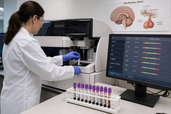
A pituitary hormone test panel evaluates several hormone systems controlled by the pituitary gland. It commonly includes ACTH with cortisol, TSH with free T4, LH and FSH with estradiol or testosterone, prolactin, and IGF-1 as a marker of growth hormone activity. A random growth hormone level may also be listed, but it is rarely useful by itself because GH is released in pulses. The panel can identify hormone deficiency, hormone excess, medication effects, and patterns that suggest a pituitary or hypothalamic disorder. Results must be interpreted as paired signals: a pituitary hormone may appear “normal” yet be inappropriately low when its target-gland hormone is deficient. Time of day, menstrual status, pregnancy, age, illness, nutrition, and medicines can change the values. A panel does not replace dynamic testing, pituitary MRI, or visual-field assessment when those are indicated.
- Pituitary tests are interpreted in hormone pairs, not as isolated numbers.
- Morning cortisol is essential because ACTH deficiency can be dangerous.
- Free T4 matters more than TSH alone when central hypothyroidism is possible.
- IGF-1 is more useful than random GH for screening growth hormone excess.
- Prolactin can rise from medicines, stress, pregnancy, stalk compression, or a prolactinoma.
Table of Contents
- What the Pituitary Panel Tests
- Why the Panel Is Ordered
- Preparation and Timing
- ACTH-Cortisol and TSH-Free T4 Results
- LH, FSH, Prolactin, and Sex Hormones
- GH and IGF-1 Results
- Common Panel Patterns and Pitfalls
- Follow-Up Tests and Urgent Findings
What the Pituitary Panel Tests
The pituitary is a small gland at the base of the brain that coordinates several endocrine organs. The hypothalamus sends releasing and inhibiting signals to the pituitary, and the pituitary sends hormones to the adrenal glands, thyroid, ovaries, testes, liver, and other tissues. Feedback from target-gland hormones helps regulate the system.
A typical panel may include:
- ACTH and cortisol: assess the hypothalamic-pituitary-adrenal axis.
- TSH and free T4: assess pituitary control of thyroid hormone production.
- LH and FSH: assess pituitary signaling to the ovaries or testes.
- Estradiol or testosterone: show the target-gland response to gonadotropins.
- Prolactin: screens for prolactin excess, medication effects, and stalk disruption.
- IGF-1: reflects integrated growth hormone action.
- Growth hormone: may be included, but a random value has major limitations.
Some panels also include sodium, serum and urine osmolality, copeptin, free T3, sex hormone-binding globulin, dehydroepiandrosterone sulfate, or other tests based on symptoms. There is no single universal “pituitary panel.” The clinician selects tests to answer a clinical question.
The posterior pituitary stores vasopressin and oxytocin, but these are not usually assessed with routine direct measurements. Suspected central diabetes insipidus is evaluated through water balance, sodium, osmolality, and often a diabetes insipidus hormone test panel.
Pituitary hormones should be paired with their target hormones. Low free T4 with a TSH inside the printed range can still indicate central hypothyroidism because TSH should rise when free T4 falls. The same principle applies to low testosterone with low-normal LH and low cortisol with low-normal ACTH.
Basal blood tests work well for some axes and only screen others. Prolactin, free T4, gonadotropins, sex hormones, and IGF-1 often provide useful baseline information. ACTH-cortisol and GH deficiency may require stimulation testing when basal results are indeterminate.
Why the Panel Is Ordered
A pituitary panel may be ordered because of symptoms, a known pituitary lesion, surgery or radiation, abnormal imaging, head injury, or an unexplained result in another endocrine test.
Possible symptoms include:
- Persistent fatigue, weakness, low blood pressure, or unexplained low sodium
- Weight change, cold intolerance, constipation, or dry skin
- Irregular or absent periods, infertility, low libido, or erectile dysfunction
- Low testosterone or estradiol
- Unexpected breast milk production
- Delayed or early puberty
- Poor growth in a child
- Enlarged hands or feet, facial change, sweating, or rapid growth
- Headaches, double vision, or loss of side vision
- Excessive thirst and urination
A panel is often obtained after MRI shows a pituitary incidentaloma. The purpose is to identify both hormone overproduction and loss of normal pituitary function. Even a lesion found by chance can secrete prolactin, GH, ACTH, or rarely TSH. Larger lesions are more likely to compress normal tissue and cause deficiencies.
After transsphenoidal surgery, testing assesses adrenal, thyroid, water-balance, reproductive, and growth hormone function. Cortisol and sodium may need early monitoring, while some axes are reassessed weeks later after postoperative changes stabilize.
Cranial radiation can cause pituitary deficiencies months or years later. Growth hormone is often affected first, followed over time by gonadotropins, ACTH, and TSH. Long-term surveillance may be needed even when early tests are normal.
Traumatic brain injury, subarachnoid hemorrhage, hypophysitis, infiltrative disease, genetic conditions, severe postpartum bleeding, and certain immune therapies can also damage the hypothalamic-pituitary system.
A panel is not a good indiscriminate explanation for every symptom. Fatigue, weight change, headache, low mood, and sexual concerns have many causes. Testing is most useful when the history, examination, or prior results point toward an endocrine axis.
Preparation and Timing
Timing is critical for several pituitary tests. A morning collection, commonly between 7 and 9 a.m., is often used so cortisol and testosterone can be interpreted against their expected daily rhythms.
Fasting is not required for every hormone, but a clinician may request it when glucose, insulin, lipids, or dynamic testing is included. Resting quietly before prolactin sampling can reduce stress-related elevation. Strenuous exercise should generally be avoided immediately before the draw.
Tell the clinician about all medicines and supplements. Important examples include:
- Glucocorticoids, including injections, inhalers, creams, and tablets
- Thyroid hormone and antithyroid medicines
- Estrogen, birth control, testosterone, or anabolic steroids
- Antipsychotics, antidepressants, metoclopramide, opioids, and verapamil
- Growth hormone and acromegaly medicines
- Immune checkpoint inhibitors
- Desmopressin
- High-dose biotin
Do not stop prescribed medicine without instructions. Some drugs suppress an axis; others interfere with the assay. The dose and time of the last medication may need documentation.
Menstrual cycle phase affects LH, FSH, estradiol, and progesterone. The order may specify a cycle day. Pregnancy, breastfeeding, menopause, and hormonal contraception require different reference expectations.
Acute severe illness can produce “non-thyroidal illness,” suppress gonadotropins, alter cortisol-binding proteins, and change IGF-1. Results obtained in intensive care may not reflect stable outpatient physiology. At the same time, suspected adrenal insufficiency cannot always wait; clinicians draw cortisol and ACTH promptly and treat if necessary.
Cortisol should ideally be drawn before glucocorticoid treatment when doing so does not delay emergency care. ACTH requires special chilled sample handling in many laboratories because it degrades quickly.
Free T4 and TSH can be collected together. For people taking levothyroxine, the clinician may standardize whether the sample is drawn before the daily dose. Consistency improves comparison over time.
Random GH sampling is not made reliable by fasting alone. GH pulses and low troughs mean a normal person may have a high value and a person with disease may have a low one. IGF-1 or a dynamic test is usually preferred.
ACTH-Cortisol and TSH-Free T4 Results
The adrenal axis has the highest immediate safety priority because severe cortisol deficiency can cause adrenal crisis.
ACTH and cortisol
ACTH from the pituitary stimulates the adrenal cortex to produce cortisol. A morning cortisol clearly within a robust normal range can make adrenal insufficiency unlikely, while a very low value raises concern. Intermediate results often need an ACTH stimulation test, insulin tolerance test, metyrapone test, or another locally validated assessment.
Patterns include:
- Low cortisol with low or inappropriately normal ACTH: possible central adrenal insufficiency from pituitary or hypothalamic disease.
- Low cortisol with high ACTH: possible primary adrenal insufficiency.
- High cortisol with suppressed ACTH: possible adrenal or exogenous glucocorticoid cause.
- High cortisol with detectable or elevated ACTH: ACTH-dependent Cushing syndrome may need dedicated testing.
A single morning cortisol does not diagnose Cushing disease. Screening for cortisol excess uses late-night salivary cortisol, 24-hour urinary free cortisol, or dexamethasone suppression according to the situation. ACTH is then used to classify confirmed hypercortisolism.
Central adrenal insufficiency may preserve aldosterone because the renin-angiotensin system controls it. Severe low blood pressure and low sodium can still occur. Potassium is often normal, unlike primary adrenal insufficiency.
TSH and free T4
TSH stimulates the thyroid to produce T4 and T3. In primary hypothyroidism, free T4 is low and TSH usually rises. In central hypothyroidism, free T4 is low while TSH may be low, normal, or mildly elevated but biologically inadequate.
A TSH-only screen can therefore miss central disease. The central hypothyroidism test panel relies on free T4, TSH, pituitary history, and other axes.
High free T4 or free T3 with nonsuppressed TSH raises a different question. Explanations include assay interference, thyroid hormone resistance, medication timing, or a rare TSH-secreting pituitary adenoma. Repeat testing and specialist laboratory review come before assuming a tumor.
When both cortisol and thyroid hormone are deficient, clinicians generally secure cortisol replacement before starting or increasing thyroid hormone. Thyroid replacement can increase cortisol clearance and precipitate crisis in untreated adrenal insufficiency.
LH, FSH, Prolactin, and Sex Hormones
LH and FSH are pituitary gonadotropins. Their interpretation depends on sex organs, age, menstrual status, and the paired estradiol or testosterone result.
In an adult male, low morning testosterone with high LH suggests primary testicular failure. Low testosterone with low or normal LH suggests central hypogonadism, functional suppression from illness or obesity, medication effects, or exogenous androgen use. FSH and semen analysis add information about sperm production.
In a premenopausal adult with ovaries, low estradiol and high FSH support ovarian insufficiency, while low estradiol with low-normal LH and FSH suggests hypothalamic or pituitary suppression. Menopause normally raises gonadotropins. Cycle phase, pregnancy, and hormone therapy must be considered.
Prolactin inhibits GnRH when elevated and can lower LH and FSH. Mild prolactin elevation may result from stress, pregnancy, breastfeeding, hypothyroidism, kidney disease, medicines, or stalk compression. Higher and persistent values can reflect a prolactinoma.
The degree of elevation is informative but not absolute. A large pituitary mass with only modest prolactin may be a nonfunctioning lesion causing stalk effect—or a large prolactinoma with a falsely low result from the hook effect. The laboratory can repeat prolactin after dilution.
Macroprolactin can falsely elevate total prolactin with less biological effect. A macroprolactin test separates this large molecular form from monomeric prolactin.
Low prolactin is less commonly diagnostic but may occur with extensive pituitary damage, dopamine-agonist treatment, or after surgery. Failure to lactate after severe postpartum hemorrhage can be a clue to postpartum hypopituitarism.
In children, LH and FSH are interpreted with pubertal stage, growth, bone age, and sex steroids. Basal values may be low early in puberty, so a GnRH-agonist stimulation test may be needed in suspected precocious or delayed puberty.
GH and IGF-1 Results
GH stimulates production of IGF-1, mainly in the liver. Because GH secretion is pulsatile, IGF-1 is the preferred baseline marker for most assessments.
A high age-adjusted IGF-1 result raises concern for acromegaly in adults or gigantism in children. Symptoms, repeat IGF-1, and sometimes oral glucose GH suppression testing establish the diagnosis. A pituitary MRI is usually obtained after biochemical evidence supports GH excess.
A low IGF-1 may occur with GH deficiency, but also with undernutrition, liver disease, hypothyroidism, poorly controlled diabetes, oral estrogen, and chronic illness. A normal IGF-1 does not exclude adult GH deficiency, especially in older adults or milder cases.
In children, slow growth velocity, height pattern, bone age, nutrition, IGF-1, IGFBP-3, and other screening tests guide whether GH stimulation is appropriate. A random low GH value does not diagnose deficiency.
In adults with structural pituitary disease, GH deficiency often requires a stimulation test. Options include insulin tolerance, glucagon, macimorelin, or other locally available protocols. Cutoffs depend on the agent, assay, and body mass index.
During GH replacement, IGF-1 helps adjust dosing alongside symptoms, side effects, and clinical response. During acromegaly treatment, IGF-1 is central to monitoring, but medication type matters. Pegvisomant raises measured GH while blocking GH receptors, so GH should not be used to judge control on that drug.
A random GH value may still provide context when it is very low in a treated acromegaly patient or repeatedly high, but it should not override validated testing. The growth hormone test must be matched to the diagnostic purpose: random, stimulation, or suppression.
Common Panel Patterns and Pitfalls
A panel is most informative when results are viewed as a pattern.
| Pattern | Possible interpretation | Typical next step |
|---|---|---|
| Low cortisol, low-normal ACTH, low free T4, normal TSH | Multiple central deficiencies | Urgent adrenal assessment, pituitary MRI, and broader evaluation. |
| High prolactin, low LH/FSH and sex hormones | Prolactin-mediated reproductive suppression | Repeat prolactin, medication review, macroprolactin, and imaging when indicated. |
| High IGF-1 with failure of GH suppression | GH excess | Pituitary MRI and acromegaly assessment. |
| Low free T4 with high TSH | Primary thyroid disease, not pituitary failure | Thyroid-focused evaluation. |
| Low testosterone with high LH/FSH | Primary testicular failure | Gonadal and genetic evaluation as appropriate. |
| All basal tests normal despite strong symptoms and pituitary history | Dynamic deficiency, intermittent disease, or a nonendocrine cause remains possible | Targeted stimulation testing or alternative evaluation. |
Common errors include interpreting pituitary hormones against population ranges without considering target hormones, using TSH alone, diagnosing GH deficiency from random GH, and assuming every high prolactin means prolactinoma.
Assay interference can affect nearly any immunoassay. Biotin, heterophile antibodies, macrohormones, thyroid hormone autoantibodies, and medication cross-reactivity can create implausible results. A mismatch between laboratory values and the clinical picture warrants repetition, dilution, another platform, or consultation with the laboratory.
Binding proteins also matter. Pregnancy and oral estrogen raise cortisol-binding globulin and total cortisol. Changes in thyroxine-binding globulin alter total T4 more than free T4. SHBG influences total testosterone. The correct free or calculated measure may be needed.
Pituitary function can evolve. A normal panel after radiation or with a growing lesion does not guarantee future normal function. Follow-up intervals depend on lesion size, imaging, symptoms, treatment, and current consensus guidance.
How the panel changes in common clinical settings
After pituitary surgery, the immediate priorities differ from routine outpatient screening. Morning cortisol or perioperative steroid coverage addresses ACTH deficiency, while urine output, thirst, sodium, and serum or urine osmolality help detect diabetes insipidus. Several days later, delayed hyponatremia can occur. Free T4, reproductive hormones, IGF-1, and formal dynamic tests are often reassessed after the acute postoperative period because early illness and medication effects can obscure the long-term baseline.
Radiation injury may develop gradually. GH is often the first axis affected, followed over time by gonadotropins, ACTH, and TSH, although the sequence varies. A normal panel one year after cranial radiation does not guarantee lifelong normal function. Long-term scheduled surveillance is particularly important after childhood cancer treatment because deficiencies may appear during growth, puberty, pregnancy planning, or adulthood.
Pregnancy raises prolactin, total cortisol, and thyroid-binding proteins and suppresses the usual reproductive hormone cycle. Nonpregnant ranges are inappropriate. In the postpartum period, failure to lactate, persistent amenorrhea, severe fatigue, low blood pressure, or hyponatremia after major bleeding can point to pituitary infarction. New headache and vision change during pregnancy or soon after delivery deserve urgent evaluation for a sellar mass or hypophysitis.
Critical illness can create a temporary “low hormone” pattern without permanent pituitary failure. TSH and T3 may fall, testosterone and gonadotropins may be suppressed, IGF-1 may decrease, and prolactin or cortisol may rise. Glucocorticoids, opioids, dopamine, and nutrition changes add further effects. Nonurgent abnormalities are often repeated after recovery, but suspected adrenal crisis or severe diabetes insipidus requires immediate treatment rather than delayed confirmation.
A panel is also time sensitive. Morning cortisol and testosterone are usually most informative early in the day; prolactin can rise with stress; GH is pulsatile; and menstrual-cycle phase changes LH, FSH, and estradiol. Recording collection time, fasting status, cycle day, pregnancy status, and last medication dose makes future comparisons much more reliable.
A result can also be “normal” yet physiologically inappropriate. Normal TSH does not reassure when free T4 is low, and normal ACTH does not reassure when cortisol is clearly deficient. Normal LH or FSH may be inadequate when estradiol or testosterone is low. Clinicians look for the expected compensatory response from the pituitary, not merely whether each number sits inside its printed interval.
When several borderline abnormalities appear together, repeating the panel under standardized conditions may be more informative than treating each one separately. Persistent multi-axis changes increase concern for structural pituitary or hypothalamic disease and usually justify endocrinology review.
Follow-Up Tests and Urgent Findings
Abnormal panel results may lead to targeted dynamic testing, imaging, and examination rather than repetition of the entire panel.
Possible follow-up includes:
- ACTH stimulation, insulin tolerance, or metyrapone testing for cortisol reserve
- Dexamethasone suppression, late-night salivary cortisol, or urine cortisol for suspected excess
- Repeat free T4 and TSH with assay-interference review
- Morning testosterone, SHBG, estradiol, semen analysis, or pubertal testing
- Repeat prolactin with macroprolactin and sample dilution
- IGF-1 confirmation and oral glucose GH suppression
- GH stimulation testing
- Pituitary MRI with a dedicated protocol
- Formal visual-field testing
- Sodium, serum osmolality, urine osmolality, and copeptin for water-balance symptoms
A pituitary MRI finding should not be interpreted without hormones, and hormone results should not be interpreted without the lesion’s size and location when a lesion is known. Multidisciplinary review may involve endocrinology, neurosurgery, neuroradiology, ophthalmology, radiation oncology, and reproductive specialists.
Seek emergency care for sudden severe headache, new vision loss or double vision, vomiting, confusion, fainting, severe weakness, very low blood pressure, or signs of adrenal crisis. Pituitary apoplexy can cause abrupt bleeding or infarction in a pituitary lesion and may rapidly impair ACTH secretion and vision.
After pituitary surgery, very high urine output with intense thirst can indicate diabetes insipidus, while headache, nausea, confusion, and low sodium can indicate delayed hyponatremia. Both need prompt assessment.
Hormone replacement order matters. Suspected cortisol deficiency is addressed before thyroid replacement. Sex hormones and GH are added only after urgent axes are secure and contraindications are reviewed.
The goal of a pituitary panel is not to make every number sit in the middle of a range. It is to determine whether each gland responds appropriately, whether an urgent deficiency is present, and which focused test will resolve remaining uncertainty.
References
- An Update on Advances in Hypopituitarism 2024 (Review)
- Management of pituitary incidentalomas 2024 (Review)
- Pitfalls in the lab assessment of hypopituitarism 2024 (Review)
- Pituitary incidentaloma: a Pituitary Society international consensus guideline statement 2025 (Consensus Statement)
- Consensus guideline for the diagnosis and management of pituitary adenomas in childhood and adolescence: Part 1, general recommendations 2024 (Consensus Guideline)
Disclaimer
Pituitary hormone results depend on timing, paired target hormones, medicines, illness, and assay method. This article is educational and cannot diagnose pituitary failure or hormone excess. Suspected cortisol deficiency, sudden neurologic symptoms, and postoperative water-balance changes require prompt clinical assessment.