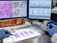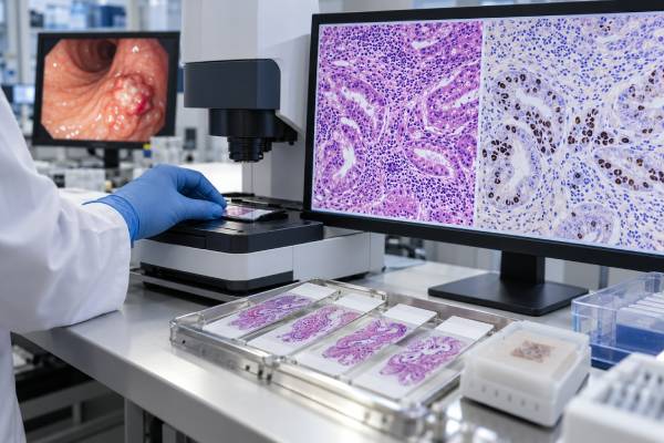
The EBV test used to classify gastric cancer is usually EBER in situ hybridization (EBER-ISH) performed on tumor tissue. It detects abundant Epstein-Barr virus-encoded small RNAs inside malignant cells and identifies an EBV-associated gastric cancer molecular subtype. This is very different from an EBV antibody blood test or a general PCR test for viral DNA. Most adults have been infected with EBV at some point, so positive blood antibodies do not show that a gastric tumor is EBV-driven. EBER-ISH localizes the virus to the cancer cells and is considered the standard tissue method. EBV-associated gastric cancers account for roughly 8%–10% of gastric cancers worldwide, with variation by population. They often show prominent immune-cell infiltration, PD-L1-related biology, PIK3CA mutations, and extensive DNA methylation. EBV status can add molecular and prognostic context, but it is not currently a stand-alone companion diagnostic that automatically determines immunotherapy. HER2, PD-L1, MSI/MMR, CLDN18.2, stage, and treatment setting still guide clinical decisions.
- EBER in situ hybridization on tumor tissue is the standard test for identifying EBV-associated gastric cancer.
- A positive EBV antibody blood test does not prove that a gastric tumor is EBV-positive because most adults have prior EBV exposure.
- EBV-associated gastric cancer represents about 8%–10% of cases overall, although prevalence varies geographically and by tumor site.
- EBV-positive tumors often have immune-rich biology and higher PD-L1 pathway activity, but EBV alone is not an approved universal immunotherapy selector.
- A negative EBER-ISH result means the tested tumor does not meet the tissue definition of EBV-associated gastric cancer.
Table of Contents
- What EBV-Associated Gastric Cancer Is
- How EBER-ISH Testing Works
- Positive and Negative Result Meaning
- EBV as a Molecular Subtype
- EBV and Immunotherapy
- Limitations and Wrong Test Types
- How EBV Fits With Other Biomarkers
What EBV-Associated Gastric Cancer Is
Epstein-Barr virus is a herpesvirus that infects most people during their lifetime and remains latent in the body. In a small subset of gastric cancers, EBV is present clonally within the malignant epithelial cells and contributes to tumor biology. These cancers are called EBV-associated gastric cancers (EBVaGC).
The important word is “associated.” A patient does not develop this tumor simply because an EBV antibody test is positive. Prior infection is extremely common, while EBV-associated gastric cancer is uncommon. The defining finding is evidence of viral RNA inside the tumor cells.
Large genomic studies established EBV-positive gastric cancer as a distinct molecular subgroup. Characteristic features include widespread CpG island DNA hypermethylation, frequent PIK3CA mutations, alterations affecting chromatin regulation, and immune-signaling changes. Some tumors show amplification of chromosome 9p regions containing genes related to PD-L1 and PD-L2.
Clinically, EBV-positive cancers are reported more often in men and in tumors involving the proximal stomach or gastric body, although they can occur elsewhere. They may show prominent lymphoid stroma under the microscope.
EBV status is one component of a broader gastric cancer molecular profile. It does not replace standard histologic diagnosis or staging.
How EBER-ISH Testing Works
EBER stands for Epstein-Barr virus-encoded small RNA. EBER1 and EBER2 are produced in very high copy numbers in latently infected cells, making them excellent tissue targets.
In situ hybridization uses a labeled nucleic-acid probe that binds EBER within formalin-fixed, paraffin-embedded tissue. Under the microscope, the pathologist can see whether the signal is located in malignant epithelial nuclei rather than only in background lymphocytes.
That localization is the key advantage over a generic PCR assay. A gastric tumor contains many immune cells, including B lymphocytes that may harbor latent EBV. PCR can detect viral DNA from those bystander cells and create a positive result without proving that the carcinoma itself is EBV-associated.
A high-quality EBER-ISH run includes appropriate positive and negative controls. Poor fixation, extensive necrosis, or an inadequate biopsy can make interpretation difficult, but the method is robust in routine pathology specimens.
The test is not usually reported as a numeric viral load. It is generally positive or negative based on convincing nuclear signal in tumor cells. Some pathology reports also describe the distribution or strength of staining.
No fasting or special patient preparation is required because the test is performed on tissue already collected during endoscopy, surgery, or biopsy.
Positive and Negative Result Meaning
A positive EBER-ISH result means EBV-encoded RNA is present within the gastric cancer cells, supporting classification as EBV-associated gastric carcinoma.
A negative result means the tested tumor does not show the EBER signal required for that classification. It does not mean the patient has never had EBV infection. Most EBER-negative gastric cancer patients will still have blood antibodies indicating prior EBV exposure.
| Test result | What it means | What it does not mean |
|---|---|---|
| EBER-ISH positive in tumor cells | Supports EBV-associated gastric cancer subtype | Does not automatically prescribe immunotherapy |
| EBER-ISH negative | Tumor is not classified as EBV-associated by the standard tissue method | Does not show absence of prior EBV infection |
| EBV blood antibodies positive | Usually reflects previous or current infection history | Does not establish EBV-positive gastric cancer |
| EBV PCR positive in bulk tissue | Viral DNA is detectable | May not prove virus is in the malignant epithelial cells |
EBV positivity does not provide a cancer stage. A small localized tumor and a metastatic tumor can both be EBER positive. Stage is determined by depth of invasion, lymph nodes, distant metastases, and imaging.
EBV status is also not a hereditary cancer result. Family members do not need germline testing simply because a tumor is EBER positive.
EBV as a Molecular Subtype
Modern gastric cancer classification recognizes biologically distinct tumor groups rather than treating every adenocarcinoma as the same disease. EBV-associated cancer is one of the best-defined subtypes.
Its molecular features often include:
- extensive promoter hypermethylation;
- recurrent PIK3CA mutations;
- alterations in chromatin-remodeling genes such as ARID1A;
- increased immune-cell infiltration;
- elevated expression of immune checkpoint-related pathways; and
- characteristic histologic patterns in some cases.
These features help explain why EBV-positive tumors have attracted interest as candidates for immune checkpoint inhibition. They also provide insight into tumor development and potential future targets.
At the population level, some studies suggest EBV-positive gastric cancers can have a more favorable prognosis than certain EBV-negative subtypes, particularly when localized. Prognosis still depends heavily on stage, resectability, treatment, age, and other tumor factors. EBV should not be used to give an individual survival estimate.
Pathologists can integrate EBER-ISH with mismatch repair IHC, p53 staining, and other assays to create simplified molecular classifications even when full genomic sequencing is not available. This can be useful for research and sometimes clinical discussion, although routine treatment decisions remain centered on validated predictive biomarkers.
EBV and Immunotherapy
EBV-positive gastric cancers often show strong immune activation. Many have increased PD-L1 expression on tumor or immune cells, and small prospective studies have reported striking responses to PD-1 blockade in selected EBV-positive patients.
These findings are biologically compelling but should be interpreted carefully. EBV status has not replaced validated clinical biomarkers such as PD-L1 combined positive score and MSI/MMR in routine treatment algorithms. Small studies can overestimate benefit, and not every EBV-positive tumor responds to immune checkpoint therapy.
For an individual patient, immunotherapy decisions depend on the treatment setting, approved regimen, PD-L1 CPS, MSI/MMR status, HER2 status, prior treatment, and overall health. An EBV-positive result can strengthen the biological rationale but should not be treated as an automatic prescription.
A patient with EBV-positive, MSI-high disease would have two immune-related features, but MSI-high status has a more established predictive role. Likewise, a HER2-positive tumor may require HER2-directed treatment regardless of EBV status.
This is an example of why molecular testing is additive rather than competitive: one tumor can carry several informative biomarkers at the same time.
Limitations and Wrong Test Types
The most common interpretation error is using the wrong EBV test for the clinical question.
EBV serology measures antibodies such as viral capsid antigen or EBV nuclear antigen. It is useful for diagnosing certain patterns of acute or prior infection, but it cannot determine whether gastric cancer cells contain EBV.
Plasma EBV DNA is useful in some EBV-driven cancers, particularly nasopharyngeal carcinoma, but it is not the standard defining test for EBV-associated gastric cancer.
Bulk-tissue PCR can detect EBV DNA from tumor-infiltrating B cells. Without localization, a positive PCR can overcall tumor association.
EBER-ISH solves this problem by showing where the virus is located.
Another limitation is sampling. A very small biopsy can contain limited viable tumor, and poor fixation can compromise nucleic-acid preservation. If the assay is indeterminate, repeating it on another tumor block may be appropriate.
Unlike a quantitative serum marker, EBER-ISH is generally not useful for serial monitoring. It does not rise and fall with tumor burden in a way that CEA or CA 19-9 might. Once the tumor is classified, repeated EBER testing is rarely needed unless a new lesion raises a diagnostic question.
How EBV Fits With Other Biomarkers
Advanced gastric cancer treatment increasingly depends on several biomarkers assessed in parallel.
HER2 identifies tumors that may benefit from HER2-directed therapy. HER2 testing in gastric cancer uses IHC and, when appropriate, in situ hybridization.
PD-L1 is reported as a combined positive score and helps estimate benefit from adding immune checkpoint therapy in relevant settings.
MSI/MMR identifies mismatch repair-deficient tumors with strong immunotherapy sensitivity. A gastric cancer MSI test can be performed by PCR or NGS, while MMR deficiency can be assessed by IHC.
CLDN18.2 is an IHC target used for zolbetuximab selection in HER2-negative advanced disease when the validated expression threshold is met. It is independent of EBER status.
A complete pathology discussion should distinguish tests that are established companion diagnostics from those that mainly provide subtype or emerging predictive information. EBV currently sits closer to the latter category in routine gastric cancer care.
The main value of EBER-ISH is precision: it identifies a real biological subtype using a test that localizes the virus to malignant cells. That is much more informative than simply knowing that the patient has encountered a common virus at some point in life.
Why EBER localization is more important than viral quantity
In EBV-associated gastric cancer, the central diagnostic question is not “How much EBV is present?” but “Is EBV present inside the malignant epithelial cells?” EBER-ISH answers that localization question directly. A strong nuclear signal in the cancer cells supports the EBV-associated subtype even if no blood viral-load test was performed.
This differs from diseases in which plasma EBV DNA is used quantitatively for monitoring. Gastric cancer does not have an established routine strategy in which a specific EBV DNA copy number replaces tissue EBER testing or serves as a standard recurrence marker.
The localization principle also explains why PCR can be misleading. A gastric tumor may contain many EBV-infected lymphocytes that are not malignant. PCR from bulk tissue can amplify DNA from those cells and produce a positive result even when the carcinoma itself is EBER negative. EBER-ISH lets the pathologist see which cells contain the viral RNA.
Histologic clues can suggest EBV but cannot replace testing
Some EBV-associated gastric cancers have dense lymphocytic infiltration and a lymphoepithelioma-like appearance. Others are conventional adenocarcinomas without striking clues. Tumor location in the proximal stomach, male sex, or a prominent immune infiltrate may raise suspicion, but none is specific enough to classify the tumor.
This is important because visual pattern recognition can be excellent for deciding when to test, but molecular subtyping requires the actual assay. Conversely, a pathologist should not reject a positive EBER result simply because the histology is not classic.
The same principle applies to PD-L1. EBV-positive tumors often show increased PD-L1 pathway activity, but the PD-L1 combined positive score must still be measured separately when it is needed for treatment planning.
EBV-positive does not mean an active contagious infection is driving current symptoms
Patients sometimes interpret an EBV-positive tumor report as evidence that they currently have mononucleosis or that they are highly contagious. That is not what the pathology result means. EBV-associated gastric cancer reflects latent viral involvement in the tumor cells, usually years after the original infection.
Routine gastric cancer management does not involve antiviral treatment aimed at clearing EBV from the body. Standard care is based on cancer stage, surgery when appropriate, chemotherapy, immunotherapy, targeted therapy, and other oncologic strategies. Research is exploring EBV-directed immune approaches, but these are not equivalent to treating an acute viral infection.
Family members also do not need gastric cancer screening merely because a relative’s tumor is EBER positive. EBV exposure is widespread, and gastric cancer risk depends on many other factors including Helicobacter pylori, smoking, diet, age, inherited susceptibility, and gastric mucosal disease.
How EBV status can be useful even when it does not select a drug by itself
A biomarker can be clinically informative without being a formal companion diagnostic. EBV status can explain why a tumor has a heavily inflamed microenvironment, add molecular subtype information, and support enrollment in clinical trials designed for EBV-associated cancers or immune-rich gastric tumors.
It can also help pathologists and researchers separate biologically different cancers that would otherwise look similar under the microscope. This improves interpretation of prognosis studies and can reveal why one tumor has a different pattern of genomic alterations from another.
The limitation is that such information should not be upgraded into a stronger claim than the evidence supports. A positive EBER result does not guarantee immunotherapy response, and a negative result does not mean immunotherapy will fail. That decision still depends on validated treatment biomarkers and the overall clinical setting.
Why EBV status is usually stable once the tumor is classified
EBV-associated gastric cancer is thought to arise from clonal infection early in tumor development, so EBER positivity is generally a stable subtype feature rather than a marker expected to switch on and off with each treatment cycle. This is another reason repeated EBER testing is rarely useful for routine monitoring.
If a new lesion has very different morphology, EBER-ISH can still help determine whether it is related to the original gastric cancer or represents another process. In that diagnostic setting, retesting answers an identity question rather than a disease-burden question.
Bottom-line interpretation
For gastric cancer, the correct EBV question is answered in tissue: are the malignant epithelial cells EBER positive? Once that is established, the tumor can be classified as EBV-associated and its immune-rich biology can be considered alongside other biomarkers. Blood antibodies, viral load, or a history of mononucleosis do not substitute for this tissue definition. Treatment still depends on the full gastric cancer profile rather than EBV status alone.
For pathology reports, the wording matters. A clear result should identify the assay as EBER in situ hybridization and state whether EBER signals are present in the malignant epithelial cells. If the report only says “EBV positive” without naming the method or cellular location, it is reasonable to ask for clarification. That detail separates true EBV-associated gastric carcinoma from incidental EBV in background lymphocytes and keeps the molecular subtype assignment reproducible across institutions.
References
- Advances and challenges in gastric cancer testing: the role of biomarkers 2025 (Review)
- Unlocking the Potential: Epstein-Barr Virus (EBV) in Gastric Cancer and Future Treatment Prospects, a Literature Review 2024 (Review)
- Implementing an integrated molecular classification for gastric cancer from endoscopic biopsies using on-slide tests 2024 (Review)
- Antibodies Against Epstein–Barr Virus as Disease Markers of Gastric Cancer: A Systematic Review 2023 (Systematic Review)
Disclaimer
EBER-ISH classifies a gastric tumor as EBV-associated but does not determine stage or automatically select a specific treatment. Results should be interpreted with pathology, HER2, PD-L1, MSI/MMR, CLDN18.2, imaging, and the current treatment setting. Blood EBV antibodies or PCR results should not be substituted for tumor-localizing EBER-ISH when the clinical question is gastric cancer subtype.