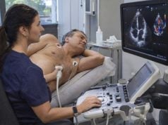
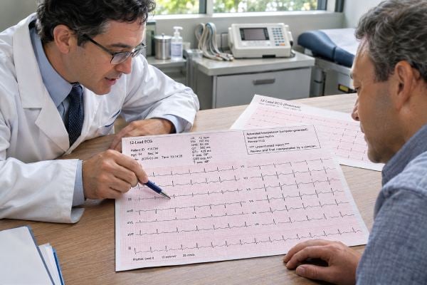
An EKG can show whether your heart rhythm is regular, how fast electrical signals travel through the heart, and whether there are patterns that suggest an arrhythmia, conduction problem, chamber enlargement, electrolyte issue, medication effect, or reduced blood flow to the heart. Many EKG findings are harmless or nonspecific, especially when they appear in someone without symptoms. Others need prompt attention. The wording on an automated EKG report can sound alarming because the computer is designed to flag possibilities, not make a final diagnosis. Terms such as “sinus bradycardia,” “nonspecific ST-T abnormality,” “possible left ventricular hypertrophy,” or “incomplete right bundle branch block” must be interpreted with your age, symptoms, medical history, medications, and prior tracings. A normal EKG is reassuring but does not rule out every heart condition, and an abnormal EKG does not automatically mean heart disease. The most useful interpretation comes from understanding the specific finding, whether it is new, and whether it fits what you are feeling.
Table of Contents
- What an EKG Measures
- Common Rhythm Findings
- Conduction and Interval Findings
- ST-Segment and T-Wave Changes
- Voltage and Chamber Patterns
- When an Abnormal EKG Is Urgent
- What Happens After an Abnormal EKG
What an EKG Measures
An electrocardiogram—called an EKG or ECG—is a short recording of the heart’s electrical activity from electrodes placed on the skin. A standard 12-lead EKG looks at the same heartbeat from multiple electrical angles. It does not directly show the coronary arteries, heart valves, or pumping strength, but it can provide clues about all of them.
The tracing is divided into waves and intervals:
- P wave: electrical activation of the atria, the heart’s upper chambers.
- PR interval: time for the signal to travel from the atria through the AV node toward the ventricles.
- QRS complex: electrical activation of the ventricles, the main pumping chambers.
- ST segment and T wave: parts of ventricular recovery, or repolarization.
- QT interval: total time for ventricular activation and recovery. Because QT changes with heart rate, clinicians usually interpret a corrected value called QTc.
The report also gives a heart rate and often electrical axes. Computer software may estimate whether the rhythm is sinus, whether conduction intervals are prolonged, and whether voltage or ST-T patterns meet preset criteria.
A key limitation is that the machine interpretation is only a screening interpretation. False-positive statements are common, particularly for “possible infarct,” “possible left atrial enlargement,” or “left ventricular hypertrophy.” Lead placement, body shape, movement, breathing, and normal variants can all affect the tracing.
Clinicians therefore compare the automated statement with the actual waveform. If a prior EKG exists, comparison is valuable. A stable pattern present for years usually carries a different meaning from a new change in a person with chest pain.
The table below summarizes several common phrases:
| EKG wording | What it usually means | Why context matters |
|---|---|---|
| Normal sinus rhythm | Electrical impulse starts in the sinus node with a regular pattern | Does not rule out structural heart disease or intermittent arrhythmias |
| Sinus bradycardia | Sinus rhythm slower than about 60 beats/min | Can be normal in fit people or due to medication, sleep, or conduction disease |
| Sinus tachycardia | Sinus rhythm faster than about 100 beats/min | Often reflects fever, pain, dehydration, anxiety, anemia, or another cause |
| Nonspecific ST-T changes | Repolarization looks different from textbook normal | May be benign, medication-related, metabolic, or associated with heart disease |
| Bundle branch block | Ventricular conduction is delayed through a right or left bundle | Meaning depends on side, whether it is new, symptoms, and structural disease |
Clinicians also look at the electrical axis, which describes the general direction of ventricular activation. A mild leftward or rightward axis can be a normal variant, especially when it has been stable for years. More marked axis deviation can accompany conduction disease, ventricular enlargement, lung disease, or prior heart injury. Like voltage criteria, axis is a clue rather than a diagnosis by itself.
Heart rate should also be interpreted at the moment the tracing was recorded. A rate of 105 after rushing into the clinic may represent normal sinus tachycardia, while the same rate at rest with fever or anemia points toward a different cause. Similarly, a resting rate of 48 may be expected in a trained athlete but more concerning in a person taking AV-node-blocking medication who has dizziness.
Artifact is another common source of confusion. Muscle tremor, loose electrodes, movement, electrical interference, or misplaced chest leads can imitate rhythm or ST-T abnormalities. When a computer interpretation does not fit the clinical picture, repeating the EKG with careful lead placement is often the simplest and most useful first step.
Common Rhythm Findings
Normal sinus rhythm
“Normal sinus rhythm” means each heartbeat is being initiated by the sinus node, the heart’s normal pacemaker, and the rate and conduction pattern fit expected limits. It is a reassuring finding, but it does not mean the heart is proven healthy in every respect. An EKG lasts only seconds, so intermittent arrhythmias can be missed.
Sinus bradycardia
Sinus bradycardia usually means a sinus rhythm below about 60 beats per minute. In an endurance athlete, during sleep, or in a relaxed healthy adult, this can be completely normal. Beta blockers and some calcium-channel blockers can also slow the sinus rate.
Bradycardia becomes more important when it is associated with fainting, near-fainting, severe fatigue, exercise intolerance, chest pain, or low blood pressure. The concern is not the number alone but whether the heart is delivering enough blood for the body’s needs. Low heart rate and bradycardia warning signs help explain when a slow pulse deserves evaluation.
Sinus tachycardia
Sinus tachycardia means the sinus node is pacing the heart faster than about 100 beats per minute. It is often a normal response rather than a primary heart-rhythm disorder. Common causes include exercise, dehydration, fever, infection, pain, anxiety, anemia, hyperthyroidism, pregnancy, stimulant use, and certain medications.
The EKG can identify that the rhythm is sinus, but it cannot always identify why the rate is fast. Treatment focuses on the cause rather than simply slowing the heart.
Premature beats
Premature atrial contractions and premature ventricular contractions are early beats that arise outside the normal timing sequence. One or two on an EKG can occur in healthy people. Caffeine, alcohol, sleep deprivation, stress, and electrolyte disturbances can increase them.
Frequent premature beats, symptoms, or a history of structural heart disease may lead to ambulatory monitoring or an echocardiogram. For PVCs specifically, see causes and risks of skipped beats.
Atrial fibrillation
Atrial fibrillation, or AFib, produces chaotic atrial electrical activity and usually an irregular ventricular rhythm. The 2023 ACC/AHA guideline describes characteristic EKG features including irregular R-R intervals, absence of distinct P waves, and irregular atrial activity. A clinician should visually confirm the diagnosis rather than relying solely on an automated or wearable algorithm.
New AFib matters because it can increase stroke risk and may require assessment of heart structure, thyroid function, kidney function, electrolyte levels, symptoms, and the need for anticoagulation. AFib symptoms and warning signs can help put a new irregular-rhythm finding into context.
Conduction and Interval Findings
Conduction findings describe how the electrical signal travels from the atria through the AV node and then through the ventricles.
First-degree AV block
First-degree atrioventricular block means the PR interval is longer than normal, usually more than 200 milliseconds, but every atrial impulse still reaches the ventricles. It often causes no symptoms and may be seen in healthy people, athletes, older adults, or people taking medications that slow AV-node conduction.
When the PR interval is very long or the finding occurs with other conduction abnormalities, clinicians may look more closely at medication effects and conduction-system disease.
Second- and third-degree AV block
Higher-grade AV block is more significant. In second-degree block, some atrial impulses fail to conduct to the ventricles. In complete, or third-degree, heart block, atrial and ventricular activity become dissociated because normal conduction through the AV node or His-Purkinje system fails.
These findings can produce dangerously slow heart rates and may require urgent assessment, particularly with fainting, dizziness, low blood pressure, or heart failure symptoms. Some forms require a pacemaker.
Right bundle branch block
Right bundle branch block, or RBBB, means conduction through the right-sided ventricular pathway is delayed. An incomplete RBBB can be a normal variant, especially in younger people. A complete RBBB may also occur without major structural disease, but it can be associated with right-heart strain, pulmonary disease, congenital heart disease, or other conditions.
The importance depends on whether the finding is new, whether symptoms are present, and whether other EKG or imaging abnormalities exist.
Left bundle branch block
Left bundle branch block, or LBBB, produces a wider QRS and an altered sequence of left-ventricular activation. It deserves more attention than an isolated incomplete RBBB because LBBB is more often associated with underlying structural heart disease, cardiomyopathy, prior injury, or conduction-system degeneration.
A 2023 review emphasized that LBBB is not merely an electrical label; it can affect diagnosis, mechanical synchrony, prognosis, and decisions about cardiac resynchronization or conduction-system pacing. A new LBBB does not by itself prove a heart attack, but new conduction changes in someone with ischemic symptoms require clinical evaluation.
Prolonged QTc
A prolonged QTc means ventricular electrical recovery takes longer than expected. Causes include inherited long-QT syndromes, certain medications, low potassium, low magnesium, low calcium, bradycardia, and acute illness.
A markedly prolonged QTc can increase risk for torsades de pointes, a potentially dangerous ventricular arrhythmia. Clinicians interpret QTc using the actual number, sex, heart rate, medication list, electrolyte values, symptoms, and family history. One computer-generated “prolonged QT” statement should be verified because automated measurements can be wrong.
ST-Segment and T-Wave Changes
ST-segment and T-wave abnormalities are among the EKG phrases most likely to cause anxiety because they can be associated with ischemia or heart attack. They can also occur for many non-emergency reasons.
The 2025 acute coronary syndrome guideline emphasizes that new ST elevation in the right clinical setting can indicate an acute coronary occlusion requiring immediate treatment. ST depression or significant T-wave inversion can also suggest ischemia. But EKG interpretation is pattern-based: the amount of change, which leads are involved, whether the changes are new, and what symptoms are occurring all matter.
“Nonspecific ST-T abnormality” is different from a diagnostic ischemic pattern. It means the repolarization pattern does not fit classic normal criteria but also does not point cleanly to one cause. Possible contributors include electrolyte abnormalities, medications, left ventricular hypertrophy, bundle branch block, heart strain, and normal variation.
A normal EKG also does not completely rule out acute coronary syndrome. The 2025 guideline notes that some patients with non-ST-elevation acute coronary syndrome have a normal or nonspecific EKG, and serial tracings may be needed when symptoms persist.
That is why symptoms override false reassurance from a printed “normal” result. If you have new chest pressure, shortness of breath, sweating, nausea, or pain spreading to the arm, jaw, back, or upper abdomen, medical evaluation should not be delayed because one EKG looks normal. Emergency chest-pain warning signs explains when to seek immediate care.
Early repolarization is another common phrase. It often appears as mild ST elevation in younger healthy people and has historically been considered a normal variant. Its significance depends on the exact pattern and clinical setting, so it should not be self-diagnosed from the report wording alone.
Voltage and Chamber Patterns
An EKG can suggest chamber enlargement or thickened heart muscle, but it does not directly measure chamber size or wall thickness.
Left ventricular hypertrophy
“Voltage criteria for LVH” means the electrical voltages meet a formula associated with a thicker left ventricular wall. High blood pressure is a common cause of true left ventricular hypertrophy, but EKG voltage is imperfect. Thin or athletic people can meet voltage criteria without pathologic thickening, while some people with true LVH do not meet the EKG criteria.
If LVH is clinically important, an echocardiogram can directly assess ventricular wall thickness and structure. Long-term blood-pressure control matters because true LVH reflects the heart working against increased pressure. Normal blood-pressure ranges and categories can help you interpret the readings that may contribute to LVH.
Atrial enlargement
P-wave shape can suggest left or right atrial enlargement, but these computer-generated statements often need confirmation. An echocardiogram is better suited to measuring atrial size.
“Possible old infarct”
Automated reports sometimes state “possible anterior infarct,” “possible inferior infarct,” or “age undetermined.” This may reflect Q-wave patterns, poor R-wave progression, or lead-placement differences. It does not automatically mean you previously had a heart attack.
A clinician may compare prior EKGs, repeat the tracing with careful lead placement, review symptoms and history, and use echocardiography or other imaging if the possibility is clinically plausible. This is a classic example of why the computer interpretation should not be read in isolation.
When an Abnormal EKG Is Urgent
Urgency depends more on the combination of the EKG, symptoms, and vital signs than on one phrase in the report.
Seek emergency care when an abnormal EKG is associated with:
- New chest pressure, squeezing, or pain
- Severe or worsening shortness of breath
- Fainting or near-fainting
- A sustained very fast or very slow heart rate with weakness or low blood pressure
- New neurologic symptoms such as facial droop, weakness, or difficulty speaking
- A newly diagnosed serious ventricular arrhythmia
- High-grade AV block with symptoms
- Marked QT prolongation plus fainting or a ventricular rhythm problem
If you have no symptoms and the EKG was obtained as part of a routine visit, many abnormalities can be evaluated without an emergency visit. Examples include stable first-degree AV block, isolated premature beats, a long-standing bundle branch block, or nonspecific ST-T changes.
The key question is whether the finding is new and clinically connected to symptoms. A tracing should be compared with previous EKGs whenever possible.
What Happens After an Abnormal EKG
The next test depends on what the EKG suggests.
A repeat EKG may be enough if lead placement or artifact is suspected. Blood tests can check potassium, magnesium, thyroid function, anemia, kidney function, or cardiac troponin depending on the situation.
An echocardiogram may be ordered when the EKG suggests chamber enlargement, cardiomyopathy, valve disease, pulmonary hypertension, or heart failure. It shows heart structure and pumping function rather than electrical activity. Echocardiogram results explains what those measurements mean.
Ambulatory monitoring is useful when the office EKG is normal but symptoms are intermittent. A Holter monitor, patch monitor, or event recorder can capture palpitations, AFib, pauses, or premature beats over a longer period.
A stress test or coronary imaging may be used when symptoms suggest coronary disease but the resting EKG does not answer the question. The exact test depends on exercise ability, baseline EKG abnormalities, age, risk, and local expertise.
The most useful questions to ask after receiving an EKG report are:
- Is this finding definitely present, or is it only the automated interpretation?
- Is it new compared with prior EKGs?
- Could medication, electrolytes, exercise training, or another condition explain it?
- Does it match my symptoms?
- Do I need a repeat EKG, blood tests, monitoring, imaging, or no further testing?
An EKG is a powerful first-line test because it is fast and inexpensive, but it is a snapshot rather than a complete cardiac evaluation. A “normal” tracing does not exclude every heart problem, and an “abnormal” one often needs context before it means disease.
References
- 2025 ACC/AHA/ACEP/NAEMSP/SCAI Guideline for the Management of Patients With Acute Coronary Syndromes: A Report of the American College of Cardiology/American Heart Association Joint Committee on Clinical Practice Guidelines 2025 (Guideline)
- 2023 ACC/AHA/ACCP/HRS Guideline for the Diagnosis and Management of Atrial Fibrillation: A Report of the American College of Cardiology/American Heart Association Joint Committee on Clinical Practice Guidelines 2023 (Guideline)
- 2024 AHA/ACC/AMSSM/HRS/PACES/SCMR Guideline for the Management of Hypertrophic Cardiomyopathy: A Report of the American Heart Association/American College of Cardiology Joint Committee on Clinical Practice Guidelines 2024 (Guideline)
- 2022 ESC Guidelines for the management of patients with ventricular arrhythmias and the prevention of sudden cardiac death 2022 (Guideline)
- Left Bundle Branch Block: Characterization, Definitions, and Recent Insights into Conduction System Physiology 2023 (Review)
- 2021 AHA/ACC/ASE/CHEST/SAEM/SCCT/SCMR Guideline for the Evaluation and Diagnosis of Chest Pain: A Report of the American College of Cardiology/American Heart Association Joint Committee on Clinical Practice Guidelines 2021 (Guideline)
Disclaimer
This article is for general education and cannot interpret an individual EKG without the tracing, symptoms, history, and clinical context. Seek urgent medical care for chest pain, fainting, severe shortness of breath, new neurologic symptoms, or a sustained very fast or very slow heartbeat with weakness or dizziness.