A “skipped beat” often is not a missing heartbeat at all. One common cause is a premature ventricular contraction, or PVC—an early beat that starts in the heart’s lower chambers. Because the early beat may pump less blood, it can be followed by a pause and then a stronger normal beat. That sequence may feel like a flip, thump, flutter, or momentary stop. PVCs are common and, in many people with a structurally normal heart, they are not dangerous. Their importance changes when they are very frequent, cause major symptoms, occur with exercise or fainting, or appear in someone with heart disease. A high PVC burden can sometimes weaken the heart over time, and PVCs can occasionally be a marker of an underlying electrical or structural problem. The goal is not to label every extra beat as harmless or dangerous. It is to identify the pattern, measure how often it happens, look for heart disease, and match treatment to risk and symptoms.
Table of Contents
- What PVCs Are and Why They Feel Like a Skipped Beat
- Common Causes and Triggers
- When PVCs Are More Concerning
- PVC Burden and PVC-Induced Cardiomyopathy
- How PVCs Are Evaluated
- Treatment: From Reassurance to Ablation
- What You Can Do Now
What PVCs Are and Why They Feel Like a Skipped Beat
A normal heartbeat usually begins in the sinus node, the heart’s natural pacemaker in the right upper chamber. Electrical activity then travels through the atria, the AV node, and the ventricles in an organized sequence. A PVC starts early from an electrical focus in a ventricle instead of following that usual route.
Because the ventricles contract before they have filled normally, the premature beat may produce a weaker pulse that you barely feel. The heart often pauses briefly afterward while the normal rhythm resets. The next beat has more filling time and can feel unusually forceful. Many people interpret that pause-and-thump pattern as a skipped beat.
PVCs can occur singly, every few beats, in pairs, or in repeating patterns such as bigeminy, where every other beat is premature. Some people feel every PVC; others have thousands per day without noticing them. Symptoms can include fluttering, pounding, a dropping sensation in the chest, brief breathlessness, or an urge to cough. Frequent PVCs may cause fatigue, reduced exercise tolerance, lightheadedness, or a general sense that the heart rhythm is unstable.
Not every palpitation is a PVC. Premature atrial contractions, atrial fibrillation, supraventricular tachycardia, sinus tachycardia, and other rhythms can feel similar. A pulse or smartwatch notification may suggest irregularity but cannot reliably identify every rhythm. A 12-lead ECG or ambulatory rhythm recording is often needed to know what is actually happening.
For a broader overview of rhythm sensations and warning signs, see heart palpitations: causes and warning signs.
Common Causes and Triggers
PVCs can appear in healthy hearts, and sometimes no specific trigger is found. They can also become more noticeable when the nervous system is stimulated or when the heart is under physiologic stress.
Commonly reported triggers include stress, anxiety, poor sleep, nicotine, alcohol, stimulant drugs, and some medications. Dehydration, fever, illness, or intense exertion may increase ectopic beats in susceptible people. Electrolyte abnormalities—particularly significant disturbances in potassium or magnesium—can promote ventricular arrhythmias, although taking supplements without documented deficiency is not a safe universal solution.
Caffeine deserves a nuanced answer. Some people clearly notice more palpitations after coffee or high doses of caffeine, while population data do not show that ordinary coffee intake universally causes dangerous ventricular arrhythmias. If symptoms consistently follow caffeine, a short reduction or elimination trial can be informative. Energy drinks are a separate concern because they may deliver large stimulant doses rapidly and can contain multiple active ingredients. If palpitations follow energy drinks or pre-workout stimulants, stopping those products is a sensible first step. More context is available in energy drinks, stimulants, and arrhythmia risk.
Alcohol can increase ectopy in some people, especially after heavier intake, dehydration, or poor sleep. Nicotine and other stimulants increase sympathetic activity and can make the heartbeat more noticeable or irritable. Decongestants containing stimulant-like ingredients may also aggravate palpitations in sensitive individuals.
Medical conditions can contribute as well. Hyperthyroidism, anemia, sleep apnea, infection, and some lung diseases can increase cardiac stress. PVCs may occur in people with coronary artery disease, prior heart attack, cardiomyopathy, myocarditis, valve disease, or heart failure. In those settings, the PVC may be less important than the underlying heart problem that made it possible.
Exercise has a complicated relationship with PVCs. Occasional PVCs can occur during activity even in healthy people. PVCs that are new, become more frequent with exertion, occur in complex runs, or are accompanied by chest pain, fainting, or marked breathlessness deserve medical assessment. The pattern during exercise and recovery can add useful risk information.
When PVCs Are More Concerning
The majority of isolated PVCs in a person with a normal heart structure and normal ventricular function are low risk. What makes clinicians look more closely is the combination of symptoms, PVC characteristics, family history, and the condition of the heart itself.
Seek urgent or emergency care if palpitations occur with fainting, near-fainting, severe chest pressure, severe shortness of breath, new neurologic symptoms, or a sustained very fast rhythm that does not settle. These symptoms can indicate a more serious arrhythmia or another cardiovascular emergency rather than simple isolated PVCs.
Features that usually justify a more complete cardiac evaluation include:
- Known heart disease, prior heart attack, cardiomyopathy, significant valve disease, or heart failure.
- Syncope or near-syncope, particularly during exertion.
- PVCs that appear or increase substantially with exercise.
- A family history of unexplained sudden cardiac death or inherited cardiomyopathy at a young age.
- Very frequent PVCs, runs of ventricular beats, or more than one PVC morphology on monitoring.
- Evidence of reduced pumping function or abnormal heart structure.
- New PVCs after myocarditis, a major infection, or exposure to a medication or drug known to affect rhythm.
A single alarming feature does not prove that the PVCs are dangerous, but it changes the threshold for testing. The 2022 European ventricular-arrhythmia guideline emphasizes looking for structural heart disease and using additional imaging when the presentation is not typical for benign idiopathic PVCs.
The ECG pattern itself also matters. PVCs that all look alike are called monomorphic and often arise from one focus. PVCs with several different shapes are called multifocal or polymorphic and may lead clinicians to look more carefully for structural or electrical disease, especially when other risk markers are present. Coupled PVCs, short runs of ventricular tachycardia, or unusually early PVCs can also change the interpretation. These findings are not meant for self-diagnosis from a consumer device; they are features a clinician interprets together with imaging, symptoms, and history.
PVCs can occasionally trigger more serious ventricular arrhythmias, but this is uncommon in people without underlying heart disease or a specific electrical disorder. The risk discussion should therefore avoid two extremes: dismissing every PVC as harmless and treating every PVC as a warning of sudden cardiac death.
If you develop palpitations together with symptoms that could represent an acute heart problem, use the same emergency thresholds you would for chest pain. Do not wait for a wearable device to label the rhythm first.
PVC Burden and PVC-Induced Cardiomyopathy
PVC burden is the percentage of recorded heartbeats that are PVCs. If 10 out of every 100 beats on a sufficiently long monitor are premature ventricular beats, the burden is about 10%. This measurement helps clinicians judge whether frequent ectopy could be affecting heart function.
A high burden can, in some people, lead to PVC-induced cardiomyopathy—a potentially reversible weakening of the left ventricle caused or worsened by frequent premature beats. The exact threshold is not identical for everyone. Risk tends to rise as burden increases, and a burden around 10% or higher often prompts closer attention when ventricular function is reduced or symptoms are significant. Some studies find greater risk at burdens in the 15% to 20% or higher range, while occasional cases occur below those numbers.
That uncertainty matters. A 10% burden is not a switch that turns a benign rhythm into a dangerous one. PVC origin, QRS width, timing, day-to-day variability, how long the burden has been present, and individual susceptibility all influence risk. A person with 12% PVCs and a normal heart may be observed, while another person with a similar burden and declining ejection fraction may need treatment.
The encouraging part is that PVC-induced cardiomyopathy can improve when the PVCs are successfully suppressed. Recovery of ventricular function after medication or catheter ablation supports the idea that the ectopy itself was contributing to the weakness.
Burden also varies from day to day. A 24-hour monitor can miss variability, so clinicians sometimes use longer ambulatory monitoring when symptoms are intermittent or when an accurate burden estimate is important. The choice between monitor types depends on how often symptoms occur. See Holter versus event monitoring for palpitations for the basic differences.
How PVCs Are Evaluated
Evaluation starts with the story. A clinician will ask what the sensation feels like, how long it lasts, whether it happens at rest or with exertion, and whether there is chest pain, breathlessness, dizziness, or fainting. They will review medications, supplements, stimulant use, alcohol, nicotine, sleep, recent illness, and family history.
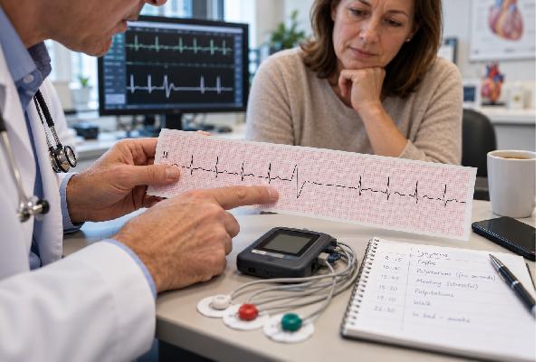
A 12-lead electrocardiogram can confirm PVCs if one occurs during the tracing and can also show conduction abnormalities, prior heart injury, long QT, or other clues. The shape of the PVC on ECG can suggest where in the ventricles it originates. Some patterns are typical of common idiopathic sites, while others raise more concern for underlying disease.
Ambulatory monitoring is used when PVCs are intermittent or when clinicians need to quantify burden. A Holter monitor may record continuously for one or more days. Patch monitors can record longer. Event monitors and other devices are useful when symptoms are less frequent. The recording can show whether symptoms match PVCs, how many occur, whether there are multiple morphologies, and whether runs of nonsustained ventricular tachycardia occur.
Echocardiography evaluates chamber size, valve function, and left ventricular ejection fraction. It is especially important when PVCs are frequent, symptoms are substantial, or there is concern for structural heart disease. Cardiac magnetic resonance imaging can provide more detailed tissue characterization and detect scar, inflammation, or cardiomyopathy that may not be obvious on an echocardiogram.
Exercise testing may be appropriate when symptoms are exertional or when clinicians need to observe how ectopy behaves with increasing workload and recovery. Coronary evaluation may be needed when symptoms or risk factors suggest ischemic heart disease.
Blood tests are individualized. Common possibilities include electrolytes, thyroid testing, a complete blood count, and tests directed at the clinical situation. The goal is not to order every possible lab for every palpitation but to identify reversible contributors and assess the heart’s structure and function.
If the initial ECG is normal, that does not mean the symptoms were imaginary or that PVCs are excluded. Intermittent ectopy may simply not occur during a short office tracing. Matching a symptom time stamp with an ambulatory recording is often the most useful way to confirm whether the sensation is actually caused by a PVC or by a different rhythm.
Treatment: From Reassurance to Ablation
Treatment depends on three questions: Is there underlying heart disease? Are the PVCs causing troublesome symptoms? Are they frequent enough to impair ventricular function or create another meaningful risk?
If evaluation shows a normal heart, the PVC burden is modest, and symptoms are mild, reassurance may be all that is needed. Knowing that the rhythm has been documented and risk-stratified can itself reduce symptom-related anxiety.
When a clear trigger exists, reducing it is reasonable. That may mean improving sleep, treating dehydration, stopping nicotine, reducing heavy alcohol intake, avoiding energy drinks, or reviewing stimulant medications with a clinician. Do not stop prescribed medication on your own. Correct documented electrolyte abnormalities under medical guidance rather than taking high-dose potassium or magnesium “just in case.”
For persistent symptomatic PVCs, beta-blockers or nondihydropyridine calcium-channel blockers are common first-line options in appropriate patients. They may reduce symptoms or PVC frequency, although response varies. Other antiarrhythmic drugs can be effective but carry more complex risks and generally require specialist selection and monitoring, especially when structural heart disease is present.
Catheter ablation targets the small area producing the PVCs. An electrophysiologist maps the abnormal electrical focus and applies energy to eliminate or isolate it. Ablation can be highly effective for suitable idiopathic PVCs and is an important option when symptoms remain significant, medications are ineffective or poorly tolerated, or PVC-induced cardiomyopathy is present. In some common idiopathic PVC locations, guidelines support ablation early because success rates are high.
Treatment success should be judged by more than whether you feel every beat. Repeat monitoring can measure PVC burden, and repeat echocardiography can show whether ventricular function improves when cardiomyopathy was present.
When PVC-induced cardiomyopathy is suspected, follow-up is especially important because improvement may take time after successful suppression. Clinicians may repeat an echocardiogram after the rhythm has been controlled to see whether ejection fraction recovers. If heart function does not improve as expected, they may reconsider whether another cardiomyopathy, coronary disease, scar, or inflammatory condition is present. This is one reason that treating the monitor number alone is not enough; the goal is to improve symptoms and protect ventricular function while making sure the PVCs are not simply a marker of another disorder.
What You Can Do Now
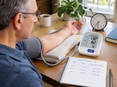
If you notice occasional skipped beats without emergency symptoms, keep a short log for several days. Record the time, what you were doing, caffeine or alcohol intake, sleep quality, exercise, medications, and any associated dizziness, chest discomfort, or breathlessness. This can reveal patterns and help a clinician choose the right monitor.
Check your pulse only long enough to characterize the episode. Repeatedly checking every few minutes can make benign sensations more intrusive without adding useful information. If you use a smartwatch with ECG capability, save a tracing captured during symptoms if the device allows it, but remember that a single-lead wearable is not a substitute for clinical evaluation. Wearables may miss ectopy, misclassify noise, or label an irregular pulse without explaining its cause. A normal-looking consumer tracing also cannot exclude an intermittent arrhythmia that was not happening during the recording.
Try basic reversible steps that are safe for you: regular sleep, adequate hydration, avoiding nicotine, and reducing stimulants that clearly correlate with symptoms. If coffee seems unrelated, there is no universal requirement to eliminate it. If palpitations repeatedly follow high-dose caffeine, energy drinks, or pre-workout products, reducing those exposures is more reasonable than assuming all caffeine is harmless. You can also review coffee, caffeine, and heart palpitations for a more detailed discussion.
Book a medical visit if PVC-like symptoms are new and recurring, are becoming more frequent, limit exercise, or cause significant distress. Evaluation is also appropriate if you have known heart disease or a family history of unexplained sudden death. Bring medication and supplement lists, saved wearable tracings, and your symptom log.
It is also useful to distinguish symptom frequency from PVC burden. A person may feel nearly every premature beat and have a relatively low burden, while another person can have thousands of PVCs with few sensations. That is one reason symptom intensity alone cannot determine risk. Ambulatory monitoring shows how often the beats occur, whether they come from one or more patterns, whether they cluster with exercise or sleep, and whether other arrhythmias are present. An echocardiogram then helps show whether heart structure and pumping function are normal. In selected cases, exercise testing or cardiac MRI can add information when symptoms, family history, ECG findings, or ventricular function raise concern.
If a clinician recommends follow-up rather than immediate treatment, ask what change should trigger reassessment. Examples may include new fainting, worsening exercise tolerance, sustained rapid heartbeat, increasing PVC burden, or a decline in ventricular function on imaging. If medication is started, the goal may be symptom relief rather than elimination of every ectopic beat. If ablation is discussed, the decision usually weighs symptom severity, PVC burden, suspected origin, heart function, procedural risks, and how well medicines have worked or been tolerated. Clear goals make it easier to judge whether a treatment is actually helping.
Most PVCs do not require aggressive treatment. The important exceptions are frequent ectopy that weakens the heart, PVCs associated with concerning symptoms, or PVCs that reveal underlying cardiac disease. Once those possibilities are assessed, management can range from reassurance and trigger reduction to medication or catheter ablation.
References
– 2022 ESC Guidelines for the management of patients with ventricular arrhythmias and the prevention of sudden cardiac death 2022 (Guideline) – To Beat or Not to Beat: When and How to Treat Premature Ventricular Complexes. 2026 (Review) – Treatment of Premature Ventricular Contractions: A Review of Medical Therapy, Catheter Ablation, and Emerging Treatments. 2026 (Review) – Premature Ventricular Contraction-Induced Cardiomyopathy: Contemporary Evidence from Risk Stratification, Pathophysiology, and Management 2024 (Review) – The Influence of Lifestyle Factors on the Occurrence and Severity of Premature Ventricular Contractions: A Comprehensive Review. 2024 (Review)
Disclaimer
This article provides general education and cannot determine whether a person’s palpitations are PVCs or another rhythm. Seek emergency care for palpitations with fainting, severe chest pain or pressure, severe shortness of breath, or a sustained rapid rhythm with significant symptoms. People with known heart disease, frequent PVCs, or worsening symptoms should discuss evaluation and treatment with a qualified clinician.