
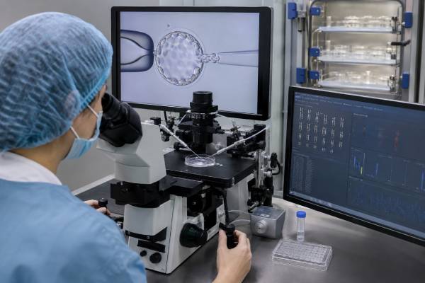
Embryo genetic testing examines a small group of cells from embryos created through in vitro fertilization. The test can look for extra or missing chromosomes, a known single-gene condition, or an unbalanced chromosome rearrangement. These uses are called preimplantation genetic testing for aneuploidy, monogenic conditions, and structural rearrangements—PGT-A, PGT-M, and PGT-SR. Although the same biopsy may sometimes support more than one analysis, each test answers a different question and has different limits. Results can help rank embryos for transfer or reduce the chance of passing on a specific condition, but they do not guarantee pregnancy, prevent every miscarriage, or prove that a future child will be unaffected by all genetic conditions. Mosaic and inconclusive results require especially careful interpretation. Before starting IVF, patients should understand why testing is being offered, how the laboratory will classify embryos, what happens when no transferable result is obtained, and why prenatal screening or diagnostic testing is still discussed after conception.
- PGT-A: Screens embryo biopsy cells for chromosome-number differences
- PGT-M: Tests for a specifically identified single-gene condition
- PGT-SR: Looks for unbalanced products of a parental chromosome rearrangement
- Sample: Usually several trophectoderm cells from a day-5 to day-7 blastocyst
- Result limits: Biopsy cells may not represent every cell in the embryo
- After pregnancy: Prenatal screening and diagnostic options remain relevant
Table of Contents
- Three Types of Embryo Genetic Testing
- From IVF Cycle to Biopsy Result
- What PGT-A Can and Cannot Do
- Designing PGT-M and PGT-SR
- Reading Euploid, Aneuploid, Mosaic, and No-Result Reports
- Choosing an Embryo for Transfer
- Accuracy, Residual Risk, and Prenatal Testing
- Questions to Settle Before Testing
Three Types of Embryo Genetic Testing
“Embryo genetic testing” is not one universal panel. The laboratory must know what clinical question it is trying to answer.
| Test | Main question | Common reason for use |
|---|---|---|
| PGT-A | Does the biopsy show the expected number of chromosomes? | Embryo selection during IVF |
| PGT-M | Did the embryo inherit a specific disease-causing gene variant or linked chromosome segment? | Known familial single-gene condition |
| PGT-SR | Is the embryo balanced or unbalanced for a known structural chromosome rearrangement? | Parent with a translocation, inversion, or other rearrangement |
PGT-A screens all or most chromosomes for aneuploidy. An embryo with the expected chromosome copy number is often reported as euploid. An embryo with a whole or segmental chromosome gain or loss may be reported as aneuploid. PGT-A is a screening test performed on placental-precursor cells, not a diagnostic test of every embryonic cell.
PGT-M is customized for a known gene condition, such as cystic fibrosis, spinal muscular atrophy, sickle cell disease, Huntington disease, or a hereditary cancer predisposition. The family’s exact pathogenic variant must usually be identified before test development begins. Depending on inheritance, results may classify embryos as affected, unaffected carriers, unaffected noncarriers, at risk, or inconclusive.
PGT-SR is used when one genetic parent carries a balanced translocation, inversion, or another structural chromosome rearrangement. The carrier may be healthy, but embryos can receive too much or too little chromosome material. PGT-SR aims to identify embryos without a clinically important imbalance. Some methods cannot distinguish an embryo with a normal chromosome arrangement from one carrying the same balanced rearrangement as the parent. That limitation should be clarified before testing.
A clinic may propose combined testing. For example, an embryo could undergo PGT-M for a familial gene variant and PGT-A for chromosome number. Adding analyses can increase cost, may create more categories of uncertain or nontransferable results, and does not necessarily improve the cumulative chance of a live birth. Each component should have its own clinical justification.
PGT for polygenic risk, sometimes called PGT-P, attempts to rank embryos by statistical risk scores for common multifactorial conditions. These conditions arise from many genetic variants plus environment and chance. Leading professional and regulatory organizations have concluded that current evidence does not support routine clinical use. Polygenic scores can perform differently across ancestry groups, have limited predictive power among siblings, and cannot promise prevention of common disease.
From IVF Cycle to Biopsy Result
Preimplantation testing requires IVF even when the people involved do not otherwise have infertility. The process begins with ovarian stimulation and egg retrieval. Eggs are fertilized, often with intracytoplasmic sperm injection, and embryos are cultured to the blastocyst stage.
A blastocyst has two visible cell groups. The inner cell mass develops mainly into the fetus. The trophectoderm contributes mainly to the placenta. Most modern PGT samples several trophectoderm cells on day 5, 6, or 7. The embryology laboratory removes the cells with a biopsy technique, places them into a labeled tube, and freezes the embryo while testing is performed.
The sequence usually includes:
- Pretest consultation. The reproductive endocrinologist and genetics team define the indication, likely embryo numbers, alternatives, and possible result categories.
- Test development when needed. PGT-M or some PGT-SR cases require parental and relative DNA samples, confirmation of variants, and construction of a family-specific assay before the IVF cycle.
- Ovarian stimulation and retrieval. The number of eggs varies with age, ovarian reserve, medications, and chance.
- Fertilization and culture. Not every egg is mature, not every mature egg fertilizes, and not every fertilized egg reaches a stage suitable for biopsy.
- Trophectoderm biopsy and vitrification. Suitable blastocysts are biopsied and rapidly frozen.
- Laboratory analysis. Methods may include next-generation sequencing, single-nucleotide polymorphism analysis, polymerase chain reaction, or linked-marker testing.
- Result review. The clinic and testing laboratory explain which embryos are eligible, lower priority, inconclusive, or not recommended for transfer.
- Frozen embryo transfer. A selected embryo is warmed and transferred in a later cycle. Not every embryo survives warming, implants, or results in live birth.
This attrition is central to counseling. A person may begin with many follicles yet finish with few or no embryos suitable for transfer. When PGT-M targets an autosomal recessive condition, for example, an expected one-quarter of embryos may be affected before accounting for aneuploidy, embryo development, or random variation in a small group. For an autosomal dominant condition, roughly half may inherit the variant. Actual results in one cycle can differ substantially from these averages.
Embryo biopsy itself is generally compatible with successful transfer in experienced laboratories, but no procedure is risk-free. Cells can be lost or damaged, the DNA amount is tiny, amplification can fail, and an embryo may receive no result. Laboratory identification and chain-of-custody systems are therefore critical.
The calendar is longer for customized testing. PGT-M assay development may take weeks to months and sometimes cannot be completed using available family samples. Patients should not assume they can start ovarian stimulation immediately after identifying a gene variant.
What PGT-A Can and Cannot Do
PGT-A is often marketed as a way to find the “best” embryo. Its actual purpose is narrower: to screen biopsy cells for chromosome gains and losses and help avoid transfer of embryos classified as aneuploid.
Aneuploidy becomes more common in embryos as egg age increases. Many aneuploid embryos do not implant, miscarry, or result in a pregnancy affected by a chromosome condition. Selecting a euploid embryo can improve the chance that a particular transfer succeeds and can reduce miscarriage per transfer in some groups. It can also reduce the number of transfers needed to identify an embryo likely to implant.
However, the outcome that matters most to many patients is the cumulative chance of a healthy live birth from all embryos created in one retrieval. Routine PGT-A has not consistently been shown to increase cumulative live-birth rates for every patient. Testing can reduce the number of embryos available, and embryos labeled mosaic or abnormal may sometimes have reproductive potential. Younger patients with few embryos may receive little benefit and may risk having no embryo recommended for transfer.
PGT-A does not evaluate:
- Most single-gene disorders
- Autism, intelligence, personality, or future athletic ability
- Most birth defects
- Every small deletion or duplication
- Epigenetic or environmental causes of disease
- Whether an embryo will implant
- Whether a pregnancy will continue to live birth
The evidence varies by outcome and patient group. Age, previous miscarriages, number of embryos, prior IVF history, and laboratory performance all influence whether PGT-A is likely to shorten time to pregnancy or reduce miscarriage. It should not be presented as automatically necessary because a patient is undergoing IVF.
Clinics should explain success rates using the correct denominator. A high live-birth rate “per euploid embryo transfer” excludes patients who never produced a euploid embryo. It is not the same as live birth per retrieval started. Patients comparing tested and untested cycles should ask for cumulative outcomes from the beginning of treatment.
The Human Fertilisation and Embryology Authority currently distinguishes outcomes: evidence may support PGT-A for reducing miscarriage in many patients, while evidence does not show that it improves the overall chance of having a baby for most patients and it may reduce treatment effectiveness in some situations. This illustrates why a single slogan such as “PGT-A works” or “PGT-A does not work” is too broad.
Designing PGT-M and PGT-SR
PGT-M begins with a confirmed molecular diagnosis. A family history alone is usually insufficient. The laboratory needs the exact gene, variant, inheritance pattern, and the relationship of available relatives.
Direct testing for the familial variant is often combined with linkage analysis. Linkage uses nearby DNA markers to track the chromosome segment inherited with the variant. This helps reduce errors caused by allele dropout, in which one copy of a gene fails to amplify from the tiny biopsy sample. It can also identify contamination or recombination that makes a result uncertain.
Test development may require DNA from:
- Both genetic parents
- An affected child or relative
- Unaffected relatives who clarify which marker pattern travels with the variant
- Stored prenatal or diagnostic samples, when ethically and legally available
Some situations are technically difficult. A de novo variant may leave no affected relative for linkage. A family may have limited relatives or unavailable samples. Recombination may occur close to the gene. Repeat expansions, mitochondrial variants, pseudogenes, or complex structural variants may require specialized methods. The laboratory should state whether testing can distinguish affected, carrier, and unaffected embryos and what residual error remains.
PGT-M can be used for childhood-onset disease, adult-onset disease, and some cancer-predisposition syndromes. The decision is personal and may involve uncertain penetrance—the chance that a person with the variant develops the condition. Counseling should discuss age of onset, severity range, available surveillance or treatment, and alternatives to embryo testing.
PGT-SR starts with a parental karyotype or another test that defines the rearrangement. A balanced reciprocal translocation can produce several possible embryo chromosome combinations. Some are balanced or normal; others contain duplications and deletions that may prevent implantation, cause miscarriage, or affect a child.
The report should answer two questions:
- Does the embryo have a clinically important imbalance?
- Can the method distinguish normal chromosomes from a balanced carrier state?
Some families want to avoid passing on the balanced rearrangement itself because it could create reproductive challenges for the future child. Not every PGT-SR platform can make that distinction. If it cannot, an embryo reported “balanced/normal” may belong to either category.
PGT-M and PGT-SR are not simply add-ons to an already completed cycle. They require coordination among the fertility clinic, embryology laboratory, genetics laboratory, and counselor. Starting test design before stimulation reduces the risk that embryos are created before a validated assay is ready.
Reading Euploid, Aneuploid, Mosaic, and No-Result Reports
PGT reports use categories that depend on laboratory thresholds and algorithms. The same underlying DNA pattern may be labeled differently by different laboratories.
Euploid means the sampled cells showed the expected chromosome copy number within the method’s resolution. It does not mean “genetically normal” in every respect. A euploid embryo may carry a gene variant not tested, develop a birth difference, fail to implant, or miscarry.
Aneuploid means the biopsy showed a whole-chromosome or segmental gain or loss. A uniform whole-chromosome aneuploid result is generally considered unlikely to lead to a healthy live birth and is usually not prioritized for transfer. Segmental findings can be more technically and biologically complex.
Mosaic means the DNA signal falls between the laboratory’s euploid and aneuploid ranges, suggesting a mixture of cells or an intermediate copy-number pattern. The test does not actually inspect each embryo cell separately. A mosaic call may reflect true biological mosaicism, the particular cells sampled, technical noise, or a combination.
Mosaic reports may specify:
- Low-level or high-level mosaicism
- Whole-chromosome or segmental mosaicism
- One chromosome or several chromosomes
- The direction of the change, such as a gain or loss
These labels are not standardized enough to support a universal ranking system. Some embryos reported as mosaic have produced healthy births. Compared with euploid embryo transfer, mosaic transfer is generally associated with lower implantation and live-birth rates and higher miscarriage rates, although outcomes vary by finding. Rarely, a pregnancy may retain the chromosome abnormality.
No result or inconclusive means the laboratory could not make a reliable call. Causes include insufficient DNA, amplification failure, contamination, poor-quality data, or sample handling problems. Re-biopsy may be possible, but warming, biopsying again, and refreezing can add stress to the embryo. The likely benefit depends on embryo quality, laboratory experience, and whether untested transfer is acceptable.
PGT-M reports may add categories such as affected, carrier, unaffected, recombinant, at risk, or inconclusive. “Carrier” may be medically different from “affected,” especially for recessive conditions. Before transfer decisions, patients should receive the laboratory’s definition of every category and the exact condition tested.
Choosing an Embryo for Transfer
Embryo selection combines genetic results with morphology, developmental timing, patient preferences, and clinic policy. Genetic testing does not produce a simple quality score that replaces all other information.
When one or more euploid embryos are available, clinics commonly prioritize them by blastocyst appearance and development. Embryo grade predicts implantation imperfectly; a lower-graded euploid embryo can still result in a healthy birth.
When only mosaic embryos remain, a specialized counseling visit is appropriate. Topics include the specific chromosome, whole versus segmental finding, level reported, whether multiple chromosomes are involved, available outcome data, alternatives, and prenatal diagnostic plans. Transfer policies differ among clinics and countries. Some clinics will transfer selected mosaic embryos after counseling; others restrict certain findings.
The decision is also different for PGT-M. A couple may have embryos that are euploid but affected with the familial condition, embryos that are unaffected but aneuploid, carriers, and embryos with no result. They should decide in advance whether they would consider transfer of:
- A carrier embryo for a recessive condition
- An embryo predicted to develop an adult-onset condition
- An embryo with reduced penetrance or uncertain severity
- A mosaic embryo that is unaffected by the targeted gene condition
- An embryo with an inconclusive PGT-M result
These are not purely laboratory questions. Reproductive autonomy, future child welfare, disease burden, available treatments, emotional beliefs, cost, and embryo availability all matter. Written consent should describe the clinic’s transfer policy before testing begins, not after results are issued.
Sex chromosome information can create additional choices. Some laboratories report chromosomal sex as part of PGT-A even when it is not medically relevant. Patients may be able to request that it be withheld. Laws and clinic policies on nonmedical sex selection vary.
Discarding, donating, storing, or transferring embryos can carry ethical, religious, financial, and emotional weight. An “abnormal” label should not be treated as self-explanatory. The actual result and its evidence should be reviewed before disposition decisions, particularly for mosaic, segmental, or uncertain findings.
Accuracy, Residual Risk, and Prenatal Testing
PGT is highly informative but not infallible. The biopsy samples a few trophectoderm cells, and the result is inferred for the embryo. Errors can arise from mosaicism, allele dropout, contamination, recombination, amplification artifacts, sample mix-up, or limitations in the test design.
A PGT-M result is limited to the targeted familial condition and any explicitly added analyses. It usually does not sequence every gene. PGT-A is limited by resolution and thresholds; it may not detect small copy-number changes, balanced translocations, low-level mosaicism, or single-gene variants. PGT-SR may detect imbalances related to the known rearrangement but not all unrelated genetic conditions.
For this reason, professional guidance recommends discussing prenatal testing after pregnancy is achieved. Options include:
- Routine prenatal screening, including ultrasound and cell-free DNA screening
- Chorionic villus sampling for earlier diagnostic testing
- Amniocentesis for diagnostic testing of amniotic-fluid cells
- Targeted testing for the familial variant after PGT-M
- Karyotype or microarray when indicated by the original PGT question
Prenatal screening is not equivalent to confirmation. A patient who wants the strongest confirmation of a PGT-M or PGT-SR result should discuss CVS or amniocentesis. After mosaic embryo transfer, amniocentesis is often preferred over CVS for fetal chromosome assessment because CVS samples placenta—the same lineage from which the embryo biopsy came. The exact recommendation depends on the mosaic finding and specialist advice.
A normal prenatal diagnosis cannot guarantee every aspect of health, just as a PGT result cannot. Ultrasound remains important for fetal anatomy. Newborn testing may be appropriate when prenatal diagnosis was declined, when a familial condition has newborn management implications, or when the child’s findings do not match expectations.
Laboratory accreditation, validation, and quality systems reduce risk but do not eliminate it. Patients should know which laboratory performs testing, whether the clinic and genetics laboratory share responsibility for result review, and how errors or amended reports are communicated.
Questions to Settle Before Testing
The best time to resolve difficult result scenarios is before the IVF cycle starts. A pretest meeting should cover medical goals, evidence, logistics, finances, and embryo disposition.
Ask the fertility and genetics teams:
- Which test—PGT-A, PGT-M, PGT-SR, or a combination—is being recommended, and why?
- What outcome is the test expected to improve for someone with my age and history?
- How many eggs and blastocysts are realistically expected?
- Does the PGT-M assay require relatives’ samples, and is test development complete?
- Can PGT-SR distinguish a normal embryo from a balanced carrier?
- How does the laboratory define euploid, mosaic, segmental, and inconclusive?
- What is the clinic’s policy on mosaic or affected embryo transfer?
- What happens if every embryo is abnormal, affected, or has no result?
- What are the costs for biopsy, testing, storage, re-biopsy, and additional cycles?
- Which prenatal test will be recommended after pregnancy?
Review success statistics per cycle started, per retrieval, and per transfer. These figures answer different questions. Also ask whether published results apply to patients with similar age, embryo number, and diagnosis.
Alternatives may include IVF without PGT, natural conception with prenatal screening or diagnosis, donor eggs or sperm, donor embryos, adoption, or not pursuing pregnancy. For a known familial condition, some people prefer prenatal diagnosis because it avoids IVF; others prefer PGT-M because it may reduce the chance of facing a decision during pregnancy. Neither choice is universally correct.
Embryo testing can provide valuable reproductive information when its purpose is precise and its limitations are understood. A high-quality plan defines the genetic target, anticipates every plausible result category, and preserves space for informed decisions after the laboratory report arrives.
References
- The use of preimplantation genetic testing for aneuploidy: a committee opinion 2024 (Committee Opinion)
- Indications and management of preimplantation genetic testing for monogenic conditions: a committee opinion 2023 (Committee Opinion)
- Clinical management of mosaic results from preimplantation genetic testing for aneuploidy of blastocysts: a committee opinion 2023 (Committee Opinion)
- Pre-implantation genetic testing for aneuploidy (PGT-A) 2025 (Regulatory Guidance)
- Good practice recommendations for preimplantation genetic testing (PGT) 2024 (Professional Guidance)
- ASRM Ethics and Practice Committees Release New Report Concluding Polygenic Embryo Screening Is Not Ready for Clinical Use 2025 (Professional Statement)
Disclaimer
This article provides general education about embryo genetic testing and does not replace individualized fertility, genetics, or prenatal counseling. Test availability, embryo-transfer policy, legal rules, laboratory thresholds, costs, and success rates vary by clinic and jurisdiction. Decisions about IVF, embryo disposition, and prenatal confirmation should be made with qualified reproductive and genetics professionals who have reviewed the complete reports.





