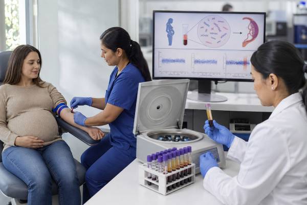
Cell-free DNA prenatal testing is a blood screening test that estimates the chance of selected fetal chromosome conditions, especially trisomy 21, trisomy 18, and trisomy 13. It analyzes short DNA fragments in the pregnant person’s bloodstream. Most of the pregnancy-related fragments come from the placenta, which usually has the same chromosome pattern as the fetus but not always. This is why cfDNA is highly accurate screening yet still not a diagnostic test. A high-chance result should be confirmed with chorionic villus sampling or amniocentesis before irreversible decisions are made. Testing can usually begin at about 10 weeks, requires no fasting, and does not increase miscarriage risk. Results often return within one to two weeks. The report may say low chance, high chance, no result, or atypical finding. Understanding the condition screened, fetal fraction, laboratory method, and pregnancy context is essential because the meaning of the same result can differ by age, ultrasound findings, twins, and the condition’s prevalence.
- cfDNA is the most sensitive screening test for trisomies 21, 18, and 13, but it cannot confirm a fetal diagnosis.
- Testing is commonly available from 10 weeks of pregnancy using a routine maternal blood sample.
- A high-chance result needs genetic counseling, detailed ultrasound, and an offer of CVS or amniocentesis.
- A low-chance result greatly lowers risk but does not exclude every chromosome or genetic condition.
- A no-call result is not automatically reassuring and may require repeat sampling, ultrasound review, or diagnostic testing.
- Routine population screening for broad microdeletion panels is not recommended by current specialist guidance.
Table of Contents
- How cfDNA screening works
- Conditions and test options
- Timing, sample, and fetal fraction
- Accuracy and positive predictive value
- Reading cfDNA results
- Twins and special situations
- Follow-up after screening
- Choosing cfDNA wisely
How cfDNA screening works
Cell-free DNA consists of small DNA fragments released into the bloodstream as cells naturally break down. During pregnancy, maternal blood contains the pregnant person’s own cell-free DNA plus fragments from the placenta. Laboratories analyze the relative amount, pattern, or sequence of fragments from selected chromosomes.
The commonly used phrase “fetal DNA” can be misleading. The pregnancy-related DNA in the sample is mainly placental. In most pregnancies, placental and fetal chromosome patterns match, so the screen performs very well. In a small number, a chromosome change is present in the placenta but not the fetus, or in the fetus but not the sampled placental cells. This biological difference explains some false-positive and false-negative results.
Laboratories use several technical approaches. Massively parallel sequencing may count fragments from across the genome. Targeted sequencing focuses on selected chromosome regions. Single-nucleotide polymorphism methods compare patterns of genetic markers and may estimate fetal fraction differently. Each approach has strengths, limitations, and its own reporting language.
The test is also called noninvasive prenatal screening, NIPS, or noninvasive prenatal testing, NIPT. “Screening” is the more accurate word because the test estimates risk. It does not directly examine a complete set of fetal chromosomes and cannot replace a diagnostic sample.
cfDNA does not assess fetal anatomy. A low-chance result cannot substitute for first-trimester or second-trimester ultrasound. Structural abnormalities may arise from conditions not included in the screen, single-gene disorders, infections, environmental factors, or causes that remain unknown.
Conditions and test options
The strongest evidence supports screening for the three common autosomal trisomies:
- Trisomy 21: Down syndrome
- Trisomy 18: Edwards syndrome
- Trisomy 13: Patau syndrome
For these conditions, cfDNA generally has higher detection rates and fewer false positives than traditional serum screening. Performance is best for trisomy 21 and somewhat lower for trisomies 18 and 13.
Many laboratories offer sex chromosome screening for monosomy X, XXY, XYY, or XXX. Current guidance treats this as an opt-in choice that requires counseling. Accuracy varies by condition, and an unexpected result can reflect the fetus, placenta, or pregnant person. For example, an apparent monosomy X result may occasionally identify maternal mosaicism or an age-related loss of an X chromosome in some maternal blood cells.
Some panels add 22q11.2 deletion and other microdeletions. These conditions are rarer than the common trisomies, so even a technically good test can have a lower positive predictive value. Current maternal-fetal medicine guidance does not recommend routine general-population screening for every microdeletion. A person who wants broad information about fetal deletions and duplications should discuss diagnostic testing with prenatal chromosomal microarray rather than assuming a cfDNA panel provides equivalent coverage.
Genome-wide cfDNA products may report rare autosomal trisomies or large copy-number changes. Evidence, thresholds, and follow-up pathways are less standardized than for trisomies 21, 18, and 13. Rare findings are more likely to arise from confined placental mosaicism and may be associated with fetal growth problems even when the fetal chromosomes are normal.
Some commercial tests report fetal sex. This result is based on detection or absence of Y-chromosome material and can be affected by early gestational age, a vanished twin, transplant history, maternal chromosome findings, or sample error. Fetal sex reporting should not be confused with a complete assessment of sex development.
cfDNA cannot routinely screen for all inherited disorders, neural tube defects, autism, cerebral palsy, or most birth defects. A separate maternal serum AFP test or ultrasound may be needed for open neural tube defect screening.
Timing, sample, and fetal fraction
Most laboratories accept samples from about 10 weeks of pregnancy. Testing too early increases the chance that there will not be enough placental DNA to produce a result. The sample is taken from a vein in the arm, and fasting is not required. Routine medications usually do not interfere, although certain medical treatments or conditions can affect interpretation.
The proportion of pregnancy-related DNA in the blood sample is called fetal fraction. Despite the name, it mainly reflects placental DNA. Many assays need a minimum fetal fraction, often around 2% to 4%, but the exact threshold differs by laboratory and method.
Fetal fraction usually rises as pregnancy progresses. It may be lower with:
- Earlier gestational age
- Higher maternal body weight
- Some chromosome conditions, particularly trisomy 18, trisomy 13, and triploidy
- Placental dysfunction
- Certain autoimmune conditions or medications
- Sample handling or technical issues
A report may provide the fetal fraction, but some validated methods do not routinely display it. A number above the laboratory cutoff does not guarantee accuracy, and a low number does not prove that the fetus has a chromosome condition. It indicates whether the laboratory believes the sample met its quality requirements.
Results commonly return in 5–10 business days, although delays occur. A sample may need to be repeated if the tube is damaged, the blood volume is insufficient, the sample is drawn too early, or the laboratory cannot generate a reliable signal.
Before blood collection, confirm gestational age, number of fetuses, whether there was a vanished twin, egg donor or surrogate arrangement, and any history of organ transplant, stem-cell transplant, cancer, or known chromosome variation. These details can change whether the laboratory accepts the sample and how the result should be interpreted.
The blood draw itself poses no pregnancy-specific procedural risk beyond ordinary bruising, lightheadedness, or infection at the needle site. The important risks are informational: false reassurance, anxiety from a high-chance or atypical result, uncertain follow-up, and decisions made before diagnosis.
Accuracy and positive predictive value
Accuracy cannot be summarized by one percentage. Detection rate, false-positive rate, specificity, and positive predictive value describe different parts of test performance.
Detection rate, or sensitivity, is the proportion of affected pregnancies that screen high chance. For trisomy 21, large studies generally report detection above 99% under study conditions. Detection is slightly lower for trisomy 18 and lower again for trisomy 13. Real-world performance varies with laboratory quality, gestational age, fetal fraction, and pregnancy type.
Specificity is the proportion of unaffected pregnancies that screen low chance. cfDNA has high specificity for the common trisomies, which means false positives are uncommon. However, uncommon does not mean impossible.
Positive predictive value, or PPV, answers the question most people ask after a high-chance result: what is the chance the fetus is actually affected? PPV depends on both test performance and how common the condition is in the tested population. It is usually higher for trisomy 21 than for trisomy 13, sex chromosome conditions, or microdeletions. It also tends to be higher when maternal age, ultrasound, or prior history raises the starting chance.
For example, suppose a rare condition affects 1 in 1,000 pregnancies. Even a test with strong sensitivity and specificity may generate a meaningful number of false positives compared with the small number of true cases. This is why a laboratory may advertise a high detection rate while an individual positive result still requires diagnostic confirmation.
A negative predictive value is generally very high for common trisomies because most pregnancies are unaffected and the test detects most affected cases. Yet a low-chance result can be false because of low placental contribution, placental-fetal differences, uncommon chromosome structures, mosaicism, or technical limits.
Do not compare laboratories using one headline accuracy number. Ask which condition the number describes, whether it came from singleton or twin pregnancies, how no-call samples were handled, and whether the study population resembled the current pregnancy.
The chance that a positive result is correct also changes as pregnancy information accumulates. A positive trisomy 21 result followed by an ultrasound showing several typical features has a different overall probability from the same screen followed by a normal detailed scan, although neither ultrasound pattern can replace diagnostic testing. Conversely, some affected fetuses have no visible ultrasound signs at the time of screening. The laboratory number and the ultrasound should be combined, not used to cancel each other out.
False-negative results deserve attention because they are less visible than false positives. They can occur when the abnormal cell line is present mainly in the fetus but not the placenta, when the placental DNA contribution is small, when a chromosome change is structurally unusual, or when the algorithm does not include the condition. A reassuring result should therefore be documented as “low chance for the screened conditions,” not “all chromosomes normal.”
Pretest probability is not limited to maternal age. A previous affected pregnancy, a parental chromosome rearrangement, a suggestive ultrasound finding, or a family history may raise the starting chance. Egg-donor age may be more relevant than the pregnant person’s age for some calculations. Accurate background information helps the laboratory and clinician provide a more meaningful interpretation.
Reading cfDNA results
Laboratories may use “low risk,” “high risk,” “screen negative,” “screen positive,” “aneuploidy detected,” “no result,” or “atypical finding.” The complete report matters more than a portal alert or phone summary.
Low chance or screen negative
A low-chance result means the observed DNA pattern did not suggest the screened condition. It substantially lowers the chance of trisomy 21, 18, or 13 but does not reduce it to zero. The result says nothing definitive about conditions outside the panel.
Routine prenatal ultrasound remains important. If a later scan finds a major anomaly, diagnostic testing should be offered even after low-chance cfDNA. Repeating cfDNA is not a substitute for diagnostic testing when ultrasound shows a significant structural concern.
High chance or screen positive
A high-chance result means the DNA pattern is associated with an increased chance of the named condition. It is not confirmation. The report may include a laboratory-specific PPV estimate, which should be interpreted using maternal age, gestational age, ultrasound, and the condition involved.
False-positive causes include confined placental mosaicism, maternal chromosome variants, a vanished twin, organ transplant, laboratory error, or rarely maternal cancer. Most positive results do not imply cancer; that possibility is considered mainly when the pattern is unusual, involves multiple chromosomes, or cannot be explained by a fetal condition.
No result or low fetal fraction
A no-call report means the laboratory could not issue a reliable risk assessment. Some no-call results are technical, but low fetal fraction can be associated with an increased chance of aneuploidy or placental problems. Current guidance supports genetic counseling, comprehensive ultrasound assessment, and an offer of diagnostic testing rather than treating the result as negative.
A repeat draw may be reasonable in selected cases, especially when the first sample was early and ultrasound is reassuring. Repeating can delay diagnosis and may fail again, particularly when maternal weight is high or a biological factor persists.
Atypical or outside-the-scope finding
An atypical result may indicate a complex DNA pattern that does not fit the standard screen. The laboratory may suspect a maternal finding, placental mosaicism, or a chromosome change not covered by its validated reporting categories. The next step should be individualized with genetics and maternal-fetal medicine input. Sending the same blood to another screening laboratory may not resolve the underlying biology.
Twins and special situations
cfDNA can be used in many twin pregnancies, but interpretation differs from singleton testing. The blood contains combined placental DNA from both fetuses, and the test may not determine which twin contributed an abnormal signal. Performance for trisomy 21 is supported by growing evidence; data for trisomies 18 and 13 are more limited because affected twin pregnancies are uncommon.
In dizygotic twins, each placenta may contribute a different amount of DNA. If one twin has a low contribution, the overall fetal fraction can appear adequate while the affected twin’s signal is weak. Some SNP-based methods attempt to estimate individual fetal fractions, but capabilities vary.
A vanished twin can release DNA into maternal blood for weeks and cause a false-positive result or fetal sex discrepancy. The timing of the loss and the testing platform matter. Diagnostic testing may be the clearest option when accurate chromosome information is needed.
Higher-order multiples have less supporting evidence, and current specialist guidance does not recommend routine cfDNA screening in triplets or more. Sex chromosome screening in multiple pregnancies is also less reliable and may not be offered.
Donor-egg pregnancies can usually be screened, but the laboratory must know that donor eggs were used because age-based calculations and SNP interpretation may differ. Gestational carrier arrangements require correct identification of the genetic and pregnant contributors. A recent bone marrow or stem-cell transplant can make the blood DNA reflect the donor and may invalidate testing. Solid-organ transplant and maternal chromosome conditions can also alter results.
Maternal cancer is rare, but tumors can release abnormal DNA fragments. An unusual pattern involving several chromosomes may trigger a maternal evaluation after fetal explanations are addressed. This is not a standard cancer screening use of cfDNA, and follow-up should be coordinated carefully to avoid unnecessary testing.
Follow-up after screening
The proper follow-up depends on the result category and the pregnancy’s ultrasound findings.
After a high-chance result, the usual sequence is:
- Review the complete laboratory report with an obstetric clinician or genetic counselor.
- Obtain or schedule a detailed ultrasound appropriate to gestational age.
- Discuss diagnostic testing with CVS or amniocentesis.
- Select the laboratory analysis that can confirm the suspected condition.
- Make pregnancy or treatment decisions only after diagnostic clarification whenever possible.
CVS is generally performed around 10–13 weeks and samples placental tissue. Because cfDNA also reflects placenta, a discordant placental result can occasionally remain uncertain. Amniocentesis, usually from 15 weeks, tests amniotic-fluid cells and may be preferred for certain results, especially when confined placental mosaicism is a concern.
After a positive screen for trisomy 21, 18, or 13, either procedure may be suitable depending on gestational age, ultrasound, and the specific chromosome. After a rare autosomal trisomy or some sex chromosome results, the clinician may recommend amniocentesis because placental mosaicism is more common.
A normal diagnostic result after positive cfDNA is not simply “the screen was wrong.” The discordance may reveal placental mosaicism that warrants growth surveillance or a maternal chromosome finding that deserves separate review. The team should explain whether any pregnancy monitoring remains appropriate.
After a low-chance result, continue standard prenatal care and ultrasound. Diagnostic testing remains available to anyone who wants it or develops a new indication. After a repeated no-call result, proceeding to amniocentesis may prevent further delay when gestational age permits.
Choosing cfDNA wisely
The best screening choice depends on what information a person wants and how they would use it. cfDNA offers excellent screening for common trisomies with one blood draw, but it does not provide the early ultrasound measurements, neural tube defect assessment, or broad diagnostic information that other tests can provide.
Before ordering, ask:
- Which chromosome conditions are included by default?
- Is sex chromosome screening optional?
- Are microdeletions or genome-wide findings included, and can they be declined?
- Does the laboratory report fetal fraction?
- How are no-call results managed?
- What is the laboratory’s PPV for each condition, not just its sensitivity?
- Does insurance cover the test and the follow-up diagnostic procedure?
- Will results be reviewed by a clinician rather than released without counseling?
Using multiple screening methods at the same time can create conflicting estimates and is generally discouraged. A person may choose cfDNA or a traditional serum-and-ultrasound pathway after counseling. Ultrasound should still be performed regardless of the screening method.
Consider whether a screening answer is sufficient. Someone with a known familial chromosome rearrangement, a major fetal anomaly, or a strong desire for comprehensive chromosome information may prefer diagnostic testing. Someone who wants to avoid invasive testing may choose cfDNA while accepting that a high-chance result will create another decision about confirmation.
No result should be interpreted in isolation. The report is one part of a larger prenatal picture that includes gestational age, family history, ultrasound, prior screening, clinical history, and personal values. Clear, individualized pretest counseling reduces the chance that a high-chance, no-call, or unexpected maternal finding arrives without a clear plan.
References
- Society for Maternal-Fetal Medicine Consult Series #74: Cell-free DNA screening for aneuploidies: Updated guidance 2025 (Guideline)
- Screening for Fetal Chromosomal Abnormalities 2026 (Practice Advisory)
- Noninvasive prenatal screening for fetal chromosome abnormalities in a general-risk population: An evidence-based clinical guideline of the American College of Medical Genetics and Genomics (ACMG) 2023 (Guideline)
- A systematic review and meta-analysis of cell-free DNA testing for detection of fetal sex chromosome aneuploidy 2023 (Systematic Review)
- Diagnostic accuracy of cell-free fetal DNA in maternal blood in detecting chromosomal anomalies in twin pregnancy: systematic review and meta-analysis 2025 (Systematic Review)
- Prenatal Genetic Screening 2025 (Review)
Disclaimer
This article is for education and does not replace individualized prenatal counseling. cfDNA is a screening test, and high-chance, atypical, or repeated no-call results require review with an obstetric professional, maternal-fetal medicine specialist, or genetic counselor. Do not make irreversible pregnancy decisions from cfDNA alone without being offered diagnostic confirmation.