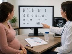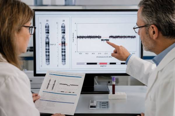
A 22q11.2 deletion syndrome genetic test looks for a missing segment of chromosome 22. The deletion can affect the heart, palate, immune system, calcium regulation, development, learning, and mental health, but the pattern differs greatly from one person to another. The condition is often called 22q11.2 deletion syndrome; older names include DiGeorge syndrome and velocardiofacial syndrome. Testing may begin after a child or adult develops suggestive features, after an ultrasound finds certain fetal abnormalities, or after prenatal screening reports an increased chance of the deletion. A screening result cannot establish the diagnosis. Confirmation usually requires chromosomal microarray or another diagnostic method performed on an appropriate sample. Results also have implications for parents, siblings, and future pregnancies because an affected person has a 50% chance of passing the deletion to each child. Genetic counseling helps families understand the result, its limits, and the medical follow-up that may be appropriate.
- The diagnostic finding is a deletion at chromosome region 22q11.2, usually detected by chromosomal microarray.
- A positive prenatal cfDNA result is not a diagnosis and should be followed by genetic counseling and diagnostic testing.
- Most typical deletions arise for the first time in the affected person, although some are inherited from a parent with mild or unrecognized features.
- A parent with the deletion has a 50% chance of passing it on in each pregnancy, but severity cannot be predicted from the parent’s symptoms.
- A negative targeted test may miss an atypical deletion if the method does not cover the relevant part of 22q11.2.
- After diagnosis, evaluation commonly includes the heart, calcium, thyroid, immune function, hearing, palate, kidneys, growth, and development.
Table of Contents
- What the 22q11.2 deletion test detects
- Who may be tested and why
- Testing methods and samples
- Prenatal screening and diagnostic testing
- How to understand the results
- Inheritance and family risk
- Medical follow-up after diagnosis
- Questions and next steps
What the 22q11.2 deletion test detects
The test looks for loss of genetic material on the long arm of chromosome 22, at a location labeled q11.2. This region contains many genes that help guide early development. Missing one copy of several genes can disrupt multiple body systems, which is why 22q11.2 deletion syndrome can appear very different from one person to another.
The most common deletion spans about 2.5 megabases on chromosomal microarray reports, although it was historically described as a 3-megabase deletion. It includes roughly 40 genes. Smaller “nested” deletions and uncommon deletions with different breakpoints also occur. TBX1 is one important gene in the region, especially for development of the heart, great vessels, thymus, and structures of the face and palate, but the syndrome results from loss of a group of genes rather than a single TBX1 change.
A laboratory report may describe the result as a pathogenic copy-number loss, a heterozygous deletion, or a deletion involving 22q11.21. “Heterozygous” means one chromosome 22 has the expected region and the other is missing it. The report may also include genomic coordinates, deletion size, genes involved, and the laboratory’s classification.
The name “DiGeorge syndrome” is still widely recognized, but 22q11.2 deletion syndrome is more precise when the chromosome deletion has been confirmed. Older diagnoses such as velocardiofacial syndrome, conotruncal anomaly face syndrome, and some cases of Cayler cardiofacial syndrome are now understood to fall within the same broad condition.
A confirmed deletion does not predict a fixed set of symptoms. Some people have a major heart defect and immune deficiency in infancy. Others have subtle speech, learning, psychiatric, endocrine, or reproductive issues and are diagnosed only after a child or other relative is tested. The result identifies the genetic cause; clinical evaluation determines how it affects the individual.
Who may be tested and why
Testing is appropriate when medical findings, family history, or prenatal information raise concern for a 22q11.2 deletion. No single feature is present in every affected person, so clinicians often recognize a pattern rather than one defining sign.
In infants and children, common reasons for testing include:
- A conotruncal heart defect, such as tetralogy of Fallot, interrupted aortic arch, truncus arteriosus, or certain ventricular septal defects
- Low calcium, seizures related to hypocalcemia, or reduced parathyroid function
- A small or poorly functioning thymus, low T-cell numbers, repeated infections, or an abnormal newborn T-cell receptor excision circle result
- Cleft palate, a submucous cleft, nasal speech, swallowing difficulty, or velopharyngeal dysfunction
- Feeding problems, poor growth, developmental delay, learning differences, or speech delay
- Kidney, skeletal, hearing, airway, or gastrointestinal abnormalities occurring with other suggestive findings
- A known deletion in a parent, sibling, or other close relative
Adolescents and adults may be evaluated because of learning problems, anxiety, attention-deficit/hyperactivity disorder, autism-related traits, psychosis, seizures, low calcium, thyroid disease, early-onset Parkinson disease, infertility, or a child with the deletion. Mild facial or palatal findings can be overlooked, and an adult may have no history of major congenital disease.
Testing may also be considered when a fetal ultrasound shows a heart defect, absent or small thymus, cleft palate, kidney anomaly, growth restriction, excess amniotic fluid, or a combination of structural findings. A normal ultrasound does not rule out the condition because many affected fetuses have no obvious prenatal structural abnormality.
A clinician choosing the broader chromosomal microarray test may identify 22q11.2 deletion syndrome even when it was not the original suspected diagnosis. This is common in children evaluated for developmental delay, congenital anomalies, or autism.
Testing should be accompanied by counseling that explains the possible results. The range of outcomes is wide, and neither deletion size nor a parent’s clinical picture can reliably forecast a child’s exact medical or developmental course.
Testing methods and samples
Chromosomal microarray is the usual first-line diagnostic method when 22q11.2 deletion syndrome is suspected but the family’s exact deletion is not already known. It surveys the genome for missing or extra DNA segments and can define the size and boundaries of the deletion more accurately than older targeted methods.
Several methods may appear on a report:
| Method | Best use | Main limitation |
|---|---|---|
| Chromosomal microarray | Initial diagnosis; defines many typical and atypical deletions | Usually does not identify balanced rearrangements and may detect unrelated copy-number findings |
| FISH | Rapid targeted testing or testing relatives for a known deletion covered by the probe | Can miss atypical deletions outside the probe location |
| MLPA or quantitative PCR | Targeted deletion confirmation and family testing | Coverage depends on the probes or targets included |
| Karyotype | Detecting large chromosome changes or a suspected translocation | Typical 22q11.2 deletions are too small to see reliably |
For a child or adult, the sample is usually blood, although saliva or a cheek swab may be accepted by some laboratories. Blood is often preferred when mosaicism, immune testing, or additional chromosome studies are being considered. Preparation is simple: fasting is not required, and medications usually do not affect the DNA result.
Prenatal diagnostic samples come from chorionic villi obtained by CVS or amniotic fluid obtained by amniocentesis. A laboratory may perform microarray directly or after cell culture. Maternal cell contamination testing may be added to confirm that the DNA result represents the pregnancy rather than maternal cells in the sample.
Turnaround time varies from several days for a rapid targeted assay to two or more weeks for a full microarray, depending on the laboratory and whether cell culture is needed. Families should ask whether the ordered method can detect atypical deletions and whether it can clarify deletion size.
A routine karyotype test can be normal in a person who has 22q11.2 deletion syndrome. This is an important source of confusion: a “normal chromosome test” from years ago may have used a method that lacked the resolution to see a microdeletion.
Prenatal screening and diagnostic testing
Prenatal testing can either estimate the chance of a 22q11.2 deletion or diagnose it. The distinction is essential because screening and diagnosis answer different questions.
Some cell-free DNA laboratories offer 22q11.2 microdeletion screening from a maternal blood sample, often from about 10 weeks of pregnancy. The test analyzes placental DNA fragments circulating in the pregnant person’s blood. It does not directly test fetal cells, and performance for 22q11.2 deletion is lower and less certain than performance for trisomy 21.
Professional guidance does not support routine population screening for all microdeletion syndromes. When 22q11.2 screening is offered, pretest counseling should cover the condition’s variable expression, the chance of false-positive and false-negative results, and the need for diagnostic confirmation. A person seeking the broadest assessment of fetal copy-number changes should discuss diagnostic testing with microarray rather than relying on prenatal microdeletion screening.
A high-chance or positive cfDNA result may reflect:
- A true fetal deletion
- A deletion confined to the placenta
- A maternal 22q11.2 deletion, sometimes previously unknown
- A technical or statistical false-positive result
- Less commonly, another maternal biological finding that changes the DNA pattern
The next step is not to make medical decisions from the screen alone. Genetic counseling, a detailed ultrasound, and diagnostic testing should be offered. Chorionic villus sampling is generally performed at about 10 to 13 weeks and samples placental tissue. Amniocentesis is usually performed from about 15 weeks and samples amniotic fluid containing fetal cells. Because CVS tests placental tissue, an unexpected or mosaic result may occasionally require amniocentesis for clarification.
When ultrasound has identified one or more fetal structural anomalies, diagnostic testing with chromosomal microarray is usually more informative than a targeted cfDNA screen. Microarray can detect the 22q11.2 deletion and many other clinically significant deletions or duplications.
A low-chance cfDNA result reduces risk but does not exclude the syndrome. Diagnostic testing remains an option when ultrasound findings or family history create concern. When a parent is known to carry the deletion, targeted prenatal diagnosis or preimplantation genetic testing may be considered because each pregnancy has a 50% chance of inheriting it.
How to understand the results
A laboratory result should be interpreted in the context of the method, sample, clinical findings, and family history. The main result categories are positive, negative, uncertain, and occasionally mosaic or technically limited.
Positive or pathogenic deletion
A positive diagnostic result means the laboratory identified a deletion judged to cause 22q11.2 deletion syndrome. The report usually states the genomic coordinates and estimated size. It may list genes within the deleted interval and recommend testing the parents.
The result confirms the genetic diagnosis, but it does not show which features will develop or how severe they will be. Even relatives with the same deletion can have very different health and learning profiles. Medical decisions should therefore be based on the person’s actual evaluations rather than assumptions from the genetic result alone.
Negative result
A negative result means the tested method did not detect the deletion it was designed to find. Its meaning depends on the assay. A negative microarray makes the common 22q11.2 deletion unlikely. A negative FISH test may be less complete because one probe can miss a smaller or differently located deletion.
If clinical suspicion remains high, a genetics professional may review the original test, order a broader microarray, consider sequencing for another syndrome, or evaluate for a different copy-number change. Conditions involving CHD7, JAG1, or other genes can produce overlapping findings.
A negative prenatal cfDNA screen is not equivalent to a negative diagnostic microarray. Screening sensitivity is not 100%, and not all deletion sizes are detected equally.
Variant or finding of uncertain significance
A microarray can occasionally find a copy-number change near 22q11.2 whose clinical meaning is unclear. This is not the same as a confirmed typical deletion. Testing both parents, reviewing databases, and reassessing the finding over time may help. Care should not be based on an uncertain result alone unless the clinical team identifies a separate reason for evaluation.
Mosaic or atypical result
Mosaicism means the deletion appears in some cells but not others. The percentage detected in blood or prenatal tissue may not match other tissues, so symptom prediction remains difficult. An atypical deletion may overlap part of the usual region or lie farther toward the end of chromosome 22. Its interpretation depends on the exact genes involved and published evidence.
Before accepting a result summary such as “positive for DiGeorge,” obtain the complete laboratory report. The method, coordinates, size, and classification often determine whether more testing is needed.
Inheritance and family risk
22q11.2 deletion syndrome follows an autosomal dominant inheritance pattern. One deleted copy of the region is enough to cause the condition. Most people with the common deletion are the first in their family to have it, but a meaningful minority inherited it from a parent.
For the typical large deletion, more than 90% of cases are new, or de novo, in the affected person. About 10% are inherited. Some smaller nested deletions are inherited more often. A parent may have mild learning problems, nasal speech, low calcium, anxiety, a heart finding, or no recognized symptoms, so appearance and medical history alone cannot reliably determine whether a parent carries the deletion.
Parental testing should use a method that can detect the child’s exact deletion. When neither parent has it in blood, recurrence risk in another pregnancy is low but not zero because rare germline mosaicism can occur. Germline mosaicism means some egg or sperm cells carry the deletion even though routine blood testing is negative.
When one parent carries the deletion, each pregnancy has:
- A 50% chance of inheriting the deletion
- A 50% chance of not inheriting it
The probabilities reset with every pregnancy. A mildly affected parent can have a child with serious congenital disease, and a more affected parent can have a child with fewer features. There is no reliable way to predict severity from the family member who transmitted the deletion.
Adult relatives may choose targeted testing after counseling. Testing can clarify personal medical surveillance and reproductive risk, but it may also uncover health concerns that were not previously suspected. Children who have no symptoms are usually tested when the result would change medical care, developmental support, or family planning.
Reproductive options may include natural conception with prenatal diagnosis, IVF with preimplantation genetic testing, donor egg or sperm, adoption, or choosing not to test. No single choice is right for every family.
Medical follow-up after diagnosis
A diagnosis should lead to a coordinated baseline assessment rather than a search for every possible complication at once. The person’s age, symptoms, prior evaluations, and deletion details guide the plan.
Common early evaluations include:
- Cardiology: echocardiogram and clinical assessment, even when no murmur is obvious
- Calcium and parathyroid function: calcium may fall during infancy, illness, surgery, puberty, or pregnancy
- Immune system: lymphocyte subsets, immunoglobulins, infection history, and vaccine planning when indicated
- Palate, feeding, and speech: evaluation for cleft palate, velopharyngeal dysfunction, swallowing problems, and hypernasal speech
- Hearing and vision: age-appropriate testing because deficits can worsen speech and learning
- Thyroid function: periodic thyroid-stimulating hormone and free thyroxine testing
- Kidneys: renal ultrasound when not previously completed
- Growth and development: developmental screening, school support, speech-language therapy, and neuropsychological assessment
- Mental health: monitoring for anxiety, attention problems, mood symptoms, psychosis, and changes in function
Immune findings deserve special attention before live vaccines. Most affected children do not have complete absence of T-cell function, but the small subgroup with severe thymic deficiency requires specialist care and protection from certain infections and blood products. Vaccine decisions should be based on immune testing, not the genetic diagnosis alone.
Low calcium may cause tingling, muscle cramps, tremor, unusual movements, or seizures. Urgent medical assessment is appropriate for a seizure, severe breathing difficulty, bluish color, fainting, signs of serious infection, or symptoms of marked hypocalcemia.
Surveillance changes with age. Children need repeated developmental, speech, hearing, dental, spine, thyroid, calcium, and immune review. Adults may need attention to psychiatric health, cardiovascular issues, endocrine problems, pregnancy-related calcium needs, seizures, and movement symptoms. A specialized 22q11.2 clinic can coordinate care, but a well-informed primary clinician and local specialists can also build an effective plan.
Questions and next steps
The most useful next step depends on whether the result came from screening, diagnostic testing, or a relative’s report.
After a positive prenatal screen, ask:
- Was this a screening test or a diagnostic test?
- What positive predictive value applies to this laboratory and pregnancy?
- Is CVS or amniocentesis more appropriate at the current gestational age?
- Will chromosomal microarray be performed on the diagnostic sample?
- Could the result reflect a maternal deletion, and should maternal testing be discussed?
After a new diagnostic result in a child or adult, ask for a copy of the complete report and a genetics referral. Confirm which baseline evaluations have already been done and which need scheduling. A written care plan is helpful because follow-up spans several specialties.
Parents should ask whether their testing must be microarray, FISH, MLPA, or another targeted method. The assay should match the affected relative’s deletion. If parental tests are negative, discuss the small residual recurrence risk rather than assuming it is exactly zero.
A result can bring relief, grief, uncertainty, or all three. The diagnosis may explain years of unrelated-seeming medical and learning concerns, but it also introduces information about future health and relatives. Genetic counseling provides space to review these implications without treating the result as a prediction of a fixed life course.
Keep the laboratory report, family testing results, cardiac records, immune studies, calcium history, and developmental assessments together. These records matter during surgery, pregnancy, emergency care, school planning, and transition from pediatric to adult services. The chromosome finding stays the same, but its medical relevance can change as a person grows.
References
- 22q11.2 Deletion Syndrome 2025 (Review)
- Updated clinical practice recommendations for managing children and adolescents with 22q11.2 deletion syndrome 2023 (Guideline)
- Updated clinical practice recommendations for managing adults with 22q11.2 deletion syndrome 2023 (Guideline)
- Clinical Practice Guidelines for the Immunological Management of Chromosome 22q11.2 Deletion Syndrome and Other Defects in Thymic Development 2023 (Guideline)
- Society for Maternal-Fetal Medicine Consult Series #74: Cell-free DNA screening for aneuploidies: Updated guidance 2025 (Guideline)
- Screening for Fetal Chromosomal Abnormalities 2026 (Practice Advisory)
Disclaimer
This information is educational and does not replace individualized care from a genetics, obstetric, pediatric, or other qualified healthcare professional. Prenatal screening cannot diagnose 22q11.2 deletion syndrome; confirmatory testing and genetic counseling are needed before making pregnancy or medical decisions. Seek urgent care for seizures, severe breathing problems, blue color, fainting, or signs of a serious infection.