
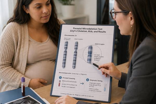
A prenatal microdeletion test can mean either a maternal blood screening test or diagnostic testing of placental or fetal cells. These approaches are not interchangeable. Cell-free DNA screening can estimate the chance of 22q11.2 deletion syndrome, but it cannot confirm that the fetus has the deletion. Chorionic villus sampling or amniocentesis followed by chromosomal microarray can establish whether the deletion is present in the tested pregnancy. The 22q11.2 deletion removes a small segment of chromosome 22 and can affect the heart, palate, immune system, calcium regulation, development, learning, and later mental health. Prenatal presentation is highly variable: some fetuses have major cardiac or other structural findings, while others have no recognizable ultrasound anomaly. Most cases arise de novo, although an affected parent can pass the deletion to each pregnancy. Screening performance is less straightforward than for trisomy 21 because the condition is uncommon and a positive signal may come from the fetus, placenta, or mother. A high-chance result needs specialist counseling and diagnostic confirmation before irreversible pregnancy decisions.
- Cell-free DNA for 22q11.2 deletion is screening; CVS or amniocentesis with microarray is diagnostic.
- Professional guidance differs: ACMG conditionally supports offering 22q11.2 screening, while SMFM advises against routine population microdeletion screening.
- A positive result may reflect a fetal, placental, or maternal deletion.
- Chromosomal microarray is generally the preferred diagnostic confirmation method.
- Ultrasound can raise suspicion but cannot identify all affected fetuses.
- A confirmed deletion has a broad outcome range that cannot be predicted precisely before birth.
Table of Contents
- What 22q11.2 deletion syndrome is
- Screening versus diagnostic testing
- When testing is considered
- How cell-free DNA results are interpreted
- Confirming a screening result
- Meaning of a confirmed deletion
- Limitations, recurrence, and next steps
What 22q11.2 deletion syndrome is
22q11.2 deletion syndrome is caused by loss of DNA on the long arm of chromosome 22. It includes conditions historically called DiGeorge syndrome and velocardiofacial syndrome. Those names described different clinical presentations before genetic testing showed that many shared the same underlying deletion.
Most affected people have a recurrent deletion of about 3 megabases, while others have a smaller nested or atypical deletion. The missing interval contains multiple genes, including TBX1, which contributes to development of the heart, great vessels, palate, thymus, and other structures. Deletion size alone does not reliably predict severity because clinical outcome also depends on the exact genes involved, background genetic variation, development, and chance.
Possible features include conotruncal heart defects, aortic arch abnormalities, cleft or submucous cleft palate, velopharyngeal dysfunction, thymic hypoplasia, immune deficiency, low calcium caused by hypoparathyroidism, feeding difficulties, kidney differences, hearing loss, growth differences, developmental delay, learning disability, and increased risk of psychiatric conditions later in life. No affected person is expected to have every feature, and two relatives with the same deletion can have very different needs.
Certain prenatal cardiac findings are particularly suggestive: tetralogy of Fallot, interrupted aortic arch type B, truncus arteriosus, and some aortic arch or ventricular outflow abnormalities. Additional clues can include a small or absent thymus, cleft palate, kidney anomalies, polyhydramnios, growth restriction, clubfoot, or multiple malformations. Yet many affected fetuses have normal or nonspecific ultrasound findings. A normal anatomy scan therefore does not exclude the syndrome.
Maternal age is not a major risk factor. Most cases arise de novo in the egg, sperm, or early embryo. A minority are inherited from a parent who may have obvious, subtle, or previously unrecognized features. If a parent carries the deletion, each pregnancy has a 50% chance of inheriting it, but the child’s severity cannot be predicted from the parent’s presentation.
A routine karyotype usually cannot see the deletion because it is below microscopic resolution. Chromosomal microarray, targeted FISH, MLPA, or another validated copy-number method is required. Microarray is usually preferred prenatally because it can define the deleted segment and assess the rest of the genome for other clinically significant gains or losses.
Screening versus diagnostic testing
A maternal blood test for 22q11.2 deletion analyzes cell-free DNA fragments. Most circulating fragments are maternal; the pregnancy-related component arises mainly from the placenta. The laboratory uses counting or SNP-based methods to estimate whether the 22q11.2 region appears underrepresented. It does not directly examine fetal cells, so it remains screening.
Diagnostic testing begins with chorionic villus sampling or amniocentesis. The laboratory then analyzes placental or fetal DNA, usually with chromosomal microarray. This can establish whether the deletion is present in the sampled cells and describe its genomic boundaries.
The distinction is especially important for a rare condition. Even a screening assay with good sensitivity and specificity can generate a meaningful proportion of false-positive results when prevalence is low. Positive predictive value—the probability that a pregnancy with a positive screen is actually affected—is therefore more useful for counseling than a sensitivity claim alone.
Commercial assays differ. Some target only the common 3-megabase deletion; others can detect selected smaller deletions. Minimum detectable size, fetal-fraction requirements, twin performance, algorithms, and handling of maternal deletions vary. Results from one platform cannot automatically be generalized to another. The exact laboratory report and technical specification matter.
Professional recommendations are not identical. The 2023 ACMG evidence-based guideline conditionally suggested offering 22q11.2 cell-free DNA screening after discussion of benefits and limitations. The 2025 SMFM consult, endorsed by ACOG, recommends against routine general-population screening for any microdeletion. It states that patients who specifically choose 22q11.2 screening should receive appropriate pretest counseling and that those seeking information about fetal copy-number variants should be offered diagnostic testing with microarray.
These positions weigh evidence, false-positive burden, access to counseling, and the potential value of early detection differently. They do not imply that the syndrome is unimportant. Patients deserve an honest explanation that professional guidance differs rather than a simplified claim that screening is universally recommended or universally inappropriate.
A diagnostic test can also be chosen without first doing a blood microdeletion screen. This may be more direct when fetal imaging is strongly suggestive, a parent carries the deletion, or a patient wants the most definitive available answer and accepts the procedural considerations.
When testing is considered
Testing may first appear as an optional part of an expanded cell-free DNA panel. Consent should separate 22q11.2 deletion from trisomies 21, 18, and 13 because prevalence, performance, confirmatory testing, and outcome prediction differ. An automatically bundled panel is not a substitute for informed choice.
A suggestive ultrasound is a stronger clinical reason to discuss diagnostic testing. Conotruncal heart disease, interrupted aortic arch, an absent or small thymus, or cardiac findings combined with palate, kidney, skeletal, or growth abnormalities may prompt chromosomal microarray. Even when 22q11.2 deletion is the leading concern, genome-wide microarray is useful because other copy-number changes can produce a similar phenotype.
Fetal echocardiography can refine the indication. A screening ultrasound may show an outflow-tract concern that later proves normal, or it may reveal a more specific aortic arch pattern. Better phenotyping improves both test selection and counseling. Fetal MRI may be considered when brain or other complex structural findings are present, but imaging cannot replace molecular diagnosis.
A known familial deletion is another indication. If a parent has a confirmed 22q11.2 deletion, targeted prenatal diagnosis can determine whether the fetus inherited it. The original laboratory report should be reviewed because an atypical deletion may require customized probe placement or genomic coordinates. If a previous child or pregnancy was affected and the parents have not been tested, parental analysis can clarify recurrence risk.
A positive, high-chance, or atypical cell-free DNA result can lead to diagnostic testing. The laboratory may sometimes suspect a maternal rather than placental source. In that situation, maternal confirmation and fetal testing may both be relevant. The sequence depends on gestational age, ultrasound findings, the reported pattern, and the patient’s preferences.
A low-chance common-aneuploidy screen does not exclude 22q11.2 deletion unless the deletion was specifically included. Even a targeted low-chance result cannot override strongly suggestive fetal findings. Diagnostic testing should remain available when ultrasound indicates a genomic disorder.
Some patients decline testing because the result would not change pregnancy management or because they prefer evaluation after birth. Others want a prenatal diagnosis to prepare for delivery near pediatric cardiology, cardiac surgery, immunology, endocrinology, or neonatal intensive care. Both choices can be reasonable when based on accurate, nondirective counseling.
How cell-free DNA results are interpreted
During pregnancy, short DNA fragments circulate in maternal plasma. The placental contribution is often called the fetal fraction, although it primarily reflects trophoblast DNA. A 22q11.2 assay looks for a statistical pattern consistent with missing material in the targeted region.
Counting-based methods compare the number of fragments mapping to 22q11.2 with expected values. SNP-based methods evaluate patterns of genetic markers and may model maternal and placental contributions differently. Their capabilities vary in twins, donor-egg pregnancies, vanished twins, and maternal copy-number changes.
A positive, high-chance, or deletion detected result means the screening pattern crossed the laboratory threshold. It does not prove a fetal deletion. The signal may reflect a fetal and placental deletion, a placental-only change, a maternal deletion, or a false-positive technical result.
The report may provide a positive predictive value. That estimate depends on prevalence assumptions, the validation population, and the specific assay. Ultrasound findings and family history change the prior probability. A patient with a characteristic fetal heart defect has a different context from a patient with a normal scan and an unselected positive screen.
A low-chance result lowers the probability of a deletion the assay is designed to detect. It does not exclude atypical breakpoints, a smaller deletion below resolution, mosaicism, low-fraction false negatives, or another genetic condition with similar fetal findings. “Low chance” should not be translated as “the fetus does not have the syndrome.”
A no-result or no-call outcome means the laboratory could not classify the sample reliably. Causes include low placental fraction, early gestation, sample quality, or an unusual genomic pattern. It is not a negative result. Options include repeat cfDNA, detailed ultrasound, maternal evaluation, or diagnostic testing, depending on gestational age and clinical risk.
An atypical or outside-the-scope result may reflect a complex maternal or placental copy-number pattern. Direct communication with the laboratory and review by a genetics professional are often more useful than automatically repeating the same assay.
A possible maternal deletion deserves sensitive counseling. Adults with 22q11.2 deletion may have congenital heart disease, palatal history, low calcium, immune issues, learning differences, anxiety, or few recognized features. Confirmatory maternal testing can guide the mother’s healthcare and clarify fetal risk. The finding should not be disclosed casually or interpreted solely through the pregnancy.
Confirming a screening result
A positive screen should be followed by an offer of diagnostic testing before irreversible pregnancy decisions. Choosing between CVS and amniocentesis requires more than comparing which can be performed earlier.
CVS analyzes placental villi. A confirmed nonmosaic deletion in CVS usually indicates fetal involvement, but confined placental mosaicism is possible. If the result is mosaic, atypical, or discordant with maternal testing and ultrasound, amniocentesis may be recommended to clarify the fetal result.
Amniocentesis analyzes cells in amniotic fluid, which more directly represent the fetus. When the screen is positive but ultrasound is normal and placental discordance is a concern, some patients and clinicians prefer amniocentesis. Waiting for a later procedure must be balanced against the value of earlier information and local gestational timelines.
Chromosomal microarray is usually the preferred diagnostic laboratory method. It can detect the deletion, define its size and gene content, and identify other pathogenic deletions or duplications. A targeted FISH test may be faster but can miss an atypical deletion if its probe lies outside the missing interval. A standard prenatal karyotype can be normal because its resolution is too low.
The sample may also undergo rapid aneuploidy testing if common trisomies remain part of the differential diagnosis. Laboratories differ in whether microarray is performed directly on uncultured cells or after culture. Maternal cell contamination testing may be needed, especially for blood-stained or low-cell samples.
If fetal microarray confirms the deletion, both parents should generally be offered targeted testing unless inheritance is already known. Whether the deletion is de novo or inherited changes recurrence counseling and can reveal medical needs in a parent. It does not reliably predict fetal severity.
If fetal diagnostic testing is normal, the screening result is discordant. Maternal testing, review of the cfDNA pattern, and placental explanations may be considered. The screening laboratory may provide additional technical interpretation, but a valid normal fetal diagnostic result should guide fetal counseling for the tested deletion.
Meaning of a confirmed deletion
A confirmed result establishes the genetic diagnosis but not an exact prognosis. Some affected newborns need early cardiac surgery, calcium treatment, infection precautions, feeding support, or airway and palate evaluation. Others have no major neonatal complication and are diagnosed because of family testing or developmental findings later in childhood.
Prenatal assessment usually includes detailed anatomy and fetal echocardiography. Serial ultrasound may monitor growth, amniotic fluid, and evolving findings. Evaluation of the thymus can be attempted but is not perfectly sensitive. Fetal MRI may help in selected pregnancies with brain or complex extracardiac anomalies.
Delivery planning follows the fetal phenotype rather than the deletion label alone. A major conotruncal defect may require birth at a tertiary center with pediatric cardiology and cardiac surgery. Anticipated immune deficiency or hypocalcemia can influence newborn testing, blood-product precautions, and vaccine planning. Observational research suggests that prenatal diagnosis can improve delivery management and reduce some morbidity, although it does not eliminate the syndrome’s risks.
After birth, evaluation may include echocardiography, calcium and parathyroid testing, immune assessment, renal ultrasound, hearing testing, feeding and palate review, thyroid testing, and developmental surveillance. The exact plan is individualized. Some immune findings are mild, while a small minority require highly specialized management.
Neurodevelopment cannot be predicted accurately from prenatal ultrasound, deletion size, or the presence of a heart defect. Learning profiles vary. Risks of attention difficulties, anxiety, autism-related features, and psychotic illness are increased, but these are susceptibilities rather than certainties. Prenatal counseling should distinguish later-life population risks from what is known about the fetus now.
Variable expressivity means an affected parent with mild features can have a child with greater medical needs, and the reverse can occur. Families may continue the pregnancy with preparation, consider palliative planning when severe anomalies are present, or consider termination where legal and personally acceptable. Counseling should include disability-informed resources and contact with affected-family organizations when desired.
Limitations, recurrence, and next steps
Cell-free DNA cannot detect every 22q11.2 deletion. Platform coverage, minimum size, placental fraction, mosaicism, maternal biology, and specimen quality all matter. A negative result should not prevent diagnostic microarray when fetal findings are strongly suggestive.
Microarray also has limits. It may miss very low-level mosaicism and does not detect most single-gene sequence variants. A fetus with a 22q11.2-like phenotype can have another chromosome disorder or monogenic condition. If microarray is normal and significant anomalies remain, prenatal exome sequencing may be considered.
If neither parent carries a confirmed fetal deletion, recurrence is low but not zero because germline mosaicism is possible. If a parent carries it, each pregnancy has a 50% chance of inheritance. Future options can include targeted prenatal diagnosis, PGT-M, donor gametes, adoption, or conception without testing.
Pretest counseling should address the possibility of discovering a maternal diagnosis, invasive follow-up after a false-positive screen, and the limited ability to predict severity even after confirmation. Post-test counseling should separate what is known, what is likely, and what remains uncertain.
The clearest question to ask about any “prenatal microdeletion test” is whether it is screening or diagnosis. A maternal blood test estimates risk from placental DNA. Microarray on CVS or amniotic fluid diagnoses a deletion in sampled cells. Keeping that distinction explicit allows families to benefit from early information without mistaking probability for certainty.
Laboratory reports and commercial materials may use overlapping terms such as microdeletion panel, genome-wide cfDNA, or copy-number screening. These labels do not define the assay’s clinical validation. Before testing, patients can ask which 22q11.2 intervals are covered, whether maternal events are reported, how twins are handled, what proportion of samples receive no result, and which diagnostic procedure the laboratory recommends after a positive screen. They can also ask whether the quoted positive predictive value comes from a prospective general population or a selected high-risk cohort. Those details influence how much weight a result should carry.
After a pregnancy with a confirmed deletion, storing the exact genomic coordinates is important. Future targeted prenatal testing or PGT-M must be designed around the family’s specific deletion rather than a generic syndrome name. A copy of the original laboratory report is more useful than a summary in a clinic note because it preserves the genome build, deletion boundaries, and testing method.
References
- Society for Maternal-Fetal Medicine Consult Series #74: Cell-Free DNA Screening for Aneuploidies: Updated Guidance (2025), clinical guideline.
- Noninvasive Prenatal Screening for Fetal Chromosome Abnormalities in a General-Risk Population: An Evidence-Based Clinical Guideline of the ACMG (2023), evidence-based guideline.
- Prenatal Screening and Diagnostic Considerations for 22q11.2 Microdeletions (2023), expert review and recommendations.
- Position Statement from the International Society for Prenatal Diagnosis on the Use of Non-Invasive Prenatal Testing for the Detection of Fetal Chromosomal Conditions in Singleton Pregnancies (2023), position statement.
- Prenatal Versus Postnatal Diagnosis of 22q11.2 Deletion Syndrome: Impact on Perinatal Management and Outcome (2024), cohort study.
- Comprehensive Prenatal and Postnatal Analysis of 22q11.2 Deletion Syndrome (2025), cohort study.
Disclaimer
This article provides general educational information and is not a substitute for prenatal diagnosis, genetic counseling, or individualized medical care. Screening panels, laboratory performance, professional recommendations, and access to confirmatory testing vary. Results should be reviewed with maternal-fetal medicine and genetics professionals using the exact report and complete pregnancy history.





