
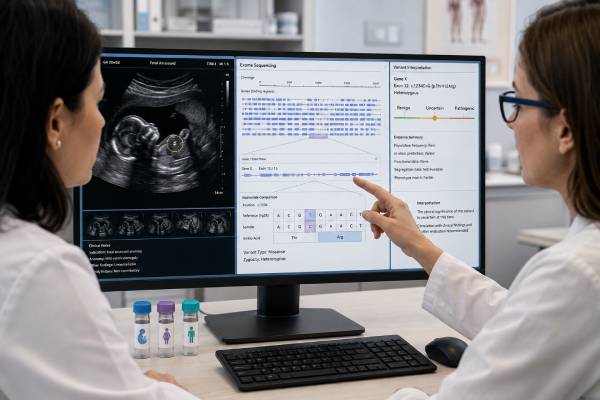
Prenatal exome sequencing is an advanced diagnostic test used when fetal ultrasound findings suggest a possible single-gene disorder, especially after chromosome testing has not found an explanation. The exome is the portion of DNA that contains most protein-coding instructions. Although it represents only a small part of the genome, it includes many variants known to cause rare genetic conditions. Testing usually analyzes fetal DNA together with DNA from both biological parents, an approach called trio exome sequencing. This comparison helps identify new fetal variants, determine inheritance, and filter thousands of normal genetic differences. Prenatal exome sequencing can provide a specific diagnosis, refine prognosis, guide pregnancy and newborn care, and clarify recurrence risk. It can also return a negative result or a finding that remains uncertain. Because fetal features are still developing and many genetic conditions are incompletely described before birth, interpretation is more difficult than simply “reading the genes.” Careful case selection, detailed ultrasound phenotyping, informed consent, and specialist review are central to obtaining useful results.
- Prenatal exome sequencing is generally considered for one or more significant fetal anomalies after chromosomal testing is nondiagnostic.
- Trio testing of the fetus and both biological parents usually improves interpretation and speed.
- A positive result may identify a single-gene disorder and change counseling, surveillance, delivery planning, or neonatal care.
- Diagnostic yield varies widely with the fetal findings; it is not a fixed percentage for every pregnancy.
- A negative result does not exclude all genetic disease because the exome and current knowledge are incomplete.
- Variants of uncertain significance, incidental findings, and parental findings require explicit consent and expert counseling.
Table of Contents
- What prenatal exome sequencing analyzes
- Who may be offered testing
- Samples, trio testing, and laboratory analysis
- Diagnostic yield and what affects it
- How results are reported
- Clinical use of a diagnosis
- Limitations, uncertainty, and next steps
What prenatal exome sequencing analyzes
The human genome contains about three billion DNA letters. The exome consists mainly of exons, the parts of genes used to make proteins. Most well-established disease-causing variants identified through clinical sequencing are located in or near these coding regions, which makes exome sequencing a practical way to examine thousands of genes at once.
The test is designed primarily to find sequence variants: single-letter substitutions and small insertions or deletions that alter a gene’s function. Some laboratories also analyze selected copy-number variants, mitochondrial DNA, or other variant types, but performance varies. Exome sequencing is therefore not automatically a replacement for prenatal chromosomal microarray testing, which is optimized for deletions and duplications, or for karyotyping, which can reveal large and balanced chromosome rearrangements.
“Whole-exome sequencing” does not mean that every exon is read perfectly. Coverage is uneven. Some genes contain repetitive, GC-rich, homologous, or technically difficult regions that cannot be analyzed reliably. Deep intronic and most regulatory variants lie outside the usual target. Repeat expansions, methylation changes, low-level mosaicism, and many structural rearrangements may be missed. The laboratory report should state the assay’s scope and technical limitations.
After sequencing, the laboratory compares the DNA sequence with a reference genome and generates a large list of variants. Most are benign differences shared by many people. Bioinformatic filtering prioritizes rare variants in genes that could plausibly explain the fetal phenotype and match an expected inheritance pattern. Laboratory scientists and clinical geneticists then evaluate population frequency, predicted molecular effect, gene-disease validity, published cases, functional evidence, inheritance, and consistency with ultrasound findings.
Prenatal interpretation is phenotype-driven. A precise description such as “bilateral enlarged echogenic kidneys with oligohydramnios” is more useful than the broad term “kidney anomaly.” Details from fetal echocardiography, neurosonography, magnetic resonance imaging, serial ultrasound, pathology, and family history can change which genes are prioritized. New findings that appear later in pregnancy may prompt reanalysis before the final report.
Some services use a virtual gene panel drawn from exome data rather than reviewing every known disease gene. This limits analysis to genes linked to the observed fetal features and may reduce unrelated findings. Other services perform broader exome-wide analysis. Neither approach is universally superior. The appropriate scope depends on the phenotype, laboratory policy, urgency, and the family’s preferences about unexpected information.
Who may be offered testing
Prenatal exome sequencing is most often offered when ultrasound identifies one or more major structural anomalies and standard diagnostic testing has not found a cause. Standard testing commonly includes a rapid assay for selected aneuploidies and a chromosomal microarray or karyotype. Exome sequencing is particularly relevant when the pattern suggests a monogenic condition: skeletal dysplasia, multiple congenital anomalies, nonimmune hydrops, severe brain malformation, suspected metabolic disease, recurrent similar fetal findings, or a phenotype associated with genetically heterogeneous disorders.
A multidisciplinary team may review the case before testing. Maternal-fetal medicine specialists, clinical geneticists, genetic counselors, fetal radiologists, laboratory scientists, and relevant pediatric subspecialists assess whether a genetic diagnosis is plausible and whether the available phenotype is sufficiently specific for interpretation. This selection process matters because diagnostic yield is much higher in some groups than others.
Testing is not generally recommended as an unselected screening test in a structurally normal fetus. Every person carries rare genetic variants, and broad sequencing without a relevant phenotype can produce uncertain or incidental findings that are difficult to interpret prenatally. Current professional statements support clinical sequencing for selected anomalous fetuses rather than routine exome analysis of all pregnancies.
An isolated soft marker is usually not enough by itself. Isolated increased nuchal translucency is a more nuanced indication: yield is relatively low when the nuchal translucency later resolves and detailed imaging remains normal, but it may rise when it is marked, persistent, or accompanied by hydrops or structural abnormalities. The team should explain evidence specific to the observed finding rather than quoting a pooled yield from dissimilar cases.
Exome sequencing may also be considered after a fetal or neonatal death, stillbirth, or pregnancy termination when anomalies suggest a genetic disorder. Postmortem imaging, examination, photographs, radiographs, and pathology can greatly improve interpretation. Testing stored fetal DNA may establish recurrence risk for future pregnancies even when it cannot affect management of the current one.
When a family has a known pathogenic variant, targeted testing is usually faster, less expensive, and easier to interpret than broad exome sequencing. Likewise, if the phenotype strongly suggests a condition requiring a specialized assay—such as a repeat-expansion, methylation, or biochemical test—the targeted method may be more appropriate.
The practical decision also depends on gestational age and turnaround. A test that takes several weeks may arrive too late to inform time-sensitive options. Rapid prenatal exome services can return results sooner, but availability and eligibility differ. Before ordering, the team should confirm sample requirements, expected reporting time, and whether preliminary or updated results are possible.
Samples, trio testing, and laboratory analysis
Fetal DNA usually comes from chorionic villi obtained by CVS or from amniotic fluid obtained by amniocentesis. The same invasive sample collected for chromosome testing can often be used for sequencing if sufficient DNA or cultured cells remain. In some cases, fetal blood, tissue after pregnancy loss, or another validated specimen is used. The laboratory checks sample identity, quantity, quality, and possible maternal cell contamination.
Most expert guidance favors trio sequencing whenever feasible: fetal DNA is analyzed alongside blood or saliva DNA from both biological parents. Trio analysis helps distinguish variants inherited from a healthy parent from variants that arose de novo in the fetus. It also identifies whether two variants in a recessive disease gene were inherited from different parents and can clarify X-linked inheritance. This information reduces the number of candidate variants and often shortens interpretation.
A duo analysis with one parent or fetal-only analysis is possible, but it generally leaves more uncertainty and may require parental follow-up. Family circumstances, donor gametes, adoption, unavailable parents, or concerns about biological relationships can affect what samples are available. These issues should be discussed respectfully before testing.
The laboratory may incidentally identify nonmaternity, nonpaternity, consanguinity, or another unexpected relationship. Policies vary on whether and how such findings are disclosed. Consent should address this possibility before samples are submitted rather than after analysis reveals it.
The workflow includes several stages. DNA is fragmented, coding regions are captured, and millions of short sequences are generated. Reads are aligned to a reference genome, variants are called, and quality filters are applied. Analysts then use phenotype terms and inheritance models to prioritize candidates. A suspected diagnosis is not reported solely because a computer predicts harm; it must be evaluated under accepted clinical classification standards.
Potentially diagnostic variants may be confirmed with another method, such as Sanger sequencing, depending on laboratory validation. Parental origin may also be confirmed. If a result has immediate implications, the fetal medicine team may ask the laboratory to review updated ultrasound findings or prioritize a particular gene.
Turnaround commonly ranges from about two to six weeks, with rapid pathways sometimes shorter and complex cases longer. A “negative” preliminary report may later change if additional analysis is completed. Laboratories should make clear whether the test is a fixed-time prenatal analysis, whether postnatal reanalysis is available, and whether a broader report will follow.
The technical result and the clinical interpretation are separate. The same variant can be classified differently as evidence evolves, and the same pathogenic variant can have different clinical significance depending on the fetus’s features and inheritance. This is why sequencing is best integrated into a clinical service rather than ordered as a stand-alone data product.
Diagnostic yield and what affects it
Diagnostic yield is the proportion of tested cases in which a pathogenic or likely pathogenic variant explains the fetal findings. It should not be confused with analytical sensitivity or the chance that a pregnancy has any genetic condition.
A widely cited 2022 systematic review and meta-analysis found that prenatal exome sequencing provided an additional diagnosis in about 31% of structurally abnormal fetuses after nondiagnostic karyotype or microarray. That number is useful as an overall research summary, but it is not a personal probability. The included studies differed markedly in case selection, fetal phenotype, gene-analysis strategy, and reporting criteria. Yields ranged from very low in broadly selected isolated findings to much higher in carefully selected phenotypes strongly suggestive of a monogenic disorder.
Body system matters. Skeletal abnormalities, recurrent multisystem malformations, hydrops, and some neurological phenotypes often have relatively high yields because many established single-gene disorders present prenatally in these ways. Isolated increased nuchal translucency with no later anomaly generally has a lower yield. Cardiac, renal, and central nervous system findings span broad categories, so the exact pattern and associated anomalies are more informative than the organ label alone.
Multiple anomalies may increase the chance of diagnosis, but the relationship is not simple. Several findings can point to a recognizable syndrome, yet severe disruption from a non-genetic cause can also affect multiple systems. Conversely, one distinctive anomaly may be highly specific for a particular gene disorder.
Case selection by a multidisciplinary team can raise yield because it identifies cases in which monogenic disease is plausible and improves phenotyping. A 2024 cohort of 629 ongoing pregnancies using exome-derived analysis reported pathogenic or likely pathogenic findings in 14% overall, with a higher rate among fetuses with multiple anomalies than among those with a single major anomaly. Other studies report different figures because their referral populations and analysis pipelines differ.
Prior testing affects yield. If chromosome analysis has already removed cases with aneuploidy and pathogenic CNVs, exome sequencing is evaluating the remaining group. A study that includes fetuses before microarray will appear to have a different distribution of diagnoses than one that sequences only after a negative array.
Consanguinity, recurrent similar pregnancies, and a family history compatible with recessive or X-linked inheritance can increase the prior probability of a monogenic diagnosis. Detailed parental examination can also reveal mild features related to an inherited variant.
A result may be “diagnostic” even when prognosis remains broad. Some gene disorders show variable expressivity, age-dependent features, or limited prenatal outcome data. Diagnostic yield therefore measures finding an explanation, not the ability to predict every future medical or developmental outcome.
How results are reported
A positive or diagnostic result usually means the laboratory identified a pathogenic or likely pathogenic variant, or a pair of variants, that matches the fetal phenotype and inheritance pattern. The report names the gene, variant, zygosity, inheritance, condition, evidence, and relevant limitations. A diagnostic result should be interpreted with the ultrasound findings rather than read as a generic disease description.
For an autosomal dominant condition, one disease-causing variant may be sufficient. It may be de novo or inherited from a parent. For an autosomal recessive condition, two disease-causing variants generally need to affect the same gene, usually one inherited from each parent. For X-linked conditions, interpretation depends on fetal sex chromosomes, the gene, the variant, and inheritance. Mitochondrial and other non-Mendelian mechanisms require additional considerations.
A negative result means no reportable variant explaining the fetal findings was identified under the laboratory’s methods and current knowledge. It does not mean that the anomalies are not genetic. The causal variant may be in a region the exome does not cover, belong to a disease gene not yet recognized, represent a variant type the pipeline misses, or be present at a mosaic level below detection. The phenotype may also have a non-genetic cause.
A variant of uncertain significance, or VUS, has insufficient or conflicting evidence for classification as disease-causing or benign. Prenatal services vary in VUS reporting. Some report only a VUS in a gene strongly linked to the fetal phenotype; others suppress most uncertain findings to reduce harm. The family should know the laboratory policy in advance. A VUS is not a confirmed diagnosis and should not be used alone for irreversible decisions.
A candidate or research finding may indicate a plausible gene or variant without enough evidence for clinical reporting. Some programs separate such findings from the clinical report and offer research follow-up. Families should understand whether research results will be returned and whether confirmation is required.
Secondary or incidental findings are medically relevant variants unrelated to the fetal anomaly. They may concern the fetus, a parent, or both. Examples can include predispositions to adult-onset cancer or cardiac disease. Prenatal programs differ in whether they deliberately analyze these genes, allow opt-in or opt-out choices, or report only findings requiring childhood action. Consent should distinguish fetal diagnostic findings from secondary findings.
The report may also reveal a parental diagnosis, such as a mild or previously unrecognized dominant condition. This can have implications for the parent’s healthcare and other relatives. Trio testing is therefore not merely a technical filter; it is potentially a genetic test of all three people.
Clinical use of a diagnosis
A specific molecular diagnosis can end a long period of uncertainty and replace a broad differential diagnosis with a more focused plan. Its value depends on what the condition means in that pregnancy and what actions are available.
During pregnancy, the result may prompt targeted imaging, serial growth assessment, fetal echocardiography, monitoring for hydrops, or evaluation of organs known to be affected. It may clarify whether a finding is likely isolated or part of a multisystem disorder. In selected conditions, it can influence eligibility for fetal therapy or identify risks relevant to an invasive procedure.
Delivery planning may change. A fetus at risk for airway compromise, severe skeletal disease, metabolic decompensation, arrhythmia, or respiratory failure may benefit from delivery at a tertiary center with specific specialists available. The diagnosis can guide neonatal testing and prevent repeated broad investigations after birth.
Prognostic counseling may improve, but it remains bounded by evidence. Prenatally diagnosed cases can be more severe than postnatal cohorts, and published reports may overrepresent unusual outcomes. Some disorders have wide clinical ranges, even among relatives with the same variant. Clinicians should clearly separate well-established risks from uncertain predictions.
A diagnosis can also inform pregnancy options. Depending on gestational age, jurisdiction, and family values, parents may continue the pregnancy with preparation, choose palliative planning, or consider termination. The genetics team’s role is to provide balanced, nondirective information, including available supports and the lived experience of affected individuals when relevant.
Inheritance establishes recurrence risk more accurately than ultrasound alone. A de novo dominant variant often implies a low but not zero recurrence risk because of possible parental germline mosaicism. A recessive diagnosis usually means a 25% recurrence chance for each future pregnancy when both parents are carriers. An inherited dominant or X-linked variant may carry a substantially higher recurrence risk. Exact counseling depends on the condition and family structure.
Future reproductive options can include targeted prenatal diagnosis, PGT-M for a known familial condition, donor gametes, adoption, or natural conception without testing. A confirmed molecular result is essential before designing most targeted tests.
The diagnosis may also benefit relatives. Cascade testing can identify family members who carry the variant or clarify whether apparently unrelated health concerns share a cause. Disclosure remains the family’s decision, supported by genetics professionals.
Limitations, uncertainty, and next steps
Prenatal exome sequencing has scientific, technical, and ethical limits. The fetal phenotype is incomplete because many features emerge late in pregnancy, after birth, or during childhood. Intellectual development, behavior, hearing, vision, immune function, and many metabolic manifestations cannot be assessed directly by prenatal ultrasound. This weakens phenotype-variant matching and makes prognosis less precise.
The exome itself is incomplete. Some coding regions have inadequate coverage, and most noncoding DNA is not analyzed. Structural variants, repeat expansions, methylation disorders, mitochondrial variants, and copy-number changes may require separate tests. A normal prenatal karyotype, microarray, and exome still cannot exclude every genetic condition.
Variant interpretation changes as knowledge grows. A negative case may become diagnosable when a new disease gene is discovered or a previously uncertain variant is reclassified. Reanalysis after birth can be particularly valuable because the child’s phenotype provides new evidence. Families should ask whether reanalysis is offered, at what interval, and whether updated samples or clinical information are needed.
A prenatal VUS can create distress without improving decision-making. The possibility should be discussed before testing, including the laboratory’s reporting threshold and whether parental or functional studies might clarify it. Uncertainty is not evidence of a poor outcome.
Secondary findings create additional choices. Some families want any medically actionable information; others prefer results limited to the fetal anomaly. Policies may restrict choice, especially when a finding has immediate implications for a parent or child. The consent process should be specific rather than relying on a general statement that “unexpected results are possible.”
Equity is another limitation. Access to fetal imaging, specialists, rapid sequencing, insurance coverage, and culturally appropriate counseling is uneven. Genomic databases also underrepresent many ancestries, which can increase uncertain classifications. These are not merely administrative problems; they affect diagnostic accuracy and the experience of families.
When the result is negative, next steps depend on the phenotype. Options may include genome sequencing, targeted assays not captured by exome, metabolic testing, infection studies, repeat imaging, postnatal evaluation, or research enrollment. When a pregnancy ends, detailed pathology and storage of DNA can preserve future diagnostic opportunities.
The most useful prenatal exome test is not necessarily the broadest one. It is the test ordered for a well-phenotyped fetus, analyzed with appropriate family samples, interpreted by an experienced laboratory, and connected to counseling that explains both the result and its limits. Used in that setting, exome sequencing can transform fetal anomalies from an unexplained observation into a clinically meaningful genetic diagnosis—while recognizing that some questions will remain unanswered.
References
- International Society for Prenatal Diagnosis Updated Position Statement on the Use of Genome-Wide Sequencing for Prenatal Diagnosis (2022), position statement.
- Guidelines for Next-Generation Sequencing Procedures Applied to Prenatal Diagnosis (2024), professional guideline.
- Diagnostic Yield of Exome Sequencing for Prenatal Diagnosis of Fetal Structural Anomalies: A Systematic Review and Meta-Analysis (2022), systematic review and meta-analysis.
- Prenatal Exome and Genome Sequencing for Fetal Structural Abnormalities (2023), clinical review.
- The High Diagnostic Yield of Prenatal Exome Sequencing Followed by 3400 Gene Panel Analysis in 629 Ongoing Pregnancies With Ultrasound Anomalies (2024), cohort study.
- Whole-Exome Sequencing for Prenatal Diagnosis of Fetal Structural Anomalies (2025), cohort study.
Disclaimer
This article is for general education and does not replace individualized medical advice, fetal imaging, or genetic counseling. Eligibility, reporting practices, turnaround time, and access to prenatal exome sequencing vary by healthcare system and laboratory. A qualified maternal-fetal medicine and genetics team should interpret results in the context of the complete pregnancy, family history, and the family’s values.





