
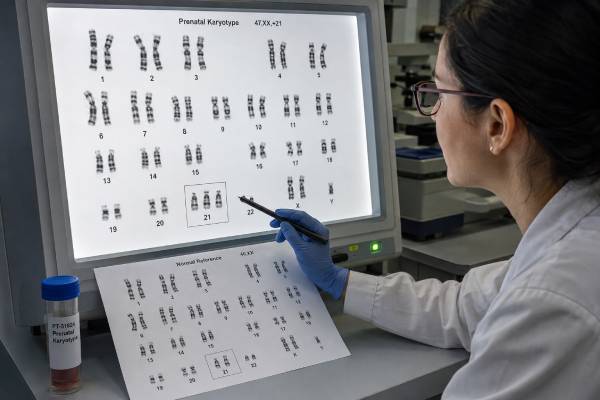
A prenatal karyotype is a diagnostic test that examines the number and large-scale structure of fetal chromosomes. It can identify whole-chromosome conditions such as trisomy 21, trisomy 18, trisomy 13, monosomy X, and many sex chromosome aneuploidies. It can also show large deletions or duplications, extra marker chromosomes, and structural rearrangements such as translocations and inversions. The test is performed on cells collected through chorionic villus sampling, amniocentesis, or, less commonly, fetal blood sampling. Unlike prenatal screening, a karyotype evaluates chromosomes in cells from the pregnancy and can establish a diagnosis for abnormalities within its resolution. Its major advantage is that it shows chromosome architecture, including balanced rearrangements that a chromosomal microarray may miss. Its major limitation is resolution: small copy-number changes and most single-gene variants are invisible. Results may be normal, abnormal, mosaic, or occasionally inconclusive. The most appropriate test—karyotype, microarray, rapid aneuploidy testing, sequencing, or a combination—depends on the screening result, ultrasound findings, family history, and the information needed for counseling.
- A prenatal karyotype is a diagnostic chromosome test, not a maternal blood screening test.
- It reliably detects extra or missing chromosomes and many large structural abnormalities.
- It can reveal balanced translocations and inversions, an important advantage over microarray.
- CVS evaluates placental cells, whereas amniocentesis generally evaluates fetal cells shed into amniotic fluid.
- A normal karyotype does not exclude small deletions, duplications, or single-gene disorders.
- Mosaic and structural results often need additional testing and genetic counseling.
Table of Contents
- What a prenatal karyotype shows
- Why karyotyping is ordered
- Sample collection and laboratory process
- Reading karyotype notation
- Aneuploidy, structural, and mosaic results
- Karyotype versus microarray and other tests
- Accuracy, limitations, and next steps
What a prenatal karyotype shows
A karyotype is an organized visual analysis of chromosomes. Most human cells contain 46 chromosomes: 22 pairs of autosomes and one pair of sex chromosomes. In the laboratory, chromosomes from dividing cells are stained to create characteristic light and dark bands, photographed, and arranged by size and banding pattern. A trained cytogenetics professional counts them and looks for visible structural differences.
This method is especially effective for aneuploidy, an extra or missing chromosome. Examples include 47,XX,+21 for a fetus with two X chromosomes and an extra chromosome 21, and 45,X for monosomy X. It can also identify triploidy, in which there are three complete chromosome sets, and tetraploidy, in which there are four, if the abnormal cell line is represented in the sample.
Karyotyping can detect large unbalanced changes, including deletions, duplications, derivative chromosomes, rings, isochromosomes, and marker chromosomes. Its practical resolution is usually measured in millions of DNA bases. The exact threshold depends on chromosome region, band quality, and laboratory standards; changes around 5–10 megabases may be visible, while smaller ones usually are not.
A distinctive strength is detection of balanced structural rearrangements. In a balanced translocation, chromosome material has moved between chromosomes without an obvious net gain or loss. A balanced inversion reverses a segment within a chromosome. These findings may not cause fetal health problems by themselves, but they can reveal inherited carrier status and affect future reproductive risk. A microarray measures DNA dosage and generally cannot see a rearrangement when no material is gained or lost.
The karyotype also shows whether an aneuploidy has a structural mechanism. Down syndrome, for example, can result from a free extra chromosome 21, a Robertsonian translocation involving chromosome 21, or mosaicism. That distinction matters because a translocation may be inherited from a balanced-carrier parent and can change recurrence counseling.
What karyotyping does not show is equally important. It does not sequence genes, detect most small copy-number variants, assess most imprinting disorders, or identify many forms of uniparental disomy. A normal chromosome picture is therefore a normal result only within the scale and cell population examined.
Why karyotyping is ordered
A prenatal karyotype may be ordered after a high-chance chromosome screening result. Cell-free DNA, first-trimester screening, or a quad screen can indicate increased probability of trisomy 21, trisomy 18, trisomy 13, or a sex chromosome condition. Because screening is not diagnostic, fetal or placental cells are needed for confirmation. Karyotyping can confirm the chromosome count and show structural details that affect counseling.
Ultrasound findings are another indication. Increased nuchal translucency, cystic hygroma, major heart defects, growth restriction with anomalies, multiple malformations, or a recognizable chromosome phenotype may lead to an offer of invasive diagnosis. When one or more major structural abnormalities are present, professional guidance commonly favors chromosomal microarray because it detects submicroscopic imbalances in addition to most large gains and losses. Karyotype may still be added when a balanced rearrangement, triploidy, marker chromosome, or visible structural mechanism is suspected.
A known parental chromosome rearrangement is a particularly clear reason for karyotyping. If one parent carries a balanced translocation or inversion, fetal karyotype can determine whether the fetus inherited a normal chromosome arrangement, the same balanced rearrangement, or an unbalanced form with missing or extra material. Microarray may identify an unbalanced gain or loss but cannot always reconstruct the chromosome architecture.
Karyotyping may also be discussed when a patient chooses diagnostic testing despite reassuring screening. Current practice supports offering diagnostic options to all pregnant patients after counseling, not only those above a maternal-age threshold. Some patients value definitive information about common chromosome abnormalities or want to avoid the residual uncertainty of screening.
A previous pregnancy or child with a chromosome abnormality can prompt testing, although the recurrence risk depends on the exact mechanism. A prior sporadic trisomy creates a different risk profile from a familial translocation. Reviewing the original laboratory report is more useful than relying on the condition name alone.
Testing may be performed after fetal death or stillbirth, but chromosomal microarray is often preferred because it does not require living dividing cells and has higher resolution. Karyotyping remains valuable when chromosome structure is central to the question or when cultured cells are available.
The choice to test is personal. A diagnosis can support preparation, pregnancy management, or reproductive decisions, but some patients prefer to avoid an invasive procedure or information they would not use. Counseling should describe both the procedure and the laboratory test because they are separate decisions.
Sample collection and laboratory process
A prenatal karyotype begins with an invasive sampling procedure. Chorionic villus sampling obtains placental villi, usually in the first trimester. Amniocentesis obtains amniotic fluid, generally from 15 weeks onward. Both are performed with ultrasound guidance by trained clinicians. Procedure-related risk is low in experienced hands but is not zero, and estimates vary with operator, technique, and clinical circumstances.
CVS offers earlier results. Because it samples placenta, it may reveal confined placental mosaicism: an abnormal cell line present in the placenta but absent from the fetus. Laboratories can analyze direct preparations and cultured villi, which represent different placental cell layers. If a mosaic or unexpected result is found, amniocentesis may be recommended to clarify the fetal chromosome complement.
Amniotic fluid contains fetal cells shed from skin, urinary tract, and other tissues. These cells are cultured so that they divide. Amniocentesis therefore more directly reflects the fetus than CVS, although tissue-limited mosaicism can still create complexity.
In the laboratory, cells are grown until sufficient cells enter metaphase, the stage when chromosomes are condensed and visible. Cell division is arrested, chromosomes are spread on slides, and a banding stain—commonly G-banding—is applied. Technologists inspect a required number of metaphase cells and prepare detailed images from selected cells. More cells may be counted when mosaicism is suspected.
Culture takes time. A full karyotype often requires about one to two weeks, sometimes longer if cell growth is slow or the sample is small. Rapid tests such as quantitative fluorescent PCR or FISH can provide preliminary information about selected chromosomes within days, but they do not replace the full structural assessment.
Maternal cell contamination is a concern, particularly when a sample is blood-stained or tissue is difficult to separate. Laboratories use sample handling, culture behavior, sex chromosome findings, or molecular comparison with maternal DNA to assess contamination. A result of 46,XX in a pregnancy expected to have XY chromosomes, for example, requires careful review rather than an automatic assumption about the fetus.
Rarely, culture fails or yields too few cells. The laboratory may request another sample, use an alternative method, or report a limited result. The report should state the number of cells analyzed, band resolution when relevant, and any technical limitations.
Reading karyotype notation
Karyotype reports use the International System for Human Cytogenomic Nomenclature. The notation is compact but follows a predictable order: total chromosome number, sex chromosome complement, then abnormalities.
46,XX and 46,XY are standard chromosome counts with two X chromosomes or one X and one Y. They do not describe every genetic characteristic or guarantee typical sexual development; they report the chromosomes seen in the analyzed cells.
A plus sign indicates extra material or a chromosome. 47,XY,+18 means 47 chromosomes, an XY complement, and an extra chromosome 18. A minus sign indicates loss: 45,X means one X chromosome and no second sex chromosome identified.
Structural notation names the rearrangement. t denotes translocation, rob a Robertsonian translocation, inv an inversion, del a deletion, dup a duplication, r a ring chromosome, i an isochromosome, and der a derivative chromosome. Numbers in parentheses identify the chromosomes and breakpoints. For example, 46,XX,t(11;22)(q23;q11.2) describes an apparently balanced translocation between chromosomes 11 and 22 at specified long-arm bands.
Square brackets may show the number of cells with each line. A mosaic result such as mos 45,X[12]/46,XX[18] indicates that 12 analyzed cells had monosomy X and 18 had a 46,XX karyotype. These counts describe the sampled cells, not an exact percentage in every fetal tissue.
A ? or other qualifier can indicate uncertainty about a chromosome or breakpoint. add means additional material of unknown origin is attached at a location. Such results often lead to FISH, microarray, or parental karyotyping to characterize the material.
The word apparently matters in a balanced result. A rearrangement can look balanced at microscopic resolution while containing a small deletion or duplication at a breakpoint. If fetal anomalies are present, microarray or sequencing may be recommended even when the karyotype appears balanced.
Parents should not have to decode notation alone. The plain-language interpretation should explain the diagnosis, evidence, inheritance questions, expected variability, and recommended follow-up. A chromosome label is the start of counseling, not the complete prognosis.
Aneuploidy, structural, and mosaic results
A full aneuploidy means the extra or missing chromosome was seen in all analyzed cells. Common examples are trisomy 21, trisomy 18, trisomy 13, monosomy X, 47,XXY, 47,XXX, and 47,XYY. Clinical outcomes vary substantially among these conditions and among individuals with the same karyotype. Ultrasound findings, mosaicism, and structural form may modify counseling but do not predict every feature.
A translocation trisomy contains extra critical chromosome material attached to another chromosome. In translocation Down syndrome, extra chromosome 21 material may be attached to chromosome 14, 21, or another chromosome. Parental karyotyping is often recommended because a healthy parent can carry the balanced form. If inherited, recurrence risk can be much higher than for a sporadic free trisomy and depends on the chromosomes involved and which parent is the carrier.
An unbalanced structural result means material is missing, duplicated, or both. The clinical significance depends on the size, chromosome region, genes involved, and whether the change is inherited. Microarray often refines the exact genomic imbalance. Parental testing can show whether the abnormal chromosome arose de novo or came from a balanced parental rearrangement.
An apparently balanced result may have no fetal effect, may be associated with gene disruption or cryptic imbalance, or may reveal carrier status with future reproductive implications. The presence of ultrasound anomalies raises concern that the rearrangement is not clinically neutral. Further molecular testing is then especially important.
A marker chromosome is an extra structurally abnormal chromosome too small or complex to identify by routine banding alone. Its effect depends on its origin, DNA content, and mosaic level. FISH and microarray can help identify it. Some markers contain little clinically significant material; others include dosage-sensitive regions.
A mosaic result shows two or more chromosome cell lines. Interpretation depends on whether the sample is CVS or amniotic fluid, how many cells are affected, whether the finding appears in independent cultures, and whether ultrasound supports the condition. CVS mosaicism may be limited to the placenta. Amniotic-fluid mosaicism is more likely to reflect the fetus but still may not predict distribution across organs.
Pseudomosaicism can arise during cell culture rather than being present in the fetus. Cytogenetic laboratories use established criteria to distinguish a single-cell artifact from true mosaicism. Follow-up may include additional cell counts, another tissue, FISH on uncultured cells, or repeat sampling.
Karyotype versus microarray and other tests
Karyotype and microarray overlap but answer different questions. Karyotype provides a low-resolution map of chromosome number and architecture. Microarray provides a high-resolution map of DNA gains and losses. If fetal structural anomalies are present, microarray is commonly the first-tier genomic test because it detects clinically important deletions and duplications that karyotype misses.
Microarray usually detects common full aneuploidies and large unbalanced rearrangements. It generally cannot identify a balanced translocation or inversion. Some SNP-based arrays detect triploidy and regions of homozygosity, whereas array-CGH platforms may not. The laboratory method matters more than the generic word “array.”
Rapid aneuploidy tests examine selected chromosomes. QF-PCR or FISH can quickly assess chromosomes 13, 18, 21, X, and Y, depending on the panel. They are useful when time is critical but may miss structural abnormalities outside the targeted regions. A normal rapid test is not a normal full karyotype.
Cell-free DNA is screening. It estimates risk from placental DNA fragments in maternal blood and cannot define chromosome structure reliably. A high-chance result should be confirmed by CVS or amniocentesis. Selecting the confirmatory method should consider placental mosaicism and the suspected condition.
FISH is a targeted method using fluorescent probes. It can confirm a suspected deletion, identify material on a marker chromosome, or clarify a rearrangement. It only evaluates the loci probed, so it is not genome-wide.
Exome sequencing evaluates coding variants in thousands of genes and may be offered when fetal anomalies remain unexplained after chromosome testing. It cannot replace the visual chromosome information of a karyotype. Genome sequencing may eventually consolidate some methods, but validation and prenatal use vary.
The best approach is often sequential rather than maximal. A suspected common trisomy may warrant rapid testing plus karyotype. Multiple fetal anomalies may warrant rapid testing plus microarray, with karyotype added for a structural question. A known parental translocation strongly favors fetal karyotype, often with microarray if an imbalance needs precise definition.
Accuracy, limitations, and next steps
For abnormalities large enough to see and present in the sampled cells, prenatal karyotyping is highly accurate. Errors can still arise from sample contamination, culture failure, low-level mosaicism, placental-fetal discordance, or rare interpretive complexity. Confirmatory studies are recommended when the result and clinical picture do not align.
Resolution is the central limitation. A fetus can have a pathogenic microdeletion, microduplication, or sequence variant despite a normal karyotype. Major structural anomalies with a normal result often lead to microarray and, in selected cases, prenatal exome sequencing.
A normal karyotype also cannot predict general health, development, pregnancy outcome, or all birth defects. It does not evaluate environmental exposures, infection, placental disease, or multifactorial causes. Ultrasound and standard obstetric care continue regardless of chromosome results.
After an abnormal result, genetic counseling should address the exact cytogenetic mechanism. Parental karyotypes may be needed for translocations, inversions, markers, or some structural abnormalities. Microarray can define copy-number content. FISH can localize material. Detailed ultrasound and condition-specific imaging can clarify the fetal phenotype.
Recurrence risk cannot be inferred from the condition name alone. A de novo free trisomy, inherited Robertsonian translocation, parental reciprocal translocation, and mosaic abnormality each require different counseling. Gonadal mosaicism means that even an apparently de novo result may carry a small residual recurrence risk.
If a mosaic result is found on CVS, amniocentesis may be recommended. If mosaicism is found on amniocentesis, postnatal blood or tissue testing may still show a different proportion. Counseling should avoid treating cultured-cell percentages as a precise map of the fetus.
Families may use a diagnosis to prepare for delivery and neonatal care, consult pediatric specialists, consider pregnancy-management options, or plan future testing. Nondirective counseling should present the range of outcomes, available supports, and uncertainty without steering the decision.
Prenatal karyotyping remains valuable because it reveals chromosome structure in a way no dosage-only test can. Its most effective use is targeted: choose it when chromosome architecture matters, combine it with higher-resolution methods when appropriate, and interpret the picture in the context of the pregnancy rather than as an isolated laboratory image.
References
- ACGS Best Practice Guidelines for Constitutional Karyotype Analysis and Targeted Chromosome Analysis (2024), laboratory guideline.
- Screening and Diagnosis of Fetal Structural Anomalies and Chromosome Conditions (C-Obs 35) (2024), clinical guideline.
- Amniocentesis and Chorionic Villus Sampling: Green-top Guideline No. 8 (2021), clinical guideline.
- An Overview of Current Prenatal Genetic Screening and Diagnostic Testing (2025), clinical review.
- Clinical Application of Chromosome Microarray Analysis in Prenatal Diagnosis (2024), cohort study.
- The Use of Chromosomal Microarray for Prenatal Diagnosis (2016), SMFM consult guideline.
Disclaimer
This article is for general educational purposes and does not replace medical advice, genetic counseling, or interpretation of an actual cytogenetic report. Procedure timing, laboratory resolution, reporting standards, and follow-up practices vary. Decisions should be made with clinicians who can review the full screening history, ultrasound findings, family history, and patient preferences.





