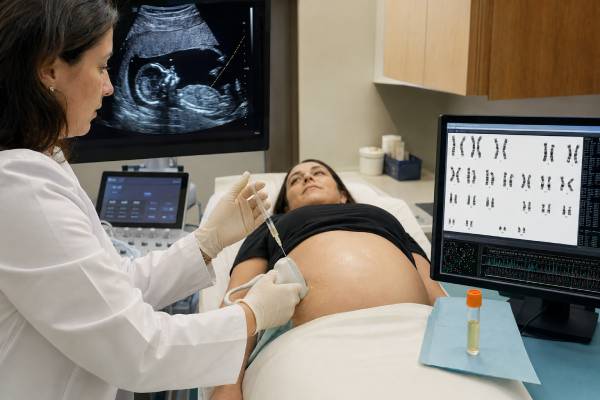
Amniocentesis is a prenatal diagnostic procedure that removes a small amount of amniotic fluid for laboratory testing. The fluid contains fetal cells and other substances that can provide direct information about chromosomes, selected genes, infections, and certain fetal conditions. Unlike a screening test, amniocentesis can often confirm whether a fetus has the condition being tested for. It is usually performed at or after 15 weeks of pregnancy with a thin needle guided by continuous ultrasound. The procedure itself commonly takes only a few minutes, although counseling, ultrasound, preparation, and observation make the appointment longer. Results may return in stages: rapid testing for common chromosome conditions can take several days, while a karyotype, chromosomal microarray, or gene test may take two weeks or longer. Amniocentesis is optional. Choosing it involves weighing the value of a diagnostic answer against a small procedure-related risk and considering how the result may affect pregnancy care, preparation, or personal choices.
- Amniocentesis is diagnostic, not a screening test, for the specific chromosome or gene conditions included in the laboratory order.
- The procedure is generally performed from 15 weeks of pregnancy onward because earlier amniocentesis has higher complication rates.
- Rapid chromosome results may be available in about 2–4 working days, while full genetic results often take 1–3 weeks.
- A normal result does not rule out every genetic disorder or birth difference; it only addresses what the selected test can detect.
- The additional miscarriage risk is small but not zero and varies with clinical circumstances, operator experience, and how risk is calculated.
- Call the maternity team urgently after the procedure for fluid leakage, heavy bleeding, fever, worsening pain, or regular contractions.
Table of Contents
- What amniocentesis can test
- Why amniocentesis is offered
- Preparation and the procedure
- Laboratory test options
- Results and their meaning
- Risks and aftercare
- Amniocentesis compared with other options
- Making an informed choice
What amniocentesis can test
Amniocentesis provides a sample of amniotic fluid, which surrounds the fetus. Fetal cells shed into the fluid can be grown or analyzed directly. The laboratory can then examine chromosomes, copy-number changes, or selected DNA sequences, depending on the clinical question and the tests ordered.
The procedure itself is only the sampling step. It is not one single genetic test. Two people can both have amniocentesis but receive very different laboratory analyses. One may have rapid testing for trisomies 21, 18, and 13. Another may have chromosomal microarray because ultrasound found structural anomalies. A family with a known inherited condition may request a targeted gene test for that specific variant.
Common uses include:
- Detecting chromosome-number conditions such as Down syndrome, Edwards syndrome, Patau syndrome, and some sex chromosome differences
- Identifying large chromosome rearrangements with a karyotype
- Detecting smaller deletions and duplications with chromosomal microarray
- Testing for a known familial gene variant or a suspected single-gene disorder
- Performing prenatal exome sequencing in selected pregnancies with fetal abnormalities when standard tests do not provide an answer
- Testing amniotic fluid for infections such as cytomegalovirus when clinically indicated
- Measuring biochemical markers in uncommon situations, including alpha-fetoprotein or enzyme studies
Amniocentesis does not automatically test for all of these. The order must specify the analyses. Before the procedure, ask exactly what will be tested, what will not be tested, whether additional testing may be added later, and how much sample the laboratory needs.
A diagnostic result can be highly reliable for the target condition, but no prenatal test can guarantee a baby without health problems. Many birth differences are not genetic, and many genetic conditions require a test that would not be part of a routine chromosome study.
Why amniocentesis is offered
Amniocentesis may be offered after screening, ultrasound, family history, or prior pregnancy information suggests an increased chance of a fetal condition. It is also available to a person who wants diagnostic information after counseling, even when screening risk is low.
Common reasons include:
- A high-chance result from cell-free DNA, first-trimester screening, or a second-trimester serum screen
- A fetal structural anomaly, growth concern, increased nuchal translucency, or another ultrasound marker
- A previous pregnancy or child with a chromosome or genetic condition
- A parent who carries a chromosome rearrangement or pathogenic gene variant
- A known carrier couple at risk for an autosomal recessive condition
- A pregnant person who carries an X-linked condition
- An inconclusive or no-call prenatal screening result when diagnostic clarification is desired
- Concern for a fetal infection that can be assessed in amniotic fluid
A positive cell-free DNA prenatal screen is a frequent reason for referral. Cell-free DNA analyzes mainly placental fragments in maternal blood, so false-positive results can occur. Amniocentesis tests cells from amniotic fluid and is often chosen to establish whether the fetus has the suspected chromosome condition.
The gestational age affects the available options. CVS can usually be performed earlier, while amniocentesis is generally delayed until at least 15 weeks. Some people prefer an earlier answer through CVS; others prefer amniocentesis because the sample is considered more directly representative of fetal cells and avoids some questions caused by placental mosaicism.
Maternal age alone is no longer the only basis for offering diagnostic testing. Current prenatal care emphasizes that all pregnant patients should receive clear information about both screening and diagnostic options and may accept or decline them according to their values.
An offer of amniocentesis does not mean that a condition is certain. For example, a screening test may report a 1 in 50 chance, which also means 49 of 50 pregnancies with that risk estimate would not have the condition. A detailed ultrasound finding may increase concern but still have several possible explanations. Diagnostic testing replaces a probability estimate with a laboratory answer for the conditions examined.
Preparation and the procedure
Most amniocentesis appointments begin with an ultrasound. The clinician confirms gestational age, fetal position, placental location, the amount of fluid, and a safe path for the needle. In a multiple pregnancy, ultrasound also helps identify which sac belongs to each fetus.
Preparation is usually simple. Food and medications generally do not need to be restricted. The clinic may advise whether the bladder should be full or empty, depending on gestational age and local practice. Bring blood-group information if available and tell the team about blood-thinning medication, fever, recent infection, HIV, hepatitis B or C, bleeding problems, allergies, and any concerns about Rh status.
The procedure commonly follows these steps:
- You lie on an examination table while the abdomen is cleaned with antiseptic.
- The clinician uses ultrasound to choose a pocket of fluid away from the fetus and umbilical cord.
- A thin needle passes through the abdominal and uterine walls into the amniotic sac under ultrasound guidance.
- A small amount of fluid, often around 15–30 mL depending on gestational age and testing needs, is withdrawn into a syringe.
- The needle is removed, and ultrasound checks the fetal heartbeat.
The needle is not inserted through the navel as a rule; the entry point depends on anatomy. The fetus usually moves away from the needle, and continuous imaging guides the operator. Most people describe pressure, stinging, or menstrual-like cramping rather than severe pain. Local anesthetic is not always used because its injection can be as uncomfortable as the brief amniocentesis needle insertion.
The fluid removed is a small fraction of the total and is replenished. The appointment may take 30–60 minutes, but the needle is generally in place for less than a few minutes. Occasionally, the first attempt does not obtain enough fluid and a second insertion is needed. In rare cases, the procedure is postponed because no safe route is available.
Rh-negative patients may need anti-D immune globulin after the procedure to reduce the chance of Rh sensitization, depending on local guidance and fetal or partner blood information.
Multiple pregnancies require extra mapping. The operator identifies each fetus, placenta, and sac before collecting fluid. Dichorionic twins may require separate samples, and the laboratory must preserve the link between each tube and the correct fetus. Families should ask whether both fetuses will receive identical analyses and what would happen if one sample were inadequate.
Laboratory test options
The value of amniocentesis depends on choosing a laboratory test that matches the reason for testing. A rapid assay, karyotype, microarray, and sequencing test differ in resolution and in the kinds of findings they can return.
| Test | What it usually detects | Typical reporting time |
|---|---|---|
| QF-PCR or rapid FISH | Common aneuploidies, usually chromosomes 13, 18, 21, X, and Y | About 2–4 working days |
| Karyotype | Extra or missing chromosomes and large structural rearrangements | About 1–3 weeks |
| Chromosomal microarray | Aneuploidies and smaller deletions or duplications | Often 1–3 weeks |
| Targeted gene testing | A known family variant or a specific suspected disorder | Several days to several weeks |
| Exome sequencing | Variants across thousands of protein-coding genes | Often several weeks |
Rapid testing gives an early answer for selected chromosome conditions but does not replace a broader study when one is indicated. A full prenatal karyotype can show a balanced or unbalanced rearrangement and the overall chromosome pattern, but it may miss copy-number changes too small for a microscope.
A prenatal chromosomal microarray has higher resolution and is usually offered when ultrasound shows one or more major fetal abnormalities. It can also be selected after counseling in a structurally normal pregnancy. Microarray may return a variant of uncertain significance, identify a finding whose health effect varies, or reveal parental relatedness. These possibilities should be discussed before testing.
Targeted testing is most efficient when the family’s exact pathogenic variant is known. The laboratory should receive a copy of the affected relative’s report. Testing only the gene name without the precise variant can lead to delays or an incomplete assay.
Prenatal exome sequencing is not a universal add-on. It is most useful when fetal anomalies suggest a genetic syndrome and karyotype or microarray has not provided a diagnosis. Trio testing, which analyzes fetal and both parental DNA, often improves interpretation. Exome testing can also identify unrelated or uncertain findings, so consent should address reporting preferences.
The team may reserve DNA or cultured cells in case another analysis becomes necessary. This is worth discussing before the procedure because some tests consume more sample than others. Insurance authorization and laboratory acceptance should be confirmed early when a specialized assay is planned.
Results and their meaning
Amniocentesis results may be reported as normal, positive, uncertain, mosaic, or unsuccessful. The wording reflects both the biological finding and the laboratory method.
Normal or negative
A normal result means the test did not find an abnormality within its detection range. For example, a normal rapid aneuploidy result makes the chromosome conditions included in that assay very unlikely. A normal microarray means no reportable chromosome loss or gain was detected, but it does not exclude every single-gene disorder, balanced rearrangement, low-level mosaic change, developmental condition, or non-genetic problem.
Always connect the word “normal” to the actual test. A normal QF-PCR result is not the same as a normal microarray or exome result. Ultrasound follow-up may still be needed when a structural concern prompted the procedure.
Positive or abnormal
A positive result identifies the condition or genetic change tested for. The report may name a trisomy, deletion, duplication, unbalanced translocation, or pathogenic gene variant. Genetic counseling should explain expected features, the range of severity, the chance that the result was inherited, and whether parental testing is useful.
A positive result can guide pregnancy monitoring, delivery location, newborn treatment, palliative planning, adoption planning, or consideration of pregnancy termination where legal and personally acceptable. The laboratory result does not make the decision; it supplies information for the pregnant person and family.
Variant of uncertain significance
A variant of uncertain significance, or VUS, means current evidence cannot determine whether a finding causes disease. Prenatal uncertainty can be especially difficult because the fetus cannot be fully examined. Testing parents may show whether the change was inherited from a healthy parent, but that does not always settle its meaning. Decisions should not be based on a VUS as though it were a confirmed diagnosis.
Mosaic result
Mosaicism means two or more cell populations were detected. The change may be present in the fetus, confined to cultured cells, or introduced during cell growth. The laboratory may recommend testing uncultured cells, repeating analysis, reviewing ultrasound, or occasionally obtaining another sample. Interpretation depends on the chromosome involved, the percentage of abnormal cells, and whether the result appears in more than one culture.
No result or test failure
Rarely, there are too few cells, culture fails, DNA quality is inadequate, or contamination interferes. The options may include repeating amniocentesis, using stored DNA for another method, relying on ultrasound and screening information, or waiting for postnatal testing. Ask whether the failure affects all ordered tests or only one component.
Results from different methods can arrive at different times and may appear contradictory when they are actually measuring different things. A rapid test can be normal while a later microarray detects a small deletion. The team should explain which result is final for each question and whether another specimen is needed.
Risks and aftercare
Amniocentesis has been used for decades and is generally safe when performed by an experienced clinician under ultrasound guidance. It still carries small risks that should be understood before consent.
Miscarriage is the concern discussed most often. Background miscarriage risk differs among pregnancies, which makes the procedure-attributable risk difficult to calculate. Published estimates vary. Many specialist services counsel an additional risk around 0.1% to 0.5%, while some patient resources use approximately 1 in 200. The individual estimate may be higher with multiple gestation, significant fetal abnormalities, bleeding, infection, difficult access, or multiple needle insertions.
The risk discussion should distinguish absolute risk from relative risk. A pregnancy may already have a higher chance of loss because of a major fetal anomaly or another complication. If a loss occurs after amniocentesis, timing alone cannot prove that the procedure caused it. Studies comparing similar pregnancies with and without testing generally estimate a smaller added risk than older figures based on all losses observed after procedures.
Other possible complications include:
- Temporary cramping or light spotting
- Leakage of amniotic fluid, which often stops but needs assessment
- Infection, generally reported as very rare
- Needle injury, minimized by continuous ultrasound
- Rh sensitization in an Rh-negative patient who does not receive indicated prophylaxis
- Transmission of certain maternal infections in uncommon circumstances
- Failure to obtain a usable sample
- Preterm contractions or membrane rupture, especially when the procedure is done later in pregnancy
Mild cramps for several hours may occur. Follow the center’s instructions about activity, exercise, sex, travel, and work. Strict bed rest has not been shown to prevent miscarriage, but many clinics suggest taking the rest of the day gently.
Contact the maternity unit promptly for persistent or severe abdominal pain, a gush or ongoing trickle of fluid, bleeding heavier than spotting, fever, chills, foul-smelling discharge, regular contractions, or reduced fetal movement when movement is normally felt. Emergency care is appropriate for severe bleeding, fainting, intense pain, breathing difficulty, or other rapidly worsening symptoms.
Amniocentesis later in pregnancy may be offered for a newly detected anomaly or infection assessment. At that stage, membrane rupture, contractions, and preterm birth become more relevant than early miscarriage. The expected reporting time should be considered so the result can still influence delivery or newborn care.
Amniocentesis compared with other options
The main alternatives are prenatal screening, CVS, or choosing no prenatal genetic test. Each offers a different balance of timing, certainty, scope, and risk.
| Option | Usual timing | Type of answer | Important limitation |
|---|---|---|---|
| Cell-free DNA screening | From about 10 weeks | Chance estimate for selected chromosome conditions | Positive results require diagnostic confirmation |
| CVS | About 10–13 weeks | Diagnostic testing on placental tissue | Placental mosaicism can complicate some results |
| Amniocentesis | From 15 weeks | Diagnostic testing on amniotic-fluid cells | Later timing and a small procedural risk |
| Ultrasound alone | Throughout pregnancy | Structural and growth information | Cannot exclude many chromosome or gene disorders |
Chorionic villus sampling may provide an earlier diagnosis. Amniocentesis may be favored when a screening result could arise from confined placental mosaicism, when an infection test requires amniotic fluid, or when the pregnancy is already beyond the usual CVS window.
Screening avoids an invasive procedure but cannot provide the same certainty. Repeating a different screening test after a positive screen may delay a diagnosis and can produce conflicting risk estimates. A diagnostic option should be discussed whenever a screen is positive or ultrasound shows a significant anomaly.
Choosing no invasive testing is also valid. Some people would not change pregnancy management and prefer to avoid the procedure; others still want diagnostic information for preparation. The decision should reflect what the result would mean to the person having the test, not pressure from family or clinicians.
Making an informed choice
Before deciding, request a counseling conversation that separates three issues: the chance that a condition is present, the ability of the proposed laboratory test to detect it, and the risks of obtaining the sample.
Useful questions include:
- What specific finding led to the offer of amniocentesis?
- What is the estimated chance of the suspected condition before diagnostic testing?
- Which laboratory tests will be performed on the fluid?
- Will rapid aneuploidy testing, karyotype, microarray, or sequencing be included?
- Could the test return uncertain or incidental findings?
- What miscarriage estimate does this center use, and how many procedures does the operator perform?
- How will twins or a placental location affect the technique?
- When should each result arrive, and who will explain it?
- Is enough sample stored to add another test if the first analysis is normal?
- What choices and specialist support would be available after an abnormal result?
Consider what you would do with a normal, abnormal, uncertain, or failed result. Some people seek certainty to make a pregnancy decision. Others want time to arrange delivery at a specialist hospital, meet pediatric teams, or prepare family members. Some decide that uncertainty from a possible VUS would outweigh the value of broader testing and choose a more targeted assay.
Consent can be withdrawn before the procedure. You can also accept amniocentesis while declining certain analyses, such as sex chromosome reporting or broad genomic testing, if the laboratory and clinical situation allow it.
Keep copies of screening reports, ultrasound findings, parental genetic reports, and the final amniocentesis result. The exact wording matters for newborn care and future pregnancies. A genetic counselor or maternal-fetal medicine specialist can translate the technical report into a clear plan without assuming that one choice is best for every family.
References
- Amniocentesis 2025 (Clinical Guidance)
- Amniocentesis 2025 (Review)
- Perinatal and Delivery Outcomes Following Amniocentesis 2025 (Study)
- Comparative analysis of obstetric, perinatal, and neonatal outcomes following chorionic villus sampling and amniocentesis 2024 (Study)
- Prenatal Genetic Testing and Screening: A Focused Review 2022 (Review)
- Screening for Fetal Chromosomal Abnormalities 2026 (Practice Advisory)
Disclaimer
This article provides general education and cannot estimate the risks or benefits of amniocentesis for an individual pregnancy. Testing choices, procedure timing, and result interpretation should be discussed with a maternal-fetal medicine clinician, obstetric professional, or genetic counselor. Seek urgent medical advice after amniocentesis for fluid leakage, significant bleeding, fever, worsening pain, contractions, or other concerning symptoms.