
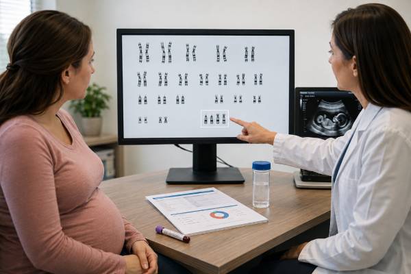
An Edwards syndrome genetic test looks for extra chromosome 18 material, a finding called trisomy 18. During pregnancy, some tests estimate the chance of trisomy 18, while others examine fetal or placental cells and can establish a diagnosis. That distinction matters because a high-chance screening result is not proof that the fetus is affected. It may be followed by chorionic villus sampling or amniocentesis, chromosome analysis, and detailed ultrasound. After birth, a blood chromosome test can confirm the diagnosis and show whether trisomy 18 is full, mosaic, or partial. Edwards syndrome is usually associated with major medical and developmental needs and a high chance of pregnancy loss or early death, but individual outcomes are not identical. Accurate counseling should address the exact chromosome result, the ultrasound findings, the family’s goals, and the full range of care choices without treating a screening result as a final answer.
- Condition tested: Extra genetic material from chromosome 18
- Screening is not diagnosis: cfDNA and combined screening estimate chance
- Confirmation methods: CVS or amniocentesis with chromosome testing
- Result forms: Full, mosaic, partial, or translocation-related trisomy 18
- Ultrasound role: Identifies structural findings and guides pregnancy planning
- Counseling focus: Prognosis, care options, recurrence, and family preferences
Table of Contents
- What Trisomy 18 Means
- How Pregnancies Are Screened
- Ultrasound Findings and Their Limits
- Confirming the Diagnosis
- Understanding the Laboratory Report
- Prognosis and Care Planning
- Recurrence and Future Pregnancies
- Next Steps After Each Result
What Trisomy 18 Means
Most cells normally contain two copies of chromosome 18. Edwards syndrome occurs when there is a third complete or partial copy. The extra genes change development across many organ systems. The condition is also called trisomy 18, written as T18 on many screening reports.
There are three major chromosome patterns:
| Pattern | Chromosome finding | Why the distinction matters |
|---|---|---|
| Full trisomy 18 | Three complete copies of chromosome 18 in the tested cells | Most common form; usually caused by a random chromosome-separation error |
| Mosaic trisomy 18 | A mixture of cells with trisomy 18 and cells with the usual chromosome number | Effects can vary, and one tissue sample may not represent every organ |
| Partial trisomy 18 | An extra segment of chromosome 18, sometimes attached to another chromosome | Features depend on the duplicated segment; parental testing may be needed |
Full trisomy 18 accounts for roughly 9 in 10 diagnosed cases. It usually begins with nondisjunction, an error when chromosome copies separate during formation of an egg or sperm. The chance of nondisjunction increases with maternal age, but trisomy 18 can occur at any reproductive age. It is generally not caused by anything a parent did before or during pregnancy.
Mosaic trisomy 18 develops when an error occurs after fertilization. Some cell lines then carry an extra chromosome 18 while others do not. The percentage of trisomic cells in blood, placenta, or amniotic fluid does not precisely predict the percentage in the heart, brain, kidneys, or other tissues. A person with a low percentage in one sample may still have important medical needs, while another person may have a less severe presentation.
Partial trisomy 18 involves only part of the chromosome. It may result from an unbalanced translocation or inversion inherited from a parent who has a balanced chromosome rearrangement. The size and location of the duplicated region strongly influence the findings. A partial result therefore cannot be interpreted simply by comparing it with full trisomy 18.
The genetic test identifies chromosome material, not a fixed life course. Even with full trisomy 18, the combination of heart, brain, kidney, airway, feeding, and growth differences varies. The chromosome result should be considered with fetal imaging and, after birth, the infant’s actual clinical condition.
How Pregnancies Are Screened
Prenatal screening can identify a pregnancy with a higher chance of trisomy 18 before a definitive procedure is performed. Screening is voluntary. A patient may choose screening, diagnostic testing, both in sequence, or neither after counseling about benefits and limitations.
Cell-free DNA screening analyzes small DNA fragments released mainly from the placenta into maternal blood. It is often available from about 10 weeks. The cell-free DNA prenatal test is the most sensitive screening approach for common trisomies, including trisomy 18, but it is not diagnostic. A placental DNA result may differ from fetal chromosomes because of confined placental mosaicism. Other causes of discordant results include a vanished twin, maternal chromosome findings, maternal illness, low fetal fraction, and technical factors.
Combined first-trimester screening uses maternal age, nuchal translucency ultrasound, pregnancy-associated plasma protein A, and human chorionic gonadotropin. In a pregnancy affected by trisomy 18, the biochemical pattern often differs from an unaffected pregnancy, but no single marker establishes the diagnosis. The first-trimester screening test provides a calculated chance for trisomy 18 and other common aneuploidies within a specific gestational window.
Second-trimester serum screening may include trisomy 18 risk assessment in some health systems. Marker levels can suggest an increased chance, but performance is lower than cfDNA screening. The exact conditions included and the cutoff for a “positive” result vary by laboratory and country.
A report may use one of several formats:
- “High risk,” “screen positive,” or “increased chance”
- “Low risk,” “screen negative,” or “decreased chance”
- A ratio such as 1 in 20 or 1 in 5,000
- A percentage or positive predictive value
- “No result,” “no call,” or “insufficient fetal fraction”
A 1-in-20 result means the screening model estimates a 5% chance, not that 5% of the fetus is affected. A high-risk cfDNA result may have a stronger predictive value than a high-risk serum result, but neither confirms trisomy 18. Positive predictive value also changes with the person’s starting chance and the presence or absence of ultrasound findings.
A low-risk result substantially reduces the chance but does not eliminate it. False-negative results can occur. A nonreportable cfDNA result is not equivalent to a negative result and deserves follow-up. Depending on timing and clinical findings, options may include repeating the blood draw, changing screening methods, proceeding to detailed ultrasound, or choosing diagnostic testing.
Using several screening tests independently can create confusing or contradictory probabilities. Clinicians usually recommend one primary aneuploidy screening strategy rather than stacking multiple calculations. Ultrasound remains important even after low-risk screening because it evaluates development that cfDNA cannot assess.
Ultrasound Findings and Their Limits
Trisomy 18 is often associated with a recognizable pattern on ultrasound, especially when several findings occur together. Possible prenatal findings include:
- Fetal growth restriction
- Increased nuchal translucency or cystic hygroma
- Congenital heart differences
- Clenched hands with overlapping fingers
- Small jaw or unusual skull shape
- Choroid plexus cysts
- Omphalocele or other abdominal-wall differences
- Kidney anomalies
- Single umbilical artery
- Foot positioning differences, including a prominent heel
- Brain or neural tube abnormalities
- Too much or too little amniotic fluid
No single ultrasound sign diagnoses Edwards syndrome. For example, an isolated choroid plexus cyst often resolves and usually has little significance when detailed anatomy is otherwise normal and aneuploidy screening is reassuring. The meaning changes when the cyst appears with growth restriction, heart disease, clenched hands, or several other findings.
Conversely, a fetus with trisomy 18 may not show every classic feature, particularly early in pregnancy. A normal first-trimester scan cannot exclude the condition. Some structural differences become easier to see during the second trimester, and growth restriction may emerge later.
After a positive screening result, a detailed ultrasound can help in several ways. It may identify findings that raise or lower the clinical suspicion, guide the timing and type of diagnostic testing, identify conditions that require fetal echocardiography, and support delivery planning. It cannot convert a screen into a diagnosis. A normal ultrasound after a positive cfDNA result reduces concern in some contexts but does not reliably rule out trisomy 18.
When multiple anomalies are present but rapid testing for trisomy 18 is negative, broader testing may be appropriate. Chromosomal microarray can detect smaller deletions or duplications. In selected cases, exome sequencing may identify a single-gene condition that resembles an aneuploidy. The testing plan should match the ultrasound pattern and family history rather than assuming every similar presentation has the same cause.
Serial ultrasound may be recommended after a confirmed diagnosis to monitor growth, amniotic fluid, fetal movement, heart function, and other concerns. The purpose of surveillance should be discussed explicitly. Some families want information for delivery preparation, while others choose a comfort-focused plan and prefer only testing that will change care.
Confirming the Diagnosis
A definitive prenatal diagnosis requires cells from the placenta or amniotic fluid. The two standard procedures are chorionic villus sampling and amniocentesis.
Chorionic villus sampling obtains a small amount of placental tissue, generally in the first trimester. It offers earlier information. The sample can be tested rapidly for common aneuploidies and then analyzed by karyotype or microarray. Because CVS tests placenta, a mosaic result may represent confined placental mosaicism rather than the fetus. The CVS genetic test article explains sampling routes, timing, and placental interpretation.
Amniocentesis obtains amniotic fluid through a needle placed under ultrasound guidance, usually at or after 15 weeks. Fetal cells in the fluid are examined for chromosome abnormalities. The amniocentesis genetic test is often recommended when a CVS mosaic finding needs clarification or when the pregnancy is beyond the typical CVS window.
Both procedures have a small complication risk, including pregnancy loss. The patient-specific risk depends on gestational age, anatomy, operator experience, and other medical factors. A discussion should include the value of a definitive answer, how the result would be used, and whether the family prefers earlier testing or waiting for amniocentesis.
Laboratory methods may include:
Rapid aneuploidy testing. FISH or quantitative fluorescent polymerase chain reaction can look for chromosome 18 and other common aneuploidies. Results may be available sooner than a full chromosome study. They are often considered preliminary until the final analysis is complete.
Karyotype. A prenatal karyotype counts chromosomes and shows large structural rearrangements. It can distinguish three separate chromosome 18 copies from an unbalanced translocation, which is important for recurrence counseling.
Chromosomal microarray. Microarray detects copy-number changes across the genome at higher resolution than karyotype. It readily detects full trisomy 18 and may define a partial duplication. However, karyotype or another structural method may still be needed to show how an extra segment is arranged and whether a balanced parental rearrangement is possible.
If CVS shows full trisomy 18 in both direct and cultured analyses and ultrasound findings are consistent, the result is generally considered diagnostic. If low-level mosaicism appears only in placental cells, amniocentesis may be offered to determine whether fetal cells are affected. A genetic counselor can explain whether the uncertainty comes from biology, sample type, or laboratory limitations.
After birth, a blood karyotype usually confirms the diagnosis. If mosaicism is strongly suspected but blood testing is normal or does not fit the clinical findings, testing another tissue may occasionally be discussed. That is a specialist decision because different tissues can contain different cell lines.
Understanding the Laboratory Report
A chromosome report may look technical, but several elements are especially useful: specimen type, method, number of cells examined, chromosome notation, and the laboratory’s interpretation.
Common full-trisomy notations include:
47,XX,+18— 47 chromosomes, two X chromosomes, and an extra chromosome 1847,XY,+18— 47 chromosomes, one X and one Y chromosome, and an extra chromosome 18
A mosaic report may show two cell lines separated by a slash. For example, it may list a trisomy 18 line and a 46-chromosome line, followed by bracketed cell counts. Those counts describe the cells examined in that sample. They are not a direct forecast of survival, development, or organ involvement.
A partial or translocation result contains additional abbreviations, such as der for derivative chromosome, dup for duplication, or t for translocation. The report may state that extra material from the long arm of chromosome 18 is attached to another chromosome. The exact breakpoints matter because they identify which genes are present in three copies.
Possible report conclusions include:
| Report wording | Practical meaning |
|---|---|
| Trisomy 18 detected | Diagnostic sample shows extra chromosome 18 material |
| No evidence of trisomy 18 | The tested method did not identify trisomy 18 |
| Mosaic trisomy 18 | More than one chromosome cell line was found |
| Suspected confined placental mosaicism | Abnormal placental cells may not represent fetal cells |
| Variant or copy-number change of uncertain significance | A finding was detected, but its health meaning is not established |
A normal rapid result does not necessarily equal a normal full chromosome study. Rapid testing targets selected chromosomes. Similarly, a normal karyotype does not exclude every small DNA change or single-gene disorder. The scope of the test must be read from the report.
When a screening report is positive but amniocentesis is normal, the pregnancy is usually considered not affected by fetal trisomy 18. The discordant cfDNA signal may have come from the placenta or another biological source. Depending on placental findings and fetal growth, the care team may recommend additional monitoring.
Families should keep the full laboratory report, not only a portal message. It may be needed for parental testing, future pregnancies, pediatric care, or review by another genetics service.
Prognosis and Care Planning
Full trisomy 18 is a serious, life-limiting condition. There is a high rate of miscarriage and stillbirth. Among liveborn infants, many die in the first days, weeks, or months because of central apnea, heart disease, breathing difficulties, infection, feeding problems, or combinations of medical complications. Some children survive for years, particularly among those with mosaic or partial forms, and a smaller number with full trisomy 18 also live beyond infancy.
Population statistics describe groups; they cannot predict an exact course for one fetus or infant. Prognosis is influenced by chromosome pattern, prematurity, heart anatomy, airway stability, neurologic function, ability to feed, kidney and gastrointestinal findings, and the level of medical intervention chosen and available.
A confirmed prenatal diagnosis should lead to individualized planning rather than a single automatic pathway. Families may consider:
- Continuing the pregnancy with standard or increased fetal surveillance
- Continuing with a perinatal palliative-care plan focused on comfort
- Planning selected medical interventions after birth
- Requesting intensive neonatal evaluation and treatment when medically appropriate
- Ending the pregnancy where legally and medically available
These choices can be emotionally and ethically complex. Counseling should be non-directive and should not imply that every family will make the same decision. It should also avoid guaranteeing either death at a specific time or long-term survival.
For a continued pregnancy, a multidisciplinary meeting can include maternal-fetal medicine, neonatology, pediatric cardiology, genetics, palliative care, nursing, social work, and spiritual care if desired. A birth plan may address fetal monitoring during labor, location and mode of delivery, resuscitation preferences, feeding support, comfort medicines, family time, photography, memory-making, and how decisions will be revisited if the infant’s condition differs from expectations.
Recent pediatric guidance emphasizes shared decision-making for infants and children with trisomy 18. Some interventions may offer meaningful benefit in selected patients, while others may add burden without achieving the family’s goals. Decisions about heart surgery, respiratory support, feeding tubes, and hospitalization should consider the individual child, not chromosome diagnosis alone.
When comfort-focused care is chosen, it is still active medical care. It may include warmth, skin-to-skin contact, relief of pain or breathlessness, feeding as tolerated, and support for parents and siblings. When intensive treatment is chosen, goals can still change if the child’s response or burdens differ from what was expected.
Recurrence and Future Pregnancies
Most full trisomy 18 results are sporadic nondisjunction events. They are not usually inherited. After one affected pregnancy, the chance in a future pregnancy is somewhat higher than the age-related chance alone, often discussed as approximately 0.5% to 1% or the maternal-age risk, whichever is greater. A genetics professional should provide the estimate because it depends on the precise result and reproductive history.
Partial trisomy 18 or an unbalanced translocation changes the evaluation. Both biological parents are commonly offered karyotyping to look for a balanced rearrangement. A balanced carrier has all or nearly all expected chromosome material but arranged differently, so the carrier may be healthy. During egg or sperm formation, however, the rearrangement can produce embryos with missing or extra chromosome material.
If a parent carries a balanced translocation or inversion involving chromosome 18, recurrence can be substantially higher and depends on the exact chromosomes, breakpoints, carrier’s sex, and reproductive history. General population figures are not adequate for that family. A laboratory geneticist or genetic counselor may calculate a case-specific range.
Mosaic trisomy 18 is usually a post-fertilization event and is generally not inherited. Rare germline mosaicism can make recurrence greater than expected even when parental blood karyotypes are normal, but this is uncommon.
Options for a later pregnancy include early cfDNA screening, combined screening, CVS, amniocentesis, or a direct diagnostic approach. When a familial rearrangement is known, targeted prenatal testing can look specifically for the unbalanced chromosome pattern. Some families consider in vitro fertilization with preimplantation genetic testing for structural rearrangements. Embryo testing reduces but does not eliminate uncertainty, and prenatal confirmation is generally discussed.
The most useful preparation is to obtain and preserve the original fetal or child chromosome report. The term “trisomy 18” alone does not show whether the case was full, mosaic, partial, or translocation-related.
Next Steps After Each Result
A result should lead to a next step that fits both its certainty and the family’s goals.
After a low-chance screen: Continue routine prenatal care and the recommended anatomy ultrasound. Reconsider diagnostic testing if new structural findings appear, growth becomes concerning, or greater certainty is desired.
After a high-chance screen: Arrange genetic counseling and a detailed ultrasound. Review the positive predictive value and decide whether CVS or amniocentesis would provide useful confirmation. Avoid making irreversible decisions from screening alone.
After a no-call cfDNA result: Ask why the sample failed and whether a repeat is likely to succeed. Discuss ultrasound and diagnostic testing, especially when gestational timing is important or fetal fraction is persistently low.
After mosaic CVS findings: Clarify whether the result was seen in direct testing, cultured cells, or both. Amniocentesis may help determine whether the fetus has trisomy 18 or the finding is limited to the placenta.
After a confirmed prenatal diagnosis: Review the full karyotype, detailed anatomy, fetal echocardiogram, and expected delivery issues. Meet relevant pediatric and palliative-care teams. Discuss all legally and medically available pregnancy options without pressure.
After a postnatal diagnosis: Confirm with blood chromosome analysis, assess breathing and feeding, evaluate the heart and kidneys, and create a care plan aligned with the family’s priorities. Ask whether parental karyotypes are needed.
Questions that often clarify the situation include:
- Was the result from screening or diagnostic testing?
- Does it show full, mosaic, or partial trisomy 18?
- Which ultrasound findings are present, and which are absent?
- Could placental mosaicism explain the result?
- What information would another test add?
- What are the realistic care choices at this hospital?
- Should either parent have a karyotype?
- Who can help us plan for delivery, comfort care, or neonatal treatment?
The most responsible interpretation combines the exact laboratory finding with the fetus or child’s actual medical features. This approach preserves diagnostic accuracy while allowing families to make values-based decisions with clear, compassionate support.
References
- Society for Maternal-Fetal Medicine Consult Series #74:Cell-free DNA screening for aneuploidies: Updated guidance 2025 (Guideline)
- Guidance for Caring for Infants and Children With Trisomy 13 and Trisomy 18: Clinical Report 2025 (Clinical Report)
- Edwards syndrome (trisomy 18) 2025 (Clinical Resource)
- Trisomy 18 (Edwards Syndrome) 2025 (Clinical Review)
- Trisomy 18 2025 (Medical Encyclopedia)
- Down’s syndrome, Edwards’ syndrome and Patau’s syndrome screening pathway requirements 2025 (Program Guidance)
Disclaimer
This article provides general education about Edwards syndrome screening, diagnosis, and chromosome results. It cannot predict an individual pregnancy or child’s course and does not replace counseling from maternal-fetal medicine, neonatology, pediatrics, clinical genetics, or a genetic counselor. Procedure risks, treatment options, pregnancy choices, and local legal availability vary and should be discussed with the treating team.





