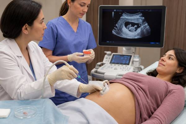
Chorionic villus sampling, or CVS, is an early prenatal diagnostic procedure that collects a small sample of placental tissue for chromosome or DNA testing. It is usually performed between 10 and 13 weeks of pregnancy, allowing a diagnostic answer earlier than amniocentesis. The sample contains chorionic villi, tiny projections from the placenta that generally carry the same genetic information as the fetus. CVS can diagnose common chromosome conditions, identify smaller deletions or duplications with chromosomal microarray, and test for a known inherited gene variant. It does not routinely assess open neural tube defects, and a placental result can occasionally differ from the fetal chromosome pattern because of confined placental mosaicism. The procedure may be done through the abdomen or cervix under continuous ultrasound guidance. Results often arrive in stages, with rapid chromosome testing in several days and broader analysis in one to three weeks. CVS is optional and should follow counseling about test scope, uncertainty, miscarriage risk, and possible follow-up with amniocentesis.
- CVS is a diagnostic procedure, not a screening test, for the chromosome or gene conditions specifically ordered.
- It is commonly performed at 10–13 weeks, providing earlier diagnostic information than amniocentesis.
- The sample comes from the placenta, so rare placental-fetal differences can complicate interpretation.
- Rapid results may return in 2–4 working days, while karyotype, microarray, or gene testing usually takes longer.
- CVS does not directly screen for open neural tube defects; later AFP testing and ultrasound may still be needed.
- Urgent review is needed after CVS for heavy bleeding, fluid leakage, fever, severe pain, or persistent contractions.
Table of Contents
- What CVS can test
- Why CVS is offered
- How CVS is performed
- Laboratory analysis of chorionic villi
- Interpreting CVS results
- Placental mosaicism and discordant findings
- Risks, recovery, and warning signs
- Choosing CVS or amniocentesis
What CVS can test
CVS obtains placental cells that can be used for several different genetic analyses. The procedure itself does not determine which conditions are tested; the laboratory order does. Two patients may undergo the same sampling procedure but receive very different reports.
Common analyses include:
- Rapid testing for trisomy 21, trisomy 18, trisomy 13, and selected sex chromosome differences
- Karyotype to examine chromosome number and large structural rearrangements
- Chromosomal microarray to detect smaller deletions and duplications
- Targeted testing for a known familial gene variant
- Biochemical or enzyme testing for selected inherited metabolic disorders
- Prenatal exome sequencing in carefully selected pregnancies with fetal anomalies
CVS is particularly useful when a parent carries a known pathogenic variant or balanced chromosome rearrangement. The laboratory can test the placenta for the specific familial change early in pregnancy. A copy of the relative’s original report is often needed so the prenatal laboratory can confirm the exact variant, coordinates, or rearrangement.
CVS does not automatically provide a broad genetic “all clear.” A normal rapid aneuploidy test does not exclude smaller copy-number changes. A normal microarray does not rule out most single-gene disorders. A targeted gene test answers only the family-specific question it was designed to answer.
The sample cannot directly measure fetal anatomy. Ultrasound remains necessary, including the first-trimester scan and the detailed anatomy scan later in pregnancy. CVS also does not routinely test amniotic-fluid alpha-fetoprotein, so maternal serum AFP screening and ultrasound may still be offered for neural tube defect assessment.
When a fetal structural anomaly is already visible, chromosomal microarray is often more informative than karyotype alone. If microarray is normal and the anomaly pattern suggests a monogenic disorder, a specialist may discuss prenatal exome sequencing using DNA from CVS and parental samples.
Why CVS is offered
CVS may be offered after a screening test, ultrasound finding, family history, or parental genetic result raises the chance of a fetal condition. Some people also choose diagnostic testing because they want a definitive chromosome result rather than a probability estimate.
Typical reasons include:
- A high-chance cell-free DNA result for trisomy 21, 18, 13, or another chromosome condition
- Increased nuchal translucency, cystic hygroma, or an early structural anomaly
- A previous pregnancy or child with a genetic condition
- A parent with a balanced translocation, inversion, or other chromosome rearrangement
- Both genetic parents carrying variants for the same autosomal recessive condition
- A pregnant person carrying an X-linked condition
- A known familial pathogenic variant with a significant recurrence risk
- A request for early diagnostic testing after informed counseling
CVS can provide results weeks earlier than amniocentesis. Earlier timing may matter for emotional preparation, specialist planning, pregnancy management, or access to pregnancy termination where chosen and legally available. Timing should not replace careful test selection, however. An early answer is useful only if the correct laboratory analysis is ordered and the possibility of placental mosaicism is understood.
A high-chance NIPT result remains screening information. CVS may confirm the suspected condition, but because both cfDNA and CVS primarily assess placental material, amniocentesis may sometimes be preferred for findings known to have a higher chance of confined placental mosaicism.
The ultrasound and chromosome involved help guide the procedure choice. A positive trisomy 21 screen with matching fetal findings may be approached differently from a rare autosomal trisomy or monosomy X result with a normal ultrasound. Genetic counseling should explain why CVS, amniocentesis, or occasionally either procedure is reasonable.
CVS is optional. Declining it does not mean declining all prenatal care. A person may choose continued ultrasound surveillance, another screening test, amniocentesis later, or postnatal testing depending on the clinical question and personal priorities.
How CVS is performed
Before CVS, an ultrasound confirms fetal viability, gestational age, placental location, number of fetuses, and a safe sampling route. The clinician also reviews blood group, Rh status, infections, medications, allergies, bleeding history, and any prior uterine or cervical procedures.
CVS should not be performed before 10 weeks because very early procedures were historically associated with limb and jaw abnormalities. Many centers schedule it from 11 weeks, while others begin at 10 weeks when experienced operators and appropriate indications are present.
There are two main approaches.
Transabdominal CVS
A thin needle passes through the abdominal and uterine walls into the placenta under ultrasound guidance. Suction collects a small amount of chorionic villi. The experience is often described as pressure, stinging, or cramping. Local anesthetic may be used, although practice varies.
Transabdominal CVS is commonly selected when the placenta is accessible through the abdomen. It resembles amniocentesis in the route of entry, but the needle samples placental tissue rather than amniotic fluid.
Transcervical CVS
A thin catheter or sampling instrument passes through the vagina and cervix to the placenta under ultrasound guidance. The clinician collects villi through gentle suction. This route may be chosen for a placenta that is easier to reach through the cervix.
Transcervical CVS can cause light vaginal bleeding or cramping. It may not be appropriate with active genital infection, heavy bleeding, cervical anatomy that limits access, or certain placental locations.
The procedure route is based on safety and access, not patient preference alone. Neither route is universally superior when performed by an experienced clinician. The actual collection often takes only a few minutes, but the entire appointment may last 30–60 minutes.
The sample is examined immediately to confirm that chorionic villi are present. If the amount is insufficient, the operator may make another pass or the laboratory may later request a repeat procedure. In multiple pregnancies, each placenta or relevant sampling site must be mapped and labeled carefully so results are assigned to the correct fetus.
Twin pregnancies require a precise plan before the first instrument enters the uterus. The clinician determines chorionicity, meaning whether the twins share a placenta, and maps the placental tissue for each fetus. In dichorionic twins, separate placental samples are usually collected and labeled. In monochorionic twins, the interpretation depends on whether they are expected to be genetically identical and on the specific reason for testing. A vanishing twin or fused placentas can make assignment more difficult.
The laboratory requisition should identify each sample consistently with the ultrasound map, such as fetus A and fetus B. If one twin has an ultrasound abnormality, the team must make sure the relevant placental site is linked to that fetus. Couples should ask whether the planned genetic test will be performed on both samples, whether separate maternal-cell contamination studies are needed, and what happens if one sample is inadequate.
Placental location can also change procedural complexity. A posterior placenta may be easier to reach transcervically in some cases, while an anterior placenta may favor a transabdominal route. Fibroids, uterine position, bowel, bladder, prior cesarean scars, and cervical anatomy may influence access. The route can change on the day of the procedure if the ultrasound shows that the original plan is no longer the safest.
If the first sample is small, the operator balances the value of another pass against the added procedural manipulation. A laboratory experienced in prenatal specimens may be able to prioritize rapid testing, culture, or DNA extraction based on the available tissue. This is another reason the desired analyses should be decided before CVS rather than after the sample reaches the laboratory.
Rh-negative patients may receive anti-D immune globulin after CVS according to local guidance. Fasting is usually unnecessary, and most medications can be continued unless the procedure team gives specific instructions about anticoagulants or other drugs.
Laboratory analysis of chorionic villi
Chorionic villi contain an outer trophoblast layer and an inner mesenchymal core. These cell populations can have different chromosome patterns, which is important when mosaicism appears. Laboratories may use direct preparations, cultured cells, DNA extraction, or a combination.
Rapid methods such as quantitative fluorescent PCR or FISH can provide an early result for selected chromosomes. A rapid result may be available within a few working days, but it does not replace broader testing when karyotype or microarray has also been ordered.
| Analysis | What it detects | Important limitation |
|---|---|---|
| QF-PCR or rapid FISH | Selected common aneuploidies | Does not assess all chromosomes or small copy-number changes |
| Karyotype | Chromosome number and large rearrangements | Lower resolution than microarray and may require culture |
| Chromosomal microarray | Aneuploidies and smaller deletions or duplications | May identify uncertain findings and generally misses balanced rearrangements |
| Targeted molecular test | A specific familial variant or repeat expansion | Answers only the defined genetic question |
| Exome sequencing | Variants in thousands of coding genes | Can return uncertain or unrelated findings and may take several weeks |
Maternal cell contamination is a concern because maternal decidual tissue lies close to the chorionic villi. Laboratories dissect the sample and may compare maternal DNA markers to ensure the report reflects placental cells rather than maternal tissue. A maternal blood sample may therefore be requested.
For single-gene testing, the laboratory may need parental samples to establish phase, confirm sample identity, or interpret a fetal variant. For PGT-confirmed pregnancies, prenatal testing is still offered because embryo testing and CVS use different samples and have different technical limitations.
Turnaround depends on the test, need for cell culture, and whether the sample is adequate. Rapid results may precede the final report. Patients should know whether a phone call communicates a preliminary result or the complete laboratory interpretation.
Interpreting CVS results
CVS results can be normal, abnormal, uncertain, mosaic, or unsuccessful. The report should be interpreted with the ultrasound, screening history, test method, and whether the finding appeared in direct or cultured placental cells.
Normal result
A normal result means no abnormality was found within the scope of the ordered test. A normal karyotype does not rule out small deletions, single-gene conditions, structural birth differences, or developmental disorders. A normal microarray does not exclude most sequence variants.
When CVS was performed for a known familial variant, a negative targeted result usually means the fetus did not inherit that specific variant. The laboratory should confirm that the familial change was detectable and that sample identity and contamination checks passed.
Abnormal result
A clearly abnormal result may identify trisomy, monosomy, an unbalanced rearrangement, pathogenic deletion or duplication, or a disease-causing gene variant. Counseling should cover the expected range of outcomes, whether the finding is fetal or potentially confined to placenta, and whether parental studies are needed.
A positive result can influence ultrasound surveillance, fetal echocardiography, delivery location, neonatal care, or personal pregnancy decisions. The chromosome or gene result does not predict every feature or severity level.
Variant of uncertain significance
A VUS means evidence is insufficient to classify a finding as harmful or benign. Microarray may identify a copy-number change with variable or unknown effects, while gene testing may find a sequence variant that has not been well studied. Testing parents and reviewing fetal anatomy can help, but uncertainty may remain.
A VUS should not be presented as a confirmed diagnosis. Laboratories may reclassify it as evidence accumulates, so families should keep the report and request review before future pregnancies.
No result or inadequate sample
Occasionally, too little villous tissue is collected, cells fail to grow, contamination is excessive, or DNA quality is poor. Options include repeating CVS, waiting for amniocentesis, using another method on stored material, or relying on postnatal testing. The best choice depends on gestational age and the urgency of the question.
Placental mosaicism and discordant findings
Mosaicism means two or more chromosome cell lines are present. In CVS, an abnormal cell line may be limited to the placenta while the fetus has a normal chromosome pattern. This is called confined placental mosaicism and occurs in roughly 1% to 2% of CVS samples, depending on definitions and laboratory methods.
Placental mosaicism develops because the placenta and fetus arise from related but distinct early cell lineages. A chromosome error can occur after these lineages begin to separate, leaving the change in one tissue but not the other.
When mosaicism is reported, the laboratory considers:
- Which chromosome is involved
- Whether the finding appears in trophoblast, mesenchymal core, or both
- The percentage and distribution of abnormal cells
- Whether ultrasound shows fetal abnormalities
- Whether the chromosome is associated with uniparental disomy after trisomy rescue
- Whether the finding could affect placental function and fetal growth
Amniocentesis may be recommended because amniotic-fluid cells more closely represent fetal tissues. A normal amniocentesis can show that the CVS result was confined to placenta, but pregnancy follow-up may still include growth ultrasound because some placental chromosome changes increase the chance of fetal growth restriction or other placental complications.
Some chromosomes have a higher likelihood of true fetal involvement than others. Rare autosomal trisomies detected by cfDNA or CVS require chromosome-specific counseling rather than one general rule. Uniparental disomy testing may be considered for chromosomes with known imprinting disorders.
Discordance can also work in the opposite direction: a normal placental sample may rarely miss fetal mosaicism. This is one reason a normal CVS cannot guarantee that every fetal cell has the same chromosome pattern.
A result described as “mosaic” should not be interpreted from the percentage alone. Culture conditions can change the apparent proportion of cell lines, and one small placental biopsy may not represent the entire placenta.
Risks, recovery, and warning signs
CVS carries a small procedure-related risk of pregnancy loss. The exact additional risk is difficult to measure because many patients undergoing CVS already have ultrasound or genetic findings associated with miscarriage. Modern estimates from experienced centers are generally lower than older figures and often fall around 0.2% to 0.5%, though individual services may counsel differently.
Risk can be influenced by gestational age, operator experience, placental access, bleeding, multiple pregnancy, fetal abnormalities, and the number of needle or catheter passes. Ask the center for its current local estimate and whether it distinguishes background loss from procedure-attributable loss.
Other possible complications include:
- Cramping and light spotting
- Heavier vaginal bleeding
- Infection, which is uncommon
- Rupture of membranes or fluid leakage
- Rh sensitization
- Inadequate sample requiring another procedure
- Rare transmission of certain maternal infections
After CVS, mild cramps or spotting may last several hours. Follow the clinic’s guidance on exercise, lifting, sex, work, and travel. Bed rest has not been proven to prevent miscarriage, but a quiet remainder of the day may be advised for comfort.
Contact the maternity team urgently for heavy or increasing bleeding, fluid leakage, fever, chills, foul-smelling discharge, severe or persistent pain, regular contractions, dizziness, or feeling acutely unwell. Emergency assessment is appropriate for severe bleeding, fainting, or rapidly worsening symptoms.
The emotional period after CVS can be as difficult as the physical recovery. Ask before the procedure who will call with rapid and final results, whether results are released electronically first, and what support is available if the finding is abnormal or uncertain.
Choosing CVS or amniocentesis
CVS and amniocentesis are both diagnostic sampling procedures, but they differ in timing and tissue source. CVS provides an earlier result from placenta. Amniocentesis is generally performed from 15 weeks and tests cells in amniotic fluid.
| Feature | CVS | Amniocentesis |
|---|---|---|
| Usual timing | About 10–13 weeks | From about 15 weeks |
| Sample | Placental villi | Amniotic fluid containing fetal cells |
| Main advantage | Earlier diagnosis | Lower concern for confined placental mosaicism |
| Special limitation | Placental mosaicism may require clarification | Later timing |
| Neural tube assessment | Not directly assessed | Amniotic-fluid AFP may be measured when indicated |
CVS may be favored when early diagnosis is important or when testing a known family variant. Amniocentesis may be favored after a result prone to placental discordance, when gestational age is already 15 weeks or more, or when amniotic-fluid infection testing is needed.
Before choosing, ask which procedure best answers the exact question, not simply which is earlier. Questions to discuss include the suspected condition, chance of mosaicism, laboratory method, local operator experience, miscarriage estimate, turnaround time, and what would happen if the result is uncertain.
The decision is personal. Some people value the earliest possible answer; others prefer to wait for amniocentesis to reduce placental ambiguity. Either choice should be supported without pressure, including the choice to decline invasive testing.
References
- Prenatal Genetic Diagnostic Tests 2025 (Clinical Guidance)
- Chorionic Villus Sampling 2025 (Review)
- Amniocentesis and Chorionic Villus Sampling 2021 (Guideline)
- Placental mosaicism in prenatal diagnosis 2026 (Review)
- Placental mosaicism for autosomal trisomies 2024 (Review)
- An overview of current prenatal genetic screening and diagnostic testing 2025 (Review)
Disclaimer
This article provides general education and does not replace individualized counseling from a maternal-fetal medicine specialist, obstetric clinician, or genetic counselor. CVS timing, procedure risk, laboratory selection, and the need for amniocentesis after a mosaic result depend on the specific pregnancy. Seek urgent care after CVS for heavy bleeding, fluid leakage, fever, severe pain, fainting, or persistent contractions.