
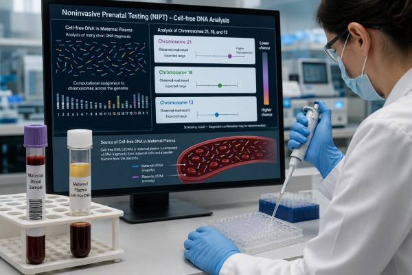
Noninvasive prenatal testing (NIPT), also called prenatal cell-free DNA screening, estimates the chance that a pregnancy has trisomy 21, trisomy 18, or trisomy 13 from a maternal blood sample. It can usually be performed from 9–10 weeks of pregnancy and is the most sensitive and specific screening method for these common chromosome conditions. Yet NIPT is not a diagnosis. Most DNA fragments measured as “fetal” actually come from the placenta, so a high-risk result can disagree with the fetus, and a low-risk result cannot exclude every chromosome or genetic condition. The usefulness of a result depends on the condition screened, the pregnancy’s starting chance, fetal fraction, ultrasound findings, and laboratory method. A high-risk or nonreportable result should lead to genetic counseling, detailed ultrasound, and discussion of chorionic villus sampling or amniocentesis. Understanding the report before making irreversible decisions protects both accuracy and personal choice.
- Timing: Most laboratories accept NIPT from 9–10 weeks, although drawing later may reduce low-fetal-fraction failures.
- Core conditions: Standard panels screen for trisomy 21, trisomy 18, and trisomy 13; added conditions vary by laboratory.
- Low risk is not zero risk: A negative result greatly lowers the chance of the screened trisomies but does not rule them out completely.
- High risk needs confirmation: Do not make permanent pregnancy decisions from NIPT alone; use CVS or amniocentesis for diagnosis.
- No result needs follow-up: A “no-call” result is not reassuring and may carry a higher chance of aneuploidy or placental complications.
- Ultrasound still matters: NIPT does not replace first-trimester assessment, anatomy ultrasound, or neural tube defect screening.
Table of Contents
- What NIPT Measures in Maternal Blood
- Choosing the Test and the Right Time
- How to Read Low-Risk, High-Risk, and No-Call Results
- Trisomy 21, Trisomy 18, and Trisomy 13 Compared
- Why a Screen Can Disagree With the Fetus
- Next Steps After an Abnormal or No-Call Result
- Twins, Vanishing Twins, and Other Special Situations
- What NIPT Does Not Replace
What NIPT Measures in Maternal Blood
NIPT analyzes short DNA fragments circulating in the pregnant person’s plasma. Most fragments come from maternal blood cells. The smaller pregnancy-related portion comes mainly from placental cells called trophoblasts, which usually share the fetus’s chromosome pattern. Laboratories often call this portion “fetal” cell-free DNA, but “placental DNA” is more biologically precise.
The test asks whether the sample contains more or less DNA than expected from particular chromosomes. An excess contribution from chromosome 21 suggests an increased chance of trisomy 21; the same principle applies to chromosomes 18 and 13. Different laboratories use different methods:
- Whole-genome counting compares millions of fragments across all chromosomes.
- Targeted sequencing focuses on selected chromosome regions.
- Single-nucleotide polymorphism analysis studies inherited DNA markers and may estimate individual fetal fractions in some twin pregnancies.
These methods can all perform well for the common trisomies, but their report formats, test-failure rules, twin capabilities, and optional panels are not identical. A result from one laboratory should be interpreted using that laboratory’s stated method and limitations.
The fetal fraction is the percentage of total cell-free DNA attributed to the pregnancy. It generally rises as pregnancy advances. Most laboratories require a minimum around 2%–4%, although the cutoff and the way fetal fraction is calculated vary. When the fraction is too low, the laboratory may issue no result rather than risk an unreliable interpretation.
NIPT is considered noninvasive because it requires a blood draw from the pregnant person and does not enter the uterus. The blood draw itself does not add a procedure-related miscarriage risk. This safety advantage is important, but it does not turn screening into diagnosis. A laboratory algorithm measures probability from mixed maternal and placental DNA; it does not directly count chromosomes in fetal cells.
Professional guidance supports offering prenatal chromosome screening and diagnostic testing options to all pregnant patients, regardless of age or baseline risk. A person may choose NIPT, another screening strategy, direct diagnostic testing, or no chromosome testing after informed counseling. Screening should be offered, not treated as mandatory.
Choosing the Test and the Right Time
NIPT is available early enough to provide information during the first trimester. Many programs begin at 10 weeks, while some validated assays accept samples at 9 weeks. Testing too early increases the chance that the placental DNA contribution is below the laboratory’s threshold. Confirm the gestational age, number of fetuses, and whether a vanished or demised twin was seen before the sample is sent.
No fasting or medication pause is normally required. The sample is collected into a special tube that preserves cell-free DNA during transport. Results often return within about one to two weeks, but timing depends on shipping, laboratory workload, repeat analysis, and the local health system.
Before testing, identify what the ordered panel includes. A basic panel usually covers trisomies 21, 18, and 13. Some laboratories automatically add fetal sex or sex chromosome aneuploidies; others require an opt-in choice. Expanded products may advertise rare autosomal trisomies, genome-wide gains or losses, selected microdeletions, or single-gene conditions. More conditions do not automatically mean a better screen. Rare conditions have lower positive predictive values, less complete validation, and a greater chance of uncertain or unexpected findings.
Current maternal-fetal medicine guidance recommends that sex chromosome screening be an opt-in choice with specific counseling. It does not recommend routine general-population screening for microdeletion syndromes or routine genome-wide screening for large deletions and duplications. Someone seeking information about fetal copy-number changes may receive clearer, broader information from diagnostic testing with chromosomal microarray than from an expanded cfDNA panel.
NIPT should usually not be layered on top of another independent aneuploidy screen simply to seek reassurance. Running multiple screening approaches can create conflicting probabilities and more false-positive results. A clinician may still recommend an ultrasound-based first-trimester assessment because ultrasound examines anatomy and pregnancy development rather than duplicating the same chromosome calculation. The first-trimester screening test can be an alternative when cfDNA is unavailable or declined, but the two should be combined only within a clearly defined clinical strategy.
Pretest counseling should cover four decisions: which conditions to include, whether fetal sex information is wanted, what the person would do with a high-risk or no-call result, and whether diagnostic certainty is preferred from the start. Direct CVS or amniocentesis may be reasonable for a known parental chromosome rearrangement, a previous affected pregnancy, a major fetal anomaly, or a person who wants a definitive answer rather than a probability estimate.
Cost and insurance coverage vary. Ask whether the quoted price includes optional panels, genetic counseling, a redraw after test failure, or diagnostic testing after a positive screen. Financial policy should not be confused with medical accuracy.
How to Read Low-Risk, High-Risk, and No-Call Results
NIPT reports commonly use “low risk,” “high risk,” “positive,” “negative,” or a numerical probability. The words can sound final, but every category needs context.
| Report category | Meaning | Appropriate response |
|---|---|---|
| Low risk or screen negative | The measured chromosome pattern is not suggestive of the screened trisomy. | Continue routine prenatal care and ultrasound; investigate any later abnormal finding independently. |
| High risk or screen positive | The DNA pattern is more consistent with an extra copy of the named chromosome. | Obtain genetic counseling, comprehensive ultrasound, and offer diagnostic confirmation. |
| No call or nonreportable | The laboratory could not issue a reliable result, often because of low fetal fraction or technical limits. | Review gestational age and clinical factors; offer counseling, ultrasound, and diagnostic testing rather than treating it as negative. |
| Atypical or outside test scope | The data do not fit the standard reporting model and may involve placental, maternal, or technical factors. | Discuss the exact laboratory comment with maternal-fetal medicine and genetics specialists. |
A low-risk result greatly lowers the chance of the condition screened. The residual chance is not zero because false negatives can occur, especially with low placental DNA contribution, mosaicism, unusual chromosome structure, or a biological difference between placenta and fetus. A normal NIPT result should never override a major ultrasound abnormality.
A high-risk result is best understood through positive predictive value, or PPV: the chance that the fetus truly has the condition after the screen is positive. PPV is not the same as sensitivity. Sensitivity describes how often a test detects affected pregnancies; PPV asks how often a positive result is confirmed in the population being tested.
PPV rises when the condition is more common. A positive trisomy 21 result generally has a higher PPV than a positive trisomy 13 result because trisomy 21 is more common and the screen performs especially well for it. Maternal age, gestational age, ultrasound findings, prior history, and the laboratory’s performance all change the estimate. A report that gives one PPV without explaining the assumptions may not match an individual pregnancy.
A no-call result is not a neutral blank. Across studies, nonreportable rates vary widely by platform and population; a recent evidence review estimated roughly 0.85%, while individual studies have reported higher rates. Low fetal fraction is the most common reason. Early sampling, higher maternal weight, twin pregnancy, assisted reproduction, some autoimmune conditions, low-molecular-weight heparin use, placental mosaicism, and certain aneuploidies can contribute. A repeat sample produces a result in many cases—often about 75%–80%—but repeating can delay diagnosis and is not always the best choice.
Trisomy 21, Trisomy 18, and Trisomy 13 Compared
The three core conditions all involve an extra chromosome, but they differ in frequency, screening performance, ultrasound patterns, survival, and medical effects. NIPT identifies a chromosome signal; it does not predict the full clinical outcome for an individual fetus.
| Condition | Chromosome finding | Screening notes | Diagnostic questions |
|---|---|---|---|
| Trisomy 21 (Down syndrome) | Extra chromosome 21 material | NIPT has its strongest overall performance for this common trisomy and usually the highest PPV. | Is it full trisomy, mosaicism, or a translocation? A karyotype helps answer this. |
| Trisomy 18 (Edwards syndrome) | Extra chromosome 18 material | Detection is high, but PPV is usually lower than for trisomy 21. Low fetal fraction and placental differences can matter. | Does diagnostic testing confirm full, mosaic, or partial trisomy 18? |
| Trisomy 13 (Patau syndrome) | Extra chromosome 13 material | The condition is rarer, so a positive result has a lower PPV than trisomy 21 in many populations. | Could placental mosaicism or another chromosome 13 rearrangement explain the signal? |
Trisomy 21 is associated with a variable degree of intellectual disability and an increased chance of congenital heart disease, hearing or vision concerns, thyroid disease, and other health needs. Many people with Down syndrome live into adulthood. NIPT cannot predict developmental ability, medical complexity, or whether a specific feature will be present. A confirmed result should be interpreted with balanced information about trisomy 21 testing and outcomes.
Trisomy 18 is associated with fetal growth restriction, heart and other structural differences, clenched hands, neurologic impairment, and a high chance of pregnancy loss or death in infancy. Some children survive longer, and outcomes vary, particularly with mosaic or partial chromosome findings and individualized treatment choices. Screening cannot distinguish these forms; diagnostic chromosome analysis is needed. The distinctions are covered more fully in trisomy 18 diagnosis and results.
Trisomy 13 can involve brain, facial, heart, limb, kidney, and other differences, with a high risk of pregnancy loss and severe medical complications. Mosaic and partial forms may differ from full trisomy 13. Ultrasound can add important information, but a normal early scan does not cancel a high-risk result.
The chromosome named on the report also affects the preferred diagnostic approach. Because NIPT samples placental DNA, CVS may reproduce a placental abnormality that is absent from the fetus. For some result patterns—particularly when ultrasound is normal and confined placental mosaicism is a concern—amniocentesis may give the clearest fetal answer. A genetics or maternal-fetal medicine specialist can match the procedure to the chromosome, gestational age, and ultrasound findings.
Why a Screen Can Disagree With the Fetus
Discordant NIPT results are not always laboratory mistakes. The test measures a biological mixture from the pregnant person and placenta, so several real conditions can produce a high-risk signal when fetal diagnostic testing is normal.
Confined placental mosaicism occurs when some or all placental cells carry a chromosome abnormality that is absent from fetal cells. Because the placenta releases the tested fragments, the screen may accurately describe the placenta but not the fetus. Confined placental mosaicism is a recognized cause of false-positive results and can sometimes be associated with placental dysfunction or fetal growth restriction.
A vanished or demised twin can release DNA for weeks after it stops developing. If that twin had aneuploidy, its DNA may create a high-risk result even when the surviving fetus has a typical chromosome pattern. The interval between the loss and blood draw, the laboratory method, and whether fetal fractions can be separated all affect interpretation.
Maternal chromosome variation can also appear. A pregnant person may have age-related mosaic loss of an X chromosome, a constitutional chromosome difference, a benign copy-number variant, or mosaicism that was never previously known. These findings are more often relevant to sex chromosome or genome-wide results than to a straightforward core-trisomy screen, but they illustrate why the maternal contribution cannot be ignored.
Maternal tumors release abnormal cell-free DNA and can rarely produce multiple unusual chromosome imbalances or an atypical profile. A single common-trisomy result is much more likely to involve the pregnancy than cancer. Maternal evaluation should be guided by the pattern, symptoms, laboratory comment, and specialist review rather than by fear from an isolated positive result.
Other explanations include a fetal mosaic condition, a chromosome translocation, maternal organ or bone marrow transplant, donor egg biology, technical noise, sample error, or an incorrect pregnancy history. A false-negative result can occur when an aneuploid placental cell line contributes too little DNA, when the fetus and placenta differ in the opposite direction, or when the laboratory method does not capture an unusual rearrangement.
This biology explains why NIPT should not be called “99% diagnostic.” A high sensitivity figure for trisomy 21 does not mean every positive result is 99% certain, and it does not apply equally to every chromosome, pregnancy, or expanded panel.
Next Steps After an Abnormal or No-Call Result
A high-risk NIPT result should trigger a structured confirmation process, not an immediate conclusion. The first steps can occur quickly while preserving time for informed choices.
- **Review the original report.** Confirm the chromosome named, fetal fraction, gestational age, singleton or twin status, laboratory method, and any comments about atypical data.
- **Meet with a genetics professional.** Discuss the individual PPV, possible discordant causes, the condition’s range, and the available diagnostic procedures.
- **Obtain a comprehensive ultrasound.** Ultrasound cannot prove or exclude a trisomy, but structural findings can change the probability and guide procedure choice.
- **Choose diagnostic testing or continued observation.** CVS and amniocentesis answer different biological questions and occur at different stages.
- **Avoid irreversible decisions before confirmation.** Screening alone cannot establish the fetal diagnosis.
Chorionic villus sampling generally samples placental tissue at about 10–13 weeks. It provides an earlier answer, but placental mosaicism can complicate interpretation. Laboratories may use rapid aneuploidy analysis, karyotyping, and chromosomal microarray. If mosaicism is found or the CVS result conflicts with ultrasound and NIPT, amniocentesis may still be recommended.
Amniocentesis is usually performed from about 15 weeks and tests cells in amniotic fluid that are more directly related to the fetus. Waiting for amniocentesis can be difficult, but it may reduce uncertainty caused by a placental-only chromosome finding. Procedure timing and risks should be discussed with the clinician who performs it.
For a no-call result, guidance recommends genetic counseling, comprehensive ultrasound, and offering diagnostic testing because the chance of aneuploidy is increased. A redraw may be reasonable when the first sample was early, ultrasound is normal, and the person accepts the possibility of another failure and delayed information. Direct diagnosis is often favored when ultrasound is abnormal, gestational age is advancing, or a definitive answer would change care.
A low-risk result followed by a major ultrasound anomaly also warrants diagnostic testing. Repeating NIPT or ordering a broader commercial screen may miss chromosome copy-number changes or single-gene disorders that microarray or sequencing can identify. The diagnostic plan should start from the ultrasound phenotype, not from the earlier reassurance.
Twins, Vanishing Twins, and Other Special Situations
NIPT can be used in twin pregnancies, but interpretation is more complex because DNA from both placentas may be present. Current guidance supports cfDNA as a first-line screening option for trisomy 21 in twins and also recommends it for trisomies 18 and 13, although affected-case numbers are smaller.
In identical twins that share the same chromosome pattern, a combined fetal fraction can perform similarly to a singleton screen. In fraternal twins, each fetus may contribute a different amount of DNA. If one affected twin contributes less than the unaffected twin, the combined sample can dilute the abnormal signal. Some single-nucleotide polymorphism methods estimate separate fetal fractions, but availability and reporting vary.
A high-risk twin result may identify the pregnancy as high risk without reliably stating which fetus is affected. Ultrasound findings, placental arrangement, and diagnostic sampling of each sac or placenta become important. Sex chromosome screening in twins and screening in triplets or higher-order multiples are not routinely recommended in current guidance because evidence is limited.
A known vanishing twin can make cfDNA results unreliable. DNA from the demised pregnancy may remain detectable and cause false-positive aneuploidy or fetal sex findings. Depending on timing and the information desired, diagnostic testing may be more informative than waiting an arbitrary number of weeks for NIPT.
Other settings require tailored counseling:
- Donor egg pregnancies: The age-related fetal aneuploidy chance follows the egg donor’s age, while maternal DNA still dominates the blood sample.
- Gestational carriers: Clinical forms must correctly separate the carrier’s health and DNA from the egg source and intended parents’ genetic history.
- Organ or bone marrow transplant: Donor-derived DNA, especially after bone marrow transplant, can interfere with fetal sex and chromosome interpretation.
- Maternal autoimmune disease or anticoagulation: These factors may increase the chance of a low fetal fraction or nonreportable result in some patients.
- High maternal weight: More maternal cell-free DNA can dilute the placental fraction; screening may still succeed, but the no-call chance is higher.
- Known parental chromosome rearrangement: NIPT may not define whether the fetus inherited an unbalanced form; diagnostic testing is usually more appropriate.
The ordering clinician should provide the laboratory with accurate pregnancy and medical details. An algorithm cannot account for a vanished twin, transplant, donor conception, or medication exposure that was never reported.
What NIPT Does Not Replace
NIPT is a focused chromosome screen, not a complete fetal health assessment. Even the broadest panel cannot rule out all genetic conditions, birth defects, pregnancy complications, or developmental differences.
It does not replace ultrasound. A first-trimester scan confirms viability, dating, number of fetuses, and major early findings. The detailed anatomy scan, commonly performed around 18–22 weeks, evaluates the brain, spine, heart, face, limbs, kidneys, placenta, fluid, and growth. Ultrasound can detect structural differences unrelated to trisomies 21, 18, and 13 and can reveal findings that require diagnostic testing despite a low-risk NIPT result.
NIPT does not screen effectively for open neural tube defects such as spina bifida. Maternal serum alpha-fetoprotein screening and ultrasound remain relevant. It also does not provide a complete assessment for balanced chromosome rearrangements, most copy-number variants, triploidy on every platform, single-gene disorders, congenital infections, autism, intellectual ability, or adult-onset disease.
A standard NIPT panel does not tell whether a confirmed trisomy is full, mosaic, or caused by a translocation. That distinction can affect recurrence counseling. Karyotyping after CVS or amniocentesis can identify the chromosome structure, while microarray can detect smaller gains and losses that karyotyping may miss. The broader fetal aneuploidy testing process combines screening, ultrasound, diagnostic sampling, and chromosome analysis rather than relying on one blood test.
It also does not eliminate the right to choose diagnostic testing later. A patient with a low-risk result may still request amniocentesis after a new ultrasound finding, a family history concern, or a desire for greater certainty. Conversely, a patient with a high-risk result may decline invasive testing and plan care based on screening and ultrasound, provided the remaining uncertainty is understood.
The strongest use of NIPT is narrow and valuable: it provides an early, highly accurate probability estimate for common trisomies without entering the uterus. Its limits are equally important. Read the exact panel, distinguish sensitivity from PPV, treat no-call results as clinically meaningful, use ultrasound throughout pregnancy, and confirm positive findings before drawing final conclusions.
References
- Society for Maternal-Fetal Medicine Consult Series #74: Cell-free DNA screening for aneuploidies: Updated guidance 2025 (Guideline)
- Noninvasive prenatal screening (NIPS) for fetal chromosome abnormalities in a general-risk population: An evidence-based clinical guideline of the American College of Medical Genetics and Genomics (ACMG) 2023 (Guideline)
- Systematic evidence-based review: The application of noninvasive prenatal screening using cell-free DNA in general-risk pregnancies 2022 (Systematic Review)
- Placental, maternal, fetal, and technical origins of false-positive noninvasive prenatal testing 2024 (Review)
- Prenatal Cell-Free DNA Screening 2024
Disclaimer
This article provides general education and cannot determine the meaning of an individual NIPT report. A high-risk, atypical, or no-call result should be reviewed promptly with an obstetric clinician, maternal-fetal medicine specialist, or genetic counselor, and screening results should not be used alone for irreversible pregnancy decisions. Testing options, procedure timing, and available panels vary by location and clinical circumstances.





