
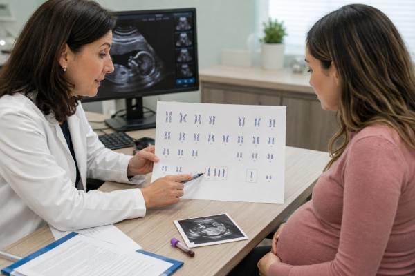
A fetal aneuploidy test evaluates whether a pregnancy may have an extra or missing chromosome. An extra copy is called a trisomy; a missing copy is called a monosomy. Common prenatal screening targets include trisomy 21, trisomy 18, trisomy 13, and selected sex chromosome differences such as monosomy X. Screening tests use maternal blood, ultrasound, or both to estimate chance. Diagnostic tests examine placental or fetal cells obtained through chorionic villus sampling or amniocentesis. The two categories should not be confused: a high-chance screen may be a false positive, while a low-chance screen cannot eliminate every chromosome condition. Results also vary in scope. Some panels examine only common aneuploidies, while diagnostic chromosome testing may detect a broader range of gains, losses, mosaic findings, or structural changes. Understanding the test method, specimen, target chromosomes, and result wording is essential before deciding whether reassurance, repeat testing, detailed imaging, or diagnostic confirmation is appropriate.
- Aneuploidy: An extra or missing chromosome in some or all cells
- Common targets: Trisomy 21, trisomy 18, trisomy 13, and sex chromosomes
- Screening tools: Cell-free DNA, serum markers, and nuchal translucency
- Diagnostic tools: CVS or amniocentesis with QF-PCR, karyotype, or microarray
- Result types: Lower chance, higher chance, no call, positive, negative, or mosaic
- Core rule: Confirm significant screening findings before irreversible decisions
Table of Contents
- Aneuploidy and the Chromosome Conditions Tested
- Screening Routes and Timing
- Why a Screening Number Is Not a Diagnosis
- Diagnostic Samples and Laboratory Methods
- Interpreting Trisomy, Monosomy, and Mosaic Results
- Special Situations That Change Accuracy
- What to Do With Each Result Category
- Questions for Informed Consent
Aneuploidy and the Chromosome Conditions Tested
Human cells usually contain 46 chromosomes arranged in 23 pairs. Aneuploidy means the cell has an atypical number of one or more individual chromosomes. It is different from polyploidy, in which an entire extra set of chromosomes is present, as in triploidy.
A trisomy occurs when there are three copies of a chromosome instead of two. A monosomy occurs when there is one copy instead of two. These changes can be present in every tested cell or in only a proportion of cells, which is called mosaicism.
Prenatal tests most commonly address:
| Condition | Chromosome finding | General testing note |
|---|---|---|
| Down syndrome | Trisomy 21 | Most accurately screened common autosomal trisomy with cfDNA |
| Edwards syndrome | Trisomy 18 | Often associated with multiple ultrasound findings and growth restriction |
| Patau syndrome | Trisomy 13 | Often associated with major brain, heart, facial, or other anomalies |
| Turner syndrome | Monosomy X | Screening has more false positives than screening for trisomy 21 |
| Klinefelter syndrome | Usually 47,XXY | May not produce prenatal structural findings |
| Triple X syndrome | 47,XXX | Often mild or variable; may be unexpected on screening |
| XYY syndrome | 47,XYY | Often not suspected on ultrasound |
Autosomal monosomies are generally not compatible with ongoing pregnancy, although mosaic or partial losses can occur. Full trisomies of many chromosomes also result in early miscarriage. Trisomies 21, 18, and 13 are the autosomal trisomies most often observed in later pregnancy and live birth.
Not every “aneuploidy panel” covers the same conditions. A basic cfDNA screen may include trisomies 21, 18, and 13. Some laboratories offer sex chromosome aneuploidy as an optional component. Others market genome-wide screening for rare autosomal trisomies or large chromosome segments. Evidence and predictive value are weaker for many rare findings, and current professional guidance does not support routine population screening for broad genome-wide copy-number changes.
The biological source also matters. Cell-free DNA in maternal blood comes mostly from the placenta. CVS also samples placenta. Amniocentesis samples cells in amniotic fluid that generally provide a closer representation of fetal chromosomes. A result may therefore describe placental aneuploidy, fetal aneuploidy, maternal chromosome variation, or a mixture requiring further investigation.
Aneuploidy testing does not assess every genetic disorder. A normal result cannot rule out most single-gene conditions, small DNA changes below the method’s resolution, congenital anomalies unrelated to chromosomes, or developmental differences that have no identifiable prenatal genetic cause.
Screening Routes and Timing
Screening estimates chance without entering the uterus. Patients should be offered information about available screening and diagnostic choices regardless of age or baseline risk, and they may accept or decline any option.
Cell-free DNA screening can usually be performed from about 10 weeks. A maternal blood sample contains the pregnant person’s DNA plus placental DNA fragments. Laboratories count or analyze chromosome-specific material to identify overrepresentation or underrepresentation. The cell-free DNA prenatal test, also called NIPT or NIPS, is the most sensitive and specific screening test for trisomies 21, 18, and 13. It remains a screen because the sampled DNA is not taken directly from fetal tissue.
First-trimester combined screening uses maternal age, blood levels of pregnancy-associated plasma protein A and human chorionic gonadotropin, and an ultrasound measurement of nuchal translucency. The first-trimester screening test calculates a chance for common trisomies within a limited gestational window. It can also identify an increased nuchal translucency that may signal heart disease or genetic conditions beyond the common aneuploidies.
Second-trimester serum screening measures several pregnancy-related substances. A quad screen primarily estimates the chance of trisomy 21 and open neural tube defects, while some regional programs include other aneuploidy calculations. It is less accurate for common trisomies than cfDNA.
Ultrasound is not a chromosome count. It can identify structural anomalies and soft markers associated with aneuploidy. A detailed scan remains important after any blood-based screen because many fetal conditions do not alter cfDNA. Ultrasound findings may also change the recommendation from screening to diagnostic testing with microarray.
The best screening strategy is usually planned rather than accumulated. Performing separate serum and cfDNA screens for the same conditions can produce discordant risk estimates. A patient who chooses cfDNA still needs routine ultrasound, but usually does not need a second independent aneuploidy blood screen.
Timing affects options. Earlier screening provides more time for counseling and diagnostic testing. CVS can often provide a diagnosis in the first trimester, while amniocentesis is generally performed from 15 weeks. A later presentation may make some serum screening windows unavailable but does not eliminate diagnostic testing.
Twin pregnancies require additional consideration. Current guidance supports cfDNA screening for common autosomal trisomies in twins, with the strongest evidence for trisomy 21. A positive result may not identify which twin is affected. Vanishing twins and higher-order multiples can make interpretation more difficult.
Why a Screening Number Is Not a Diagnosis
Screening reports describe probability. Diagnostic reports describe what was found in the tested cells. The word “positive” can appear on both, so the report heading and test method must be checked.
Four performance measures are commonly confused:
- Sensitivity: How often the test identifies affected pregnancies
- Specificity: How often the test is negative in unaffected pregnancies
- False-positive rate: How often an unaffected pregnancy screens positive
- Positive predictive value: The chance that a positive result is truly fetal aneuploidy
Positive predictive value depends on the condition’s prevalence in the tested population. Trisomy 21 is more common than many sex chromosome aneuploidies, and cfDNA performance is strongest for trisomy 21. A positive trisomy 21 screen therefore usually has a higher predictive value than a positive result for a rare chromosome condition, although individual probability still depends on age, ultrasound, laboratory method, and other factors.
A ratio such as 1 in 50 means an estimated 2% chance. A “screen positive” label may be triggered by a laboratory cutoff, even when most pregnancies with that result are unaffected. Conversely, “low risk” does not mean no risk. The residual chance may be very small, but false-negative results occur.
A cfDNA no-call or nonreportable result deserves its own category. It may arise from low fetal fraction, early gestational age, higher maternal weight, sample problems, medication, placental biology, or certain fetal chromosome conditions. It should not be interpreted as reassuring. Options include repeat sampling, ultrasound, another screening method, or diagnostic testing, chosen according to gestational age and clinical findings.
Sex chromosome results have special limitations. Placental mosaicism and maternal mosaicism are more common explanations for discordance than with trisomy 21. Age-related loss of an X chromosome in maternal blood can contribute to an apparent monosomy X signal. A positive result may lead to maternal testing as well as fetal diagnostic testing.
Rare autosomal trisomy and genome-wide cfDNA findings have lower predictive value and can be associated with placental mosaicism rather than an affected fetus. Even when fetal amniocentesis is normal, a placental abnormality may be relevant to growth and pregnancy complications. This is one reason broad panels can generate complex follow-up rather than simple reassurance.
Before acting on a screen, ask for the condition-specific positive predictive value and the laboratory’s reason for the call. A generic statement that the test is “99% accurate” is not enough because it may refer to sensitivity or specificity rather than the chance that this particular positive result is true.
Diagnostic Samples and Laboratory Methods
Diagnostic prenatal testing obtains pregnancy tissue and examines chromosomes directly. It is available after a positive screen, concerning ultrasound, known parental rearrangement, previous affected pregnancy, or patient preference for definitive information.
Chorionic villus sampling collects placental villi through the cervix or abdominal wall, usually in the first trimester. It offers earlier results but can reveal confined placental mosaicism. The CVS genetic test may require follow-up amniocentesis when placental findings are mosaic or inconsistent with ultrasound.
Amniocentesis removes a small amount of amniotic fluid using an ultrasound-guided needle, generally from 15 weeks. The fluid contains fetal cells. The amniocentesis genetic test is often preferred when the main question is whether a placental screening signal represents the fetus.
Both procedures carry a small risk of pregnancy loss and other complications. Risk estimates should come from the local clinician and account for operator experience, gestational age, placental location, multiple pregnancy, and individual medical factors.
The sample may undergo one or more tests:
QF-PCR or rapid FISH. Quantitative fluorescent polymerase chain reaction and fluorescence in situ hybridization quickly assess selected chromosomes, often 13, 18, 21, X, and Y. A rapid answer can be available before the full study, but it does not evaluate every chromosome abnormality.
Karyotype. A prenatal karyotype test counts and displays chromosomes. It detects whole-chromosome aneuploidy, large structural changes, and many translocations. Cell culture may lengthen turnaround time.
Chromosomal microarray. Microarray detects chromosome gains and losses at higher resolution. It is especially useful when ultrasound shows one or more fetal anomalies. It can identify clinically significant copy-number changes too small for karyotype, but it may also produce uncertain findings. Standard microarray does not reliably show balanced chromosome rearrangements.
Specialized testing. If aneuploidy testing is normal but the fetal pattern suggests a genetic syndrome, targeted gene testing or prenatal exome sequencing may be considered. These tests answer different questions and require separate consent.
A rapid positive result is often confirmed or characterized with a full analysis. For example, detection of trisomy 21 may be followed by karyotype to determine whether there are three separate chromosomes or a translocation. That distinction affects recurrence counseling.
Maternal cell contamination can complicate testing, especially when the DNA amount is small. Laboratories use identity markers and sample-quality checks to reduce this risk. Reports should state specimen type and method so the result is not mistakenly generalized beyond what was tested.
Interpreting Trisomy, Monosomy, and Mosaic Results
Diagnostic chromosome notation is compact. The first number is the total chromosome count, followed by sex chromosomes and the abnormality.
Examples include:
47,XX,+21— trisomy 21 in a fetus with two X chromosomes47,XY,+18— trisomy 18 in a fetus with X and Y chromosomes47,XX,+13— trisomy 13 in a fetus with two X chromosomes45,X— monosomy X47,XXY— an extra X chromosome in a fetus with a Y chromosome
A slash between two formulas indicates more than one cell line. A mosaic result might show some cells with 45,X and others with 46,XX. Bracketed numbers may indicate how many cells of each type were examined. The proportion in the sample does not precisely predict the proportion in every fetal organ.
Full aneuploidy means the abnormal chromosome count was found consistently in tested cells. The expected health effects depend on the chromosome. Full trisomy 21 is compatible with long-term survival, while full trisomy 18 and trisomy 13 are usually life-limiting. Full monosomy of an autosome generally does not result in an ongoing pregnancy. Monosomy X has a broad clinical range.
Mosaic aneuploidy can have a wide range of outcomes. Interpretation depends on which chromosome is involved, the tissue tested, the number of abnormal cells, ultrasound findings, and whether the result could be confined to placenta. CVS mosaicism often leads to amniocentesis. Amniotic-fluid mosaicism may lead to additional cell counts, ultrasound, specialist review, and sometimes testing after birth.
Partial trisomy or monosomy means a chromosome segment is duplicated or deleted. The report may use terms such as dup, del, or der. Effects depend on the exact genes and size. A parental karyotype may be needed because a balanced translocation in a parent can produce an unbalanced fetal result.
Normal result means no abnormality was found within the method’s scope. A normal QF-PCR does not exclude aneuploidy of chromosomes not tested. A normal karyotype does not exclude small copy-number changes. A normal microarray does not exclude most single-gene disorders or balanced rearrangements.
Variant of uncertain significance is usually a microarray term, not a diagnosis of aneuploidy. It means a DNA gain or loss was found but current evidence cannot determine whether it affects health. Parental testing may clarify whether it is inherited from a healthy parent, but inheritance does not always settle the question.
Special Situations That Change Accuracy
The same test may perform differently depending on pregnancy and medical context.
Vanishing twin. DNA from a demised co-twin can remain in maternal blood and cause a positive cfDNA result that does not represent the surviving fetus. Diagnostic testing may be more informative than repeating the screen.
Twins. A negative result generally applies to the pregnancy, but a positive screen may not identify the affected twin. Dizygotic twins can contribute different DNA fractions. Selective diagnostic sampling requires experienced fetal medicine care.
Donor eggs and gestational carriers. The prior chance should be based on the egg source’s age where appropriate, while the blood sample comes from the pregnant person. Laboratories need accurate reproductive history to calculate and interpret results.
Maternal chromosome variation. Maternal mosaic sex chromosome aneuploidy, benign copy-number variants, or a previously unknown chromosome condition can alter cfDNA. A result may prompt maternal karyotype or microarray after counseling.
Maternal cancer or organ transplant. Tumors can release abnormal DNA patterns, and transplanted organs can introduce donor DNA. Complex or multiple chromosome signals may not reflect the fetus. Such results require specialist review rather than routine interpretation.
Low fetal fraction. The proportion of placental DNA may be too low for reliable analysis. It is influenced by gestational age, maternal weight, placental function, and some chromosome conditions. Different laboratories use different minimum thresholds and algorithms.
Placental mosaicism. The placenta and fetus begin from the same fertilized egg but can develop different cell lines. Confined placental mosaicism can cause false-positive cfDNA or CVS results. It may also be associated with poor placental function and fetal growth restriction even when amniocentesis is normal.
Recent blood transfusion or stem-cell treatment. Donor cells can potentially complicate some genetic tests. The laboratory should receive a complete history before sample collection.
These situations do not mean testing is impossible. They mean the result may need a different method, a different specimen, or a more cautious explanation.
What to Do With Each Result Category
A practical response begins by identifying whether the result came from screening or diagnosis.
| Result | Typical next discussion |
|---|---|
| Low-chance screen | Continue routine care and anatomy ultrasound; revisit testing if new findings appear |
| High-chance screen | Genetic counseling, detailed ultrasound, and offer of CVS or amniocentesis |
| No-call cfDNA | Review cause and timing; consider repeat draw or diagnostic testing |
| Positive diagnostic result | Confirm exact chromosome pattern, prognosis, recurrence, and pregnancy-care options |
| Mosaic diagnostic result | Clarify tissue source, cell counts, ultrasound, and need for another specimen |
| Normal diagnostic result with anomalies | Consider microarray or gene-level testing based on the fetal phenotype |
After a high-chance screen, do not use another screening test merely to “confirm” it. A second screen may lower or raise probability but cannot establish the fetal karyotype. Diagnostic testing is the route to confirmation when certainty is needed.
After a low-chance screen, a major ultrasound anomaly may still justify diagnostic testing. The blood test only assessed selected chromosome conditions. Structural anomalies can result from other copy-number changes, single-gene disorders, infection, environmental causes, or unknown factors.
After a positive diagnostic result, the next steps are condition-specific. They may include fetal echocardiography, serial growth assessment, consultation with pediatric specialists, delivery planning, perinatal palliative care, or discussion of pregnancy termination where available. Counseling should be non-directive and should include the range of outcomes rather than a single assumption.
After a mosaic result, avoid predicting outcome from the percentage alone. Ask whether the sample came from placenta or amniotic fluid, whether the abnormality appeared in one or several cultures, and whether maternal cell contamination was excluded.
After a discordant result—positive cfDNA and normal amniocentesis, for example—the fetal result is usually considered reassuring for the targeted condition. The team may still monitor placental function or evaluate the pregnant person if the pattern suggests a maternal finding.
Keep copies of all reports. Exact notation can influence recurrence counseling and future testing. A portal summary such as “chromosomes normal” or “trisomy positive” is not an adequate substitute.
Questions for Informed Consent
Pretest counseling should make the possible outcomes understandable before blood is drawn or a procedure is scheduled.
Ask:
- Is this a screening test or a diagnostic test?
- Which chromosomes and conditions are included?
- Are sex chromosome results optional?
- Does the panel include rare trisomies or genome-wide changes, and what evidence supports them?
- What will the report call a positive, negative, or no-call result?
- What is the condition-specific positive predictive value for my situation?
- Could twins, donor eggs, a vanished twin, or my medical history affect accuracy?
- Which diagnostic procedure is available if the screen is positive?
- Would the diagnostic sample receive rapid testing, karyotype, microarray, or more than one test?
- What findings could be uncertain or unrelated to the original reason for testing?
A patient may value early information, maximum certainty, avoiding an invasive procedure, preparing for a child’s needs, or limiting information about conditions with variable effects. These goals can lead to different reasonable choices.
The term “noninvasive prenatal test” can create false confidence. The blood draw is noninvasive, but the result remains probabilistic and may lead to invasive confirmation. Conversely, diagnostic testing is not mandatory after a positive screen. A patient can decline it after understanding the remaining uncertainty.
Fetal aneuploidy testing is most useful when the result category, biological source, and test scope are clear. The central safety step is simple: do not treat a probability estimate as a fetal chromosome diagnosis.
References
- Society for Maternal-Fetal Medicine Consult Series #74:Cell-free DNA screening for aneuploidies: Updated guidance 2025 (Guideline)
- A Brief Guide to SMFM’s Updated Prenatal Genetic Screening Recommendations 2025 (Patient Guidance)
- Prenatal Cell-Free DNA Screening 2024 (Medical Test Guide)
- Non-invasive prenatal testing (NIPT) 2025 (Clinical Resource)
- Common aneuploidy testing (QF-PCR) 2025 (Clinical Resource)
- Karyotype Genetic Test 2025 (Medical Test Guide)
Disclaimer
This article is general education about fetal aneuploidy screening and diagnosis, not individualized medical advice. Test performance, procedure timing, laboratory methods, and follow-up options vary with pregnancy circumstances and location. Discuss personal results with an obstetric clinician, maternal-fetal medicine specialist, laboratory genetics professional, or genetic counselor before making decisions.





