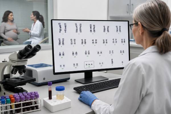
A Down syndrome genetic test can estimate the chance that a pregnancy is affected by trisomy 21 or confirm whether an extra copy of chromosome 21 is present. Those are two different purposes. Prenatal screening—including cell-free DNA, first-trimester screening, and second-trimester serum screening—does not diagnose Down syndrome. It identifies pregnancies with a higher or lower chance and helps families decide whether they want more information. Diagnostic testing uses placental tissue or amniotic fluid to examine fetal chromosomes directly. Testing after birth can confirm the diagnosis and identify the chromosome pattern, which may affect counseling about recurrence in future pregnancies. Results are most useful when they are interpreted alongside gestational age, ultrasound findings, family history, and personal values. A clear discussion with an obstetric clinician or genetic counselor can help explain what the test did, what it did not do, and which next step fits the situation.
- What it detects: Extra chromosome 21 material associated with Down syndrome
- Screening choices: Cell-free DNA, combined first-trimester screening, and serum screening
- Diagnostic choices: Chorionic villus sampling or amniocentesis with chromosome analysis
- Possible results: Lower chance, higher chance, no result, positive, negative, or mosaic finding
- Why chromosome type matters: Full trisomy, translocation, and mosaic Down syndrome have different recurrence considerations
- Best next step: Match the result to the test type before making medical decisions
Table of Contents
- What Down Syndrome Testing Detects
- Prenatal Screening Options
- Diagnostic Testing Before Birth
- How to Read Screening Results
- How Diagnostic Results Are Reported
- Inheritance and Recurrence
- Testing After Birth and Health Care
- Deciding What to Do Next
What Down Syndrome Testing Detects
Down syndrome results from extra genetic material from chromosome 21. Chromosomes are packages of DNA found in most cells. People usually have 46 chromosomes arranged in 23 pairs. In Down syndrome, cells contain an extra full or partial copy of chromosome 21. The additional genes affect development, but the medical, learning, and physical features vary widely from one person to another.
A test may look for one of three main chromosome patterns:
| Chromosome pattern | What it means | Typical testing issue |
|---|---|---|
| Full trisomy 21 | Most or all tested cells have three separate copies of chromosome 21 | Usually occurs as a random chromosome-separation event |
| Translocation Down syndrome | Extra chromosome 21 material is attached to another chromosome | A parent may sometimes carry a balanced translocation |
| Mosaic Down syndrome | Some tested cells have trisomy 21 and others have the usual chromosome number | The percentage in one tissue may not predict findings in every tissue |
Full trisomy 21 accounts for the large majority of cases. It usually begins when chromosome 21 does not separate normally during the formation of an egg or sperm. This is called nondisjunction. Translocation Down syndrome is less common. In this form, a complete or nearly complete extra chromosome 21 is joined to another chromosome, often chromosome 14 or another chromosome 21. Mosaic Down syndrome develops after fertilization when an early cell division produces two cell lines.
The phrase “Down syndrome test” can therefore describe several different laboratory approaches. A prenatal screening test may measure DNA fragments or pregnancy-related proteins that correlate with trisomy 21. A chromosome test such as a karyotype counts and displays chromosomes. Rapid tests such as fluorescence in situ hybridization, or FISH, may target chromosome 21 and several other common aneuploidies. Chromosomal microarray measures gains and losses of DNA across the genome, although a standard karyotype is often important when a translocation is possible because it shows chromosome structure.
Testing does not measure a child’s future abilities, personality, health severity, or quality of life. It identifies a chromosome finding or estimates its probability. Families benefit when technical results are separated from assumptions about what an individual life will be like.
Prenatal Screening Options
Prenatal screening is optional and should be offered in a way that allows a pregnant person to accept or decline it. Current professional guidance supports discussing both screening and diagnostic testing with every pregnant patient, regardless of age or baseline risk.
The main screening choices differ in timing and method:
Cell-free DNA screening. Also called cfDNA screening or noninvasive prenatal testing, this blood test analyzes placental DNA fragments circulating in maternal blood. It can generally be performed from about 10 weeks of pregnancy. For trisomy 21, it has a higher detection rate and lower false-positive rate than traditional serum screening, but it remains a screening test. The cell-free DNA prenatal screening result reflects DNA from the placenta, which usually—but not always—matches fetal chromosomes.
Combined first-trimester screening. This approach combines maternal age, a blood test measuring pregnancy-associated plasma protein A and free or total human chorionic gonadotropin, and an ultrasound measurement called nuchal translucency. It is usually performed in a defined window near the end of the first trimester. The first-trimester screening test estimates the chance of trisomy 21 and certain other chromosome conditions.
Second-trimester serum screening. A quad screen measures alpha-fetoprotein, hCG, unconjugated estriol, and inhibin A. It is less accurate for trisomy 21 than cfDNA screening but can provide information when first-trimester testing was not completed or cfDNA is not available. Alpha-fetoprotein also contributes to screening for open neural tube defects.
Ultrasound. Ultrasound may identify findings associated with Down syndrome, such as increased nuchal translucency, certain heart differences, absent or small nasal bone, or other markers. However, many fetuses with trisomy 21 have no clear ultrasound findings, and many isolated markers occur in pregnancies with typical chromosomes. Ultrasound is not a chromosome diagnosis.
Some patients are offered more than one type of screen, but uncoordinated screening can create conflicting estimates and unnecessary uncertainty. Clinicians generally try to use one screening strategy rather than ordering several independent aneuploidy screens at the same time. A detailed anatomy ultrasound is still recommended because it evaluates fetal development beyond chromosome risk.
Screening performance is often described using detection rate, false-positive rate, sensitivity, specificity, and positive predictive value. These terms are not interchangeable. A test can have excellent sensitivity yet still produce some false-positive results, especially when the condition is uncommon in the tested population. The chance that a positive result is truly positive depends partly on the person’s starting chance before testing.
A screening choice may also be affected by twins or higher-order multiples, a vanished twin, organ transplantation, active cancer, maternal chromosome differences, donor eggs, gestational age, and body weight. These factors do not automatically prevent testing, but they can affect accuracy or the chance of receiving a result.
Diagnostic Testing Before Birth
Prenatal diagnostic testing examines cells obtained from the pregnancy. It can confirm whether trisomy 21 is present with much greater certainty than screening. The two standard procedures are chorionic villus sampling and amniocentesis.
Chorionic villus sampling (CVS) removes a small sample of placental tissue, usually during the first trimester. The sample may be collected through the cervix or through the abdomen, depending on placental position and clinical factors. Because the tissue comes from the placenta, a rare result called confined placental mosaicism can complicate interpretation. In that situation, some placental cells have an abnormal chromosome pattern while fetal cells may not. Follow-up amniocentesis may be recommended. More detail is available in the CVS genetic test guide.
Amniocentesis removes a small amount of amniotic fluid through a thin needle placed through the abdomen under ultrasound guidance, usually from 15 weeks onward. The fluid contains fetal cells that can be used for chromosome testing. The amniocentesis genetic test generally avoids the placenta-specific uncertainty that can occur with CVS, although rare mosaic or laboratory findings may still need clarification.
Both procedures carry a small risk of pregnancy loss and other complications. The exact risk depends on the procedure, operator, gestational age, and patient-specific factors. A clinician should explain local experience and how the quoted risk applies to the individual pregnancy.
Diagnostic laboratory testing may include:
- Karyotype: Counts chromosomes and shows large structural rearrangements. It can distinguish free trisomy 21 from a translocation.
- Rapid aneuploidy testing: FISH or quantitative fluorescent polymerase chain reaction may provide a preliminary answer for chromosome 21 and selected other chromosomes within a few days.
- Chromosomal microarray: Detects smaller gains and losses of DNA than a karyotype. It may be recommended when ultrasound shows one or more structural differences. Microarray can also identify findings unrelated to Down syndrome, including results of uncertain significance.
A rapid result is often followed by a full chromosome study. A preliminary FISH result should not be treated as the only information if the final karyotype is still pending. The complete analysis helps confirm the finding and determine whether a translocation is present.
Diagnostic testing may be considered after a higher-chance screen, an ultrasound difference, a previous pregnancy or child with a chromosome condition, a known parental chromosome rearrangement, or simply because a patient wants a definitive answer. A person does not have to complete screening before choosing diagnostic testing.
How to Read Screening Results
A screening report often uses language such as “high risk,” “positive,” “increased chance,” “low risk,” or “screen negative.” These labels can sound more certain than they are. The first question should be: Is this a screening result or a diagnostic result?
A lower-chance result means the test found no strong evidence of trisomy 21. It substantially reduces risk but does not make the chance zero. False-negative results are uncommon with cfDNA screening but can occur because of placental mosaicism, low fetal fraction, sample problems, or biological factors not captured by the test.
A higher-chance or positive screening result means trisomy 21 is more likely than it was before testing. It is not a diagnosis. The report may provide a positive predictive value, which estimates the probability that the pregnancy is truly affected given the test result and other inputs. If the report states a ratio such as 1 in 10, that corresponds to a 10% estimated chance, not certainty.
For example, two people can receive the same positive cfDNA signal but have different positive predictive values because their prior chances differ. Maternal age, gestational age, ultrasound findings, and the test laboratory’s validated population can influence the estimate. Online calculators may illustrate this concept, but they should not replace interpretation of the actual report.
A no-call, nonreportable, or insufficient fetal fraction result means the laboratory could not provide a reliable answer. It is not the same as a negative result. Low fetal fraction can occur when testing is performed early, with higher maternal weight, because of sample handling, or in some pregnancies with chromosome abnormalities. Options may include repeat cfDNA screening, another screening approach, a detailed ultrasound, or diagnostic testing. The best choice depends on gestational age and why the result failed.
Traditional serum screening reports may list a calculated chance such as 1 in 100 or 1 in 2,000. Laboratories use a cutoff to label the result screen positive or screen negative. A result just above the cutoff is not biologically different from one just below it; the numbers exist on a continuum.
After a positive screen, recommended next steps usually include genetic counseling, a detailed ultrasound, and an offer of CVS or amniocentesis. Making irreversible pregnancy decisions based only on a screening result risks acting on a false positive. Diagnostic confirmation is especially important when the result would change pregnancy management.
A negative screen does not rule out all genetic conditions, birth differences, or developmental concerns. Most screening panels focus on a limited number of chromosome conditions. Even broad cfDNA panels cannot evaluate every gene or chromosome change reliably.
How Diagnostic Results Are Reported
A diagnostic chromosome report may use compact laboratory notation. Understanding the main pieces can make the result less intimidating.
A typical full trisomy 21 result may appear as:
47,XX,+21— 47 chromosomes, two X chromosomes, and an extra chromosome 2147,XY,+21— 47 chromosomes, one X and one Y chromosome, and an extra chromosome 21
A translocation result may include abbreviations such as rob, which means Robertsonian translocation, or der, which means derivative chromosome. The notation specifies which chromosomes are joined and whether extra chromosome 21 material is present. Because translocation notation can be difficult to interpret, a genetic counselor or clinical geneticist should review the exact report rather than relying on a general explanation.
A mosaic result may list two cell lines separated by a slash, such as a trisomy 21 line and a typical chromosome line. Bracketed numbers may show how many cells of each type were counted. Those numbers describe the tested sample; they do not reliably predict the percentage of affected cells in the brain, heart, or other tissues. Clinical features can range widely.
The report may say consistent with Down syndrome, trisomy 21 detected, or no evidence of trisomy 21. A negative diagnostic result for trisomy 21 is highly reassuring, but its scope still depends on the method. A rapid targeted test may not assess all chromosomes or structural changes. A normal karyotype does not rule out small DNA variants or single-gene conditions that the test was not designed to detect.
Occasionally, CVS shows mosaic trisomy 21 while amniocentesis is normal. This can reflect confined placental mosaicism. The care team may recommend additional ultrasound surveillance because placental chromosome differences can sometimes affect placental function even when the fetal chromosome result is normal.
Rarely, a laboratory may identify a structural chromosome finding whose significance is unclear until parental testing is performed. For example, a balanced rearrangement in a fetus may have little or no direct health effect but can have reproductive implications later in life. A microarray may report a copy-number variant of uncertain significance. That does not mean Down syndrome is present; it is a separate finding that requires its own interpretation.
Request a copy of the complete laboratory report. The exact testing method, specimen type, chromosome notation, and comments matter. A verbal summary such as “the chromosome test was normal” may leave out information important for future care or family counseling.
Inheritance and Recurrence
Most Down syndrome is not inherited. Full trisomy 21 usually occurs because of a random nondisjunction event in the egg or sperm. The chance rises with maternal age, but trisomy 21 can occur at any reproductive age, and most individuals did nothing to cause or prevent it.
After a pregnancy or child with full trisomy 21, the chance in a future pregnancy is higher than the age-related chance alone. Counseling often uses the person’s age at the next pregnancy together with an empiric recurrence estimate. The exact figure should come from a genetics professional because recommendations differ by age, chromosome result, and reproductive history.
Translocation Down syndrome requires more specific evaluation. A parent may have a balanced Robertsonian translocation, meaning the expected amount of genetic material is present but arranged differently. Balanced carriers are usually healthy, yet some eggs or sperm can receive an unbalanced chromosome combination. The recurrence chance depends strongly on which chromosomes are involved and which parent carries the rearrangement.
When a fetus or child has translocation Down syndrome, both biological parents are commonly offered a karyotype. If neither parent carries the translocation, it likely arose as a new event and recurrence is generally lower than when a parent is a carrier. If a parent carries a translocation involving chromosome 21, individualized counseling is essential. Certain rare rearrangements, such as a chromosome 21-to-21 translocation, can create a very high chance of an affected or nonviable pregnancy.
Mosaic Down syndrome usually results from a random early cell-division event and is not typically inherited. Even so, recurrence counseling should be based on the actual chromosome report rather than the word “mosaic” alone.
Reproductive options after a trisomy 21 diagnosis may include natural conception with prenatal screening or diagnosis, in vitro fertilization with preimplantation genetic testing when appropriate, use of donor eggs or sperm, adoption, or choosing not to pursue another pregnancy. These are personal choices, not a required sequence. Preimplantation genetic testing can reduce the chance of transferring an embryo with a targeted chromosome imbalance, but it is not perfect and prenatal testing is still discussed after pregnancy begins.
A family history of Down syndrome does not automatically mean another relative has a high chance. The most useful document is the affected person’s karyotype. Without it, clinicians may not be able to distinguish random full trisomy from an inherited translocation.
Testing After Birth and Health Care
Down syndrome may be suspected after birth because of physical findings, low muscle tone, a heart difference, feeding concerns, or prenatal test results. A blood karyotype is the standard confirmation test. It establishes whether extra chromosome 21 material is present and identifies full trisomy, translocation, or mosaicism.
If a rapid prenatal test was positive but the full chromosome analysis was not completed, postnatal karyotyping can provide definitive documentation. A prenatal cfDNA result alone should not be used as the baby’s final diagnostic record. Likewise, a clinical appearance strongly suggestive of Down syndrome should be confirmed with chromosome testing because other conditions can share some features.
The chromosome result begins—not ends—the medical evaluation. Recommended early care commonly includes:
- Echocardiography or review of a detailed fetal/newborn heart assessment
- Hearing screening and ongoing hearing surveillance
- Thyroid testing
- Evaluation for feeding, swallowing, growth, and gastrointestinal concerns
- Eye assessment
- Monitoring for sleep-disordered breathing
- Developmental support and early-intervention services
- Routine immunizations and preventive care adjusted for individual health needs
Not every person with Down syndrome will have every associated condition. Some have major congenital heart disease; others do not. Some need extensive feeding support in infancy; others feed without major difficulty. Learning profiles, communication, independence, and health needs also differ. Care should be based on the individual rather than a checklist of assumptions.
The American Academy of Pediatrics publishes health-supervision guidance for children and adolescents with Down syndrome. A primary-care clinician can use it to coordinate screenings at recommended ages while specialists address specific findings. Adults need continued attention to thyroid health, hearing, vision, sleep apnea, mental health, mobility, and other age-related needs.
Families may also benefit from balanced information from clinicians, local Down syndrome organizations, and people living with Down syndrome. Medical facts are important, but they are not a complete description of family life. Counseling should include both possible health challenges and the range of meaningful educational, social, and community experiences.
Deciding What to Do Next
The next step depends less on the word “positive” or “negative” than on which test produced it.
After a positive screening test, ask for the estimated positive predictive value, the gestational age, whether ultrasound findings change the interpretation, and which diagnostic procedure is available. A genetic counselor can explain the difference between CVS and amniocentesis and help clarify what information is needed before making decisions.
After a negative screen, continue routine prenatal care and the recommended anatomy ultrasound. Diagnostic testing remains available if new ultrasound findings appear or if a patient wants greater certainty. A lower-chance result should not be interpreted as a guarantee of a healthy baby because no prenatal screen evaluates every condition.
After a no-call result, do not assume the result is reassuring. Ask why the sample was nonreportable, whether repeating it is likely to work, and whether time-sensitive diagnostic testing should be considered.
After a confirmed prenatal diagnosis, families may consider continuing the pregnancy and preparing for the baby’s medical needs, making an adoption plan, or ending the pregnancy where legally and medically available. The care team should provide accurate, non-directive information and respect the patient’s values. Preparation may include fetal echocardiography, delivery planning at a center with appropriate newborn care, meeting pediatric specialists, and connecting with support organizations.
After a postnatal diagnosis, request the complete karyotype, schedule recommended newborn evaluations, and ask whether parental chromosome testing is needed. Early intervention can support feeding, movement, communication, and learning. Parents should also receive attention for their own emotional and practical needs.
Useful questions for the appointment include:
- Was this a screening test or a diagnostic test?
- What exactly did the laboratory measure?
- What is the chance that this result is correct for this pregnancy or child?
- Does the report show full trisomy, translocation, or mosaicism?
- Would another test change medical care or recurrence counseling?
- What health evaluations are recommended now?
- Can I meet a genetic counselor and receive a copy of every report?
Down syndrome testing can produce urgent emotions before all the facts are available. Slowing the process long enough to identify the test type, confirm uncertain screening findings, and understand the exact chromosome result helps families make informed decisions without confusing probability with diagnosis.
References
- Down Syndrome Tests 2026 (Patient Education)
- Society for Maternal-Fetal Medicine Consult Series #74: Cell-free DNA screening for aneuploidies: Updated guidance 2025 (Guideline)
- Health Supervision for Children and Adolescents With Down Syndrome 2022 (Clinical Report)
- Down Syndrome 2026 (Official Guidance)
- Down Syndrome 2023 (Clinical Review)
- Down syndrome (trisomy 21) 2025 (Clinical Resource)
Disclaimer
This article provides general education about Down syndrome screening and diagnostic testing and is not a substitute for individualized medical or genetic counseling. Test timing, accuracy, procedure risks, and next-step options depend on the pregnancy, laboratory, and health system. Discuss personal results with an obstetric clinician, maternal-fetal medicine specialist, pediatrician, or genetic counselor before making medical decisions.