
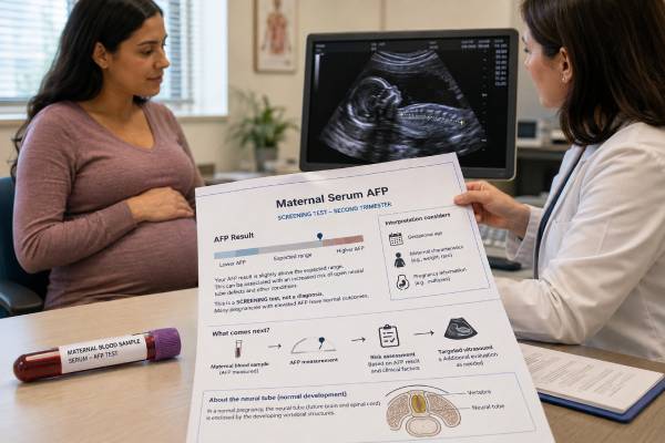
The maternal serum alpha-fetoprotein test, usually shortened to MSAFP, is a second-trimester blood screening test used mainly to estimate the chance of an open neural tube defect. It measures a fetal protein that normally reaches the pregnant person’s bloodstream in small amounts. A high result can occur with spina bifida or anencephaly, but it also has many non-genetic explanations, including incorrect pregnancy dating, twins, bleeding, abdominal wall defects, or placental problems. For that reason, MSAFP is not a diagnosis and is not technically a genetic test. Results are adjusted for gestational age and other pregnancy details, then reported as a multiple of the median, or MoM. The most useful response to an abnormal result is prompt verification of the information used in the calculation and a detailed ultrasound, not an assumption that the fetus has a serious condition.
- MSAFP is a screening blood test, not a diagnostic genetic test.
- Testing is generally performed in the second trimester, with laboratory windows often spanning about 15 to 22 weeks.
- Results are reported as multiples of the median (MoM) rather than interpreted from the raw AFP concentration alone.
- A high AFP result has several possible causes, and most screen-positive results do not by themselves prove a neural tube defect.
- A negative result lowers risk but cannot rule out every neural tube or structural defect.
- Follow-up usually starts with dating review and targeted ultrasound, with amniocentesis considered in selected cases.
Table of Contents
- What Maternal Serum AFP Measures
- Timing, Preparation, and Information the Laboratory Needs
- How AFP Results Are Calculated and Reported
- What a High Maternal Serum AFP Result Can Mean
- What Negative or Low AFP Results Mean
- Follow-Up After a Screen-Positive Result
- Neural Tube Defects, Limits, and Prevention
- Questions and Next Steps
What Maternal Serum AFP Measures
Alpha-fetoprotein is a protein produced mainly by the fetal yolk sac early in pregnancy and then by the fetal liver. AFP is present at high levels in fetal blood. Smaller amounts enter amniotic fluid and cross into the pregnant person’s bloodstream, where they can be measured in a serum sample.
Maternal blood AFP normally rises as pregnancy advances. A number that is expected at 20 weeks could be unusually high at 16 weeks, so the raw concentration is not meaningful without an accurate gestational age. Laboratories compare the measured level with the expected median for pregnancies at the same stage and adjust the interpretation for relevant maternal and pregnancy factors.
Why open neural tube defects raise AFP
The neural tube forms the brain and spinal cord and normally closes very early in pregnancy. In an open neural tube defect, fetal tissue or fluid has an open connection with the amniotic cavity. AFP can leak into amniotic fluid and then reach maternal blood in larger-than-expected amounts.
The main conditions screened for are:
- Open spina bifida, in which part of the spine and tissues covering the spinal cord do not close normally
- Anencephaly, in which major portions of the brain and skull do not develop
- Encephalocele, in which brain tissue and membranes protrude through an opening in the skull
MSAFP is better suited to open defects than closed spinal defects covered by skin. A closed defect may not release much AFP, so a normal result cannot exclude every form of spina bifida.
What the test is—and is not
MSAFP is a biochemical screen. It does not examine chromosomes, sequence DNA, or identify a specific gene variant. It may be ordered as a single-marker test or included in a quad or integrated serum screen. In a quad screen, AFP contributes to neural tube defect screening while AFP, hCG, unconjugated estriol, and inhibin A are combined to estimate chances of certain chromosome conditions.
Cell-free DNA screening is more accurate than serum screening for common fetal aneuploidies, but standard cfDNA does not replace screening for open neural tube defects. A patient who has low-risk cfDNA may still be offered MSAFP and should still be offered a second-trimester anatomy ultrasound. The tests answer different questions.
MSAFP also does not diagnose placental disease or predict pregnancy outcome with certainty. An unexplained elevation can be associated with later complications, but the result is one clue among ultrasound findings, growth measurements, medical history, and other clinical information.
Timing, Preparation, and Information the Laboratory Needs
MSAFP is collected during the second trimester. Exact accepted dates differ by laboratory and local program. A common window is 15 weeks 0 days through 22 weeks 6 days, while many clinicians prefer collection around 16 to 18 weeks because dating is usually established and there is time for follow-up.
A blood sample taken too early or too late may not be interpretable under the laboratory’s validated model. If the pregnancy is near the end of the testing window, ask when the sample must reach the laboratory rather than assuming the draw date alone is sufficient.
No fasting is usually required. Routine medications are generally continued unless the clinician gives different instructions. The blood draw does not increase miscarriage risk.
Details that affect the calculation
The laboratory needs more than the AFP concentration. Incorrect information can shift the MoM enough to change a result from screen negative to screen positive or the reverse. The requisition may request:
- Gestational age and how it was determined
- Estimated due date and ultrasound date
- Number of fetuses
- Maternal weight at collection
- Preexisting diabetes and insulin use, depending on the laboratory model
- Race or ethnicity if the laboratory still uses a validated adjustment
- Whether conception involved IVF and, in some programs, donor age
- Smoking status, depending on the screening system
- Prior pregnancy affected by a neural tube defect
- Recent vaginal bleeding, fetal procedure, or suspected fetal loss
Modern laboratories differ in which adjustments they apply. The report should state the factors incorporated. A patient should not try to recalculate the result with a general online chart because the laboratory’s assay, reference population, and software matter.
Dating matters most
Underestimated gestational age is a common explanation for apparently high AFP. If a pregnancy thought to be 16 weeks is actually 18 weeks, the measured AFP may be normal for the true stage. Conversely, overestimated gestational age can make AFP appear low.
First-trimester ultrasound dating is usually more accurate than a later estimate based only on fetal size. When a positive result arrives, the clinician compares the gestational age used by the laboratory with the best obstetric dating already established. A small difference does not always justify changing the due date; obstetric dating rules determine whether recalculation is appropriate.
Multiple gestation also raises AFP because more than one fetus produces the protein. Twin and higher-order pregnancies require a multiple-specific model. A report mistakenly calculated as a singleton can appear markedly elevated.
How AFP Results Are Calculated and Reported
The laboratory first measures AFP in units such as ng/mL. It then divides that result by the median expected value for an unaffected pregnancy at the same gestational age. The result is the multiple of the median.
For example, an AFP result equal to the median is 1.0 MoM. A result twice the median is 2.0 MoM. This does not mean the fetus has twice a normal amount of AFP or twice a particular risk. MoM is a standardized comparison used in a screening algorithm.
Screen-positive cutoffs
Many singleton screening programs use a cutoff near 2.5 MoM, but cutoffs vary. Some use 2.0 MoM, and twin-pregnancy thresholds may be higher. The correct interpretation is the one on the issuing laboratory’s report.
A typical report may contain:
| Report item | Meaning |
|---|---|
| AFP concentration | The measured amount in maternal serum |
| AFP MoM | The measured value compared with the expected median after adjustments |
| Screen status | Whether the value crosses that laboratory’s cutoff |
| Estimated neural tube defect risk | A model-based chance, when the laboratory reports one |
| Gestational age used | The pregnancy age entered for calculation |
| Assumptions | Singleton or twins, weight, diabetes status, and other factors |
| Recommendation | Repeat calculation, ultrasound, counseling, or diagnostic evaluation |
A screen-positive label is a triage threshold. It identifies a group in which further evaluation is worthwhile; it is not a statement that an affected fetus is more likely than an unaffected fetus. The actual chance depends on how high the MoM is, the accuracy of pregnancy data, ultrasound findings, prior history, and the background prevalence of open neural tube defects.
Why the MoM may be recalculated
If the laboratory used the wrong gestational age, fetal number, or maternal weight, it may issue a corrected report. Repeating the blood draw is not always necessary. A recalculation from the original specimen may be enough if the laboratory can validate the new information.
A repeat sample can be considered when the result is borderline, the specimen was collected early, or a technical issue is suspected. It should not delay a timely ultrasound when the value is clearly elevated or the testing window is closing. Repeated normal blood tests also cannot substitute for assessment of a concerning ultrasound finding.
Risk language on the report
Reports may say “screen negative,” “screen positive,” “increased risk,” or “elevated AFP.” They may provide a numerical estimate such as 1 in 100, or only a cutoff classification. Compare the number with the condition being screened. A Down syndrome risk from a quad screen and an open neural tube defect interpretation based on AFP are separate outputs.
If the portal displays only “abnormal,” request the complete report. The AFP MoM, cutoff, gestational age, fetal number, and laboratory comments are needed for meaningful interpretation.
What a High Maternal Serum AFP Result Can Mean
A high MSAFP result has a broad differential diagnosis. Neural tube defects are important, but they are not the only explanation and are not the most common explanation for every positive screen.
Pregnancy dating or fetal-number differences
The first checks are whether gestational age was underestimated and whether the pregnancy includes more fetuses than entered. Ultrasound can confirm fetal number and review dates. A vanished twin or recent fetal loss can also increase maternal serum AFP because fetal or placental material may release AFP.
Open fetal structural conditions
Elevated AFP can occur with open spina bifida, anencephaly, or encephalocele. Other fetal conditions that expose internal tissues or allow protein leakage may also raise AFP, including:
- Gastroschisis, an opening in the abdominal wall usually beside the umbilical cord
- Omphalocele, in which abdominal organs extend into a sac at the cord insertion
- Certain fetal kidney, skin, or gastrointestinal abnormalities
- Sacrococcygeal teratoma or other uncommon masses
These conditions differ greatly in prognosis and treatment. An AFP result cannot tell which condition is present. Detailed ultrasound provides the anatomical information.
Bleeding, placental, or pregnancy complications
Vaginal bleeding, placental separation, abnormal placental attachment, fetal-maternal bleeding, or fetal demise may increase AFP. An invasive procedure performed shortly before collection, such as amniocentesis, can also affect the result, which is why laboratories ask about timing.
When a careful ultrasound finds no fetal abnormality and dates are correct, the elevation is called unexplained. Unexplained high AFP has been associated in population studies with placental dysfunction, fetal growth restriction, preeclampsia, preterm birth, and stillbirth. Association does not mean any one outcome will occur. The degree of elevation and the presence of other abnormal serum markers or ultrasound findings affect concern.
There is no single follow-up schedule for every isolated elevation. A clinician may review placental appearance, uterine artery flow in some settings, fetal growth, blood pressure, and other risk factors. Current obstetric guidance does not support treating an isolated AFP value as proof that intensive surveillance is always needed when growth and other findings remain normal. Management should be individualized rather than based on the MoM alone.
Maternal causes and laboratory issues
Rarely, maternal liver disease, a germ-cell tumor, or another source can raise AFP, but prenatal screening values are usually interpreted first in relation to the pregnancy. Sample identification, assay interference, and data-entry errors are uncommon but possible. A result that is extremely discordant with the clinical picture deserves direct laboratory review.
A high result calls for systematic investigation, not diagnosis from a portal alert.
What Negative or Low AFP Results Mean
A screen-negative MSAFP result means the adjusted value is below the laboratory’s threshold for increased open neural tube defect risk. It is reassuring, but it is not a guarantee.
The test can miss:
- Closed spinal defects that do not leak much AFP
- Small open defects
- Defects present despite an AFP below the cutoff
- Structural abnormalities unrelated to AFP
- Conditions that develop or become visible later
For that reason, a second-trimester anatomy ultrasound remains important after a negative screen. ACOG recommends offering all pregnant patients an ultrasound for fetal structural defects, ideally at 18 to 22 weeks, whether aneuploidy screening was low risk, high risk, or declined.
Low AFP is not a stand-alone chromosome diagnosis
Low AFP has been associated with trisomy 21 and trisomy 18 when interpreted as part of a multiple-marker serum screen. A single-marker MSAFP test does not provide a reliable chromosome risk calculation from low AFP alone. The other analytes, maternal age, gestational age, and screening model are required.
A person who has already had low-risk cfDNA should not interpret a low AFP value as contradicting that result without reviewing the complete report. Similarly, a low AFP result does not diagnose a chromosome condition. Further testing is based on the actual screening program, ultrasound, and patient preferences.
“Normal” does not mean every pregnancy risk is absent
MSAFP targets a limited group of open structural defects. It does not screen comprehensively for heart defects, cleft lip, skeletal disorders, developmental disability, single-gene conditions, infection, or most placental problems. It also cannot promise a healthy newborn.
This limitation is not a failure of the test; it reflects the purpose of screening. Its value is identifying pregnancies that may benefit from closer anatomical evaluation.
Follow-Up After a Screen-Positive Result
Follow-up should be prompt enough to preserve options but careful enough to avoid conclusions from incomplete data. A common sequence is:
- Review the full report. Confirm AFP MoM, cutoff, gestational age, fetal number, maternal weight, diabetes status, and specimen date.
- Verify obstetric dating. Compare the laboratory entry with the best ultrasound-established due date.
- Arrange a detailed ultrasound. Evaluate the brain, skull, spine, abdominal wall, kidneys, placenta, fluid, fetal number, viability, and growth.
- Discuss results with an experienced clinician. Maternal-fetal medicine or genetic counseling may be appropriate, especially with a markedly elevated value or abnormal ultrasound.
- Consider diagnostic testing. Amniocentesis may be offered when ultrasound is unclear, when the AFP is substantially elevated, or when the patient wants additional information.
- Plan pregnancy follow-up. If no anomaly is found, decide whether growth assessment or other monitoring is appropriate based on the complete risk profile.
What ultrasound can show
High-quality ultrasound can detect most major open neural tube defects and may identify other causes of elevated AFP. Evaluation includes direct inspection of the spine and indirect brain signs that can occur with open spina bifida. The sonographer also examines the skull and brain for anencephaly or encephalocele and the abdominal wall for gastroschisis or omphalocele.
Ultrasound accuracy depends on gestational age, fetal position, maternal body habitus, equipment, and operator expertise. A technically limited study may need repeating. A normal scan substantially reduces the likelihood of a major open defect but does not make the original blood result meaningless; the care team still considers whether the elevation may relate to the placenta or pregnancy course.
Amniotic fluid AFP and acetylcholinesterase
During amniocentesis, a small amount of amniotic fluid is collected under ultrasound guidance. The fluid can be tested for AFP. If AFP is elevated, many laboratories reflex to acetylcholinesterase, or AChE. AChE in amniotic fluid can support the presence of an open fetal defect, though blood contamination and other factors can complicate interpretation.
Amniocentesis can also provide fetal cells for chromosome testing or microarray when indicated. These chromosome tests do not diagnose a neural tube defect by themselves, but they may be offered when ultrasound shows multiple abnormalities or when the patient wants broader diagnostic information.
Whether to have amniocentesis is a preference-sensitive decision. A detailed ultrasound alone may provide enough information for some patients. Others value the additional biochemical or chromosome data despite the small procedure-related risks. Counseling should explain what each test could add in that specific pregnancy.
When an abnormality is confirmed
The next steps depend on the exact condition and gestational age. Counseling may involve maternal-fetal medicine, pediatric neurosurgery, neonatology, genetics, and supportive care specialists. For open spina bifida, selected pregnancies may be evaluated at a fetal treatment center for prenatal surgery, while others plan postnatal repair. Anencephaly has a very different prognosis and is not compatible with long-term survival.
Families should receive balanced information about pregnancy continuation, fetal or neonatal treatment, adoption planning where relevant, and termination where legal and desired. AFP level alone should never be used to make these decisions without anatomical confirmation.
Neural Tube Defects, Limits, and Prevention
Neural tube closure occurs in the third and fourth weeks after conception, often before a person knows they are pregnant. By the time MSAFP is measured, the developmental event has already occurred. Taking extra folic acid after an abnormal AFP result cannot close an existing defect.
Folic acid is still important for future pregnancies and general preconception care. The CDC recommends 400 micrograms of folic acid daily for people capable of becoming pregnant. Those with a previous neural tube defect-affected pregnancy are commonly advised to take 4,000 micrograms daily beginning at least one month before conception and through the first three months, under clinician guidance. High-dose folic acid should be planned with a healthcare professional rather than added to multiple supplements without review.
Other factors associated with neural tube defect risk include poorly controlled preexisting diabetes, certain antiseizure medications, folate-antagonist medicines, high fever or overheating early in pregnancy, and some genetic or family factors. Medication should not be stopped abruptly. Preconception counseling can balance maternal disease control with fetal risk.
MSAFP does not explain cause or recurrence by itself
Most neural tube defects are multifactorial, meaning they result from a combination of genetic susceptibility and environmental factors rather than a single inherited mutation. A confirmed defect does not automatically mean a parent “carries the gene.” Genetic testing may be considered when there are additional anomalies, a family pattern, or findings suggesting a specific syndrome, but there is no one universal neural tube defect gene test.
After an affected pregnancy, a genetics or preconception visit can review the diagnosis, pathology or imaging, medication exposure, diabetes control, family history, folic acid dose, and recurrence estimate. The exact recurrence chance is higher than population risk but varies with the type of defect and family history.
Why anatomy ultrasound remains central
Screening strategies have changed as cfDNA has become common, but cfDNA primarily addresses chromosome probability. It does not provide the direct anatomical survey needed for neural tube defects. MSAFP and ultrasound may complement each other, and local programs differ in whether they routinely use both or rely more heavily on high-quality ultrasound.
A patient can accept or decline any screening after counseling. Declining MSAFP does not mean declining all prenatal care, and accepting it does not obligate a patient to undergo amniocentesis. Informed choice includes understanding the timing, possible follow-up, and how results would be used.
Questions and Next Steps
When reviewing an AFP result, use the exact laboratory report and ask focused questions:
- What was the AFP MoM, and what cutoff does this laboratory use?
- Which gestational age and due date were entered?
- Was the calculation correct for a singleton, twins, or higher-order pregnancy?
- Were maternal weight, diabetes status, IVF history, and recent bleeding entered correctly?
- Is recalculation possible, or is a new sample useful?
- How soon should the detailed ultrasound occur?
- Does the scan need a maternal-fetal medicine specialist?
- What could amniotic fluid AFP and AChE add after ultrasound?
- Is chromosome testing also indicated, or would it answer a different question?
- If the ultrasound is normal, is a later fetal-growth scan recommended?
Seek urgent pregnancy care for heavy bleeding, fluid leakage, severe abdominal pain, fever, markedly reduced fetal movement at a gestational age when movement is normally established, or other symptoms identified by the obstetric team. An AFP report itself is not an emergency, but follow-up should not be postponed for weeks without discussion.
Keep copies of the screening report and ultrasound interpretation. If an abnormality is found, the precise diagnosis is more useful for future pregnancy counseling than the phrase “high AFP.” If no cause is found, ask the clinician to document whether the elevation was isolated and what, if any, growth or placental monitoring is planned.
The key distinction is simple: MSAFP estimates risk; ultrasound and, in selected cases, amniotic fluid testing investigate the cause. Interpreting the result in sequence prevents both false reassurance from a negative screen and unnecessary alarm from a positive one.
References
- Current ACOG Guidance — 2026, professional guidance.
- Maternal Serum Screening | Test Fact Sheet — 2025, ARUP Consult.
- Alpha-Fetoprotein (AFP), Single Marker Screen, Maternal, Serum — 2026, laboratory test guidance.
- Indications for Outpatient Antenatal Fetal Surveillance — 2021, ACOG Committee Opinion.
- Neural Tube Defects — 2025, Centers for Disease Control and Prevention.
- Neural Tube Defects — 2017, ACOG Practice Bulletin.
Disclaimer
This article provides general education about maternal serum AFP screening and does not interpret an individual pregnancy result. AFP calculations, testing windows, and follow-up recommendations vary by laboratory and clinical circumstances. Discuss a positive, negative, or uncertain result promptly with the obstetric clinician or maternal-fetal medicine team managing the pregnancy.





