
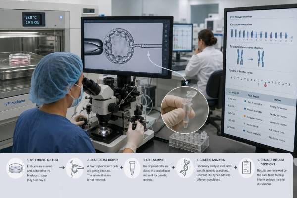
Preimplantation genetic testing, or PGT, analyzes a small sample of cells from embryos created through in vitro fertilization before an embryo is selected for transfer. The term covers several different tests. PGT-A looks for whole-chromosome gains and losses, PGT-M targets a known single-gene condition, and PGT-SR looks for chromosome imbalances related to a parental translocation or inversion. These tests answer different questions and should not be treated as interchangeable.
Most modern PGT is performed on a few trophectoderm cells removed from a day-5, day-6, or day-7 blastocyst. The embryo is then frozen while the laboratory analyzes the sample. Results can help rank embryos or reduce the chance of transferring an embryo with a specific genetic finding, but they do not guarantee implantation, a healthy pregnancy, or an unaffected child. A biopsy represents cells destined mainly to form placenta, not every cell in the embryo, and laboratory thresholds can affect how results are labeled. Careful pretest counseling is therefore as important as the test itself.
- PGT requires IVF because embryos must be created and biopsied in a laboratory before transfer.
- PGT-A screens chromosome number; PGT-M tests a known gene condition; PGT-SR tests imbalances from structural chromosome rearrangements.
- Most embryos are biopsied at the blastocyst stage and frozen while results are pending.
- A “euploid” result lowers chromosome-related risk but does not prove that an embryo is genetically or medically normal.
- Mosaic and no-result findings may require reanalysis, embryo-ranking decisions, or additional counseling rather than automatic discard.
- Prenatal screening and diagnostic testing should still be discussed after pregnancy because PGT has biological and technical limits.
Table of Contents
- Three Main Types of PGT
- From IVF Cycle to Embryo Biopsy
- Who May Consider PGT
- Reading an Embryo Test Report
- Benefits and Evidence Limits
- Transfer, Prenatal Testing, and Pregnancy
- Cost, Ethics, and Clinic Questions
Three Main Types of PGT
PGT is a group of laboratory methods, not a single universal panel. The right test depends on the reproductive question and the genetic information available before IVF begins.
PGT-A: chromosome-number screening
Preimplantation genetic testing for aneuploidy evaluates whether the biopsied cells have the expected number of chromosomes. An embryo with no detected whole-chromosome gain or loss is generally reported as euploid. An embryo with an extra or missing chromosome is reported as aneuploid. Some laboratories also report segmental changes, mosaic findings, or results below a defined confidence threshold.
PGT-A does not test every gene or every chromosome change. It is mainly an embryo-selection tool intended to identify chromosome patterns associated with implantation failure, miscarriage, or conditions such as trisomy 21. Its value depends on age, embryo number, laboratory quality, and how outcomes are measured. More information about this specific method appears in PGT-A embryo chromosome testing.
PGT-M: testing a known monogenic condition
Preimplantation genetic testing for monogenic disease targets a specific inherited condition caused by a variant in one gene. Examples include cystic fibrosis, sickle cell disease, Huntington disease, spinal muscular atrophy, or a familial cancer-predisposition syndrome. The laboratory generally needs the exact familial variant and DNA samples from the intended parents; additional relatives or an affected child may be needed to build a reliable family-specific test.
PGT-M often combines direct variant analysis with linkage, which tracks nearby DNA markers inherited with the disease-causing variant. This reduces the chance that allele dropout or a tiny DNA sample will cause a wrong call. Test development may take weeks or months and should begin before ovarian stimulation when possible. PGT-M for inherited disease has separate technical and counseling requirements from routine PGT-A.
PGT-SR: testing structural chromosome rearrangements
Preimplantation genetic testing for structural rearrangements is designed for a person who carries a balanced translocation, inversion, or another defined chromosome rearrangement. The carrier may be healthy but can produce eggs or sperm with unbalanced chromosome material. PGT-SR looks for those gains and losses in embryos.
The test may not always distinguish a chromosomally normal embryo from an embryo carrying the same balanced rearrangement as the parent. That distinction depends on the platform, breakpoints, family samples, and laboratory design. When it is clinically important, the laboratory should state whether normal-versus-balanced discrimination is possible. The detailed process is covered in PGT-SR for translocations and inversions.
An IVF cycle can include more than one PGT type. For example, a couple may request PGT-M for a familial condition and also choose chromosome screening. Adding analyses increases complexity, cost, and the chance that no embryo meets all transfer criteria.
From IVF Cycle to Embryo Biopsy
PGT begins well before the biopsy. The fertility clinic and genetics laboratory first review the indication, medical records, laboratory reports, and consent choices. PGT-M or PGT-SR may require custom test development. Treatment should not start until the clinic knows the test is feasible and understands which samples are needed.
A typical process includes:
- Ovarian stimulation: injectable medications encourage several follicles to mature during one cycle.
- Egg retrieval: a clinician removes eggs using an ultrasound-guided procedure, usually under sedation.
- Fertilization: eggs are fertilized in the laboratory. Many programs use intracytoplasmic sperm injection to reduce contamination from sperm attached to the egg, especially for PGT-M.
- Embryo culture: fertilized eggs grow for five to seven days. Not every fertilized egg reaches the blastocyst stage.
- Trophectoderm biopsy: an embryologist removes several cells from the outer layer that will contribute mainly to the placenta. The inner cell mass, which develops into the fetus, is not intentionally sampled.
- Vitrification: the embryo is rapidly frozen after biopsy.
- DNA analysis: the genetics laboratory amplifies the small amount of DNA and performs the planned test.
- Result review and transfer planning: the reproductive team ranks available embryos using genetic results, morphology, developmental timing, and the patient’s priorities.
The biopsy does not test the entire embryo. Early embryos can contain more than one chromosome cell line, and the distribution may not be uniform. A five- to ten-cell sample can therefore differ from the inner cell mass or from other trophectoderm areas. This sampling issue is central to mosaic results.
The number of embryos available at the end is usually much smaller than the number of follicles seen at the beginning. Some follicles yield no egg, some eggs are immature, some do not fertilize, and some embryos stop developing before blastocyst. Of the blastocysts tested, some may be affected, aneuploid, mosaic, or inconclusive. Patients should ask for attrition estimates based on age and clinic data rather than assuming every retrieved egg could become a transferable embryo.
Most PGT cycles use frozen embryo transfer because analysis takes time and biopsy-stage embryos are vitrified. A later cycle prepares the uterine lining with natural ovulation, medications, or both. The transfer itself is usually a brief catheter procedure without anesthesia.
Who May Consider PGT
The clearest PGT indications involve a known, substantial genetic risk that the test is designed to detect. Other uses, particularly routine PGT-A, require more individualized discussion because evidence differs among patient groups.
PGT-M may be considered when one or both intended parents have:
- A pathogenic variant for an autosomal dominant condition
- Carrier status for the same autosomal recessive condition
- An X-linked condition or carrier state
- A mitochondrial DNA variant, although embryo testing and risk interpretation can be more complex
- A disease-predisposition variant with later or variable onset
- A need for human leukocyte antigen matching in rare, carefully reviewed family circumstances
The condition’s severity, penetrance, age of onset, treatment options, and residual uncertainty should be discussed. PGT-M is not limited to childhood-lethal disease, but it should involve informed consent and nondirective genetic counseling.
PGT-SR may be considered after a balanced reciprocal or Robertsonian translocation, inversion, or another structural chromosome rearrangement is found in an intended parent. It may reduce transfers of embryos with related unbalanced gains or losses. A parental karyotype result often supplies the chromosome information needed to design the test.
PGT-A is commonly discussed for advanced maternal age, recurrent pregnancy loss, repeated implantation failure, severe male-factor infertility, embryo banking, or a desire to reduce the chance of transferring an aneuploid embryo. However, a plausible indication is not proof that testing increases cumulative live birth for that individual.
A person with few expected embryos may face a different tradeoff from someone likely to produce many blastocysts. PGT-A can reduce the number of transfers needed to find a euploid embryo, but it can also leave no embryo labeled suitable, and some potentially viable embryos may receive mosaic or uncertain labels. Outcomes should be considered per retrieval cycle, not only per embryo transfer, because per-transfer statistics exclude patients who never reach transfer.
PGT does not treat infertility. A couple may have an excellent genetic indication for PGT-M but still face age-related egg decline, low ovarian response, sperm problems, uterine factors, or other barriers. Conversely, someone fertile who wants PGT for a familial disease must undergo IVF solely to access embryo testing.
Reading an Embryo Test Report
An embryo report should be read according to the exact test type, laboratory thresholds, and clinic policy. Labels that sound definitive may describe probabilities based on a small biopsy.
| Term | Usual meaning | Important limitation |
|---|---|---|
| Euploid | No tested whole-chromosome gain or loss was detected. | Does not rule out all genetic disease, birth defects, mosaicism, or IVF failure. |
| Aneuploid | A whole chromosome or large segment appears gained or lost. | The call reflects sampled cells; technical and biological exceptions exist. |
| Mosaic | The DNA signal falls between expected euploid and aneuploid ranges. | It may reflect true mosaicism, sampling, amplification noise, or laboratory thresholds. |
| No result | The laboratory could not issue a reliable call. | Rebiopsy may produce a result but adds manipulation and another freeze-thaw cycle. |
| Unaffected | The targeted PGT-M condition was not detected under the test design. | The embryo may still carry the familial variant or have unrelated findings, depending on the inheritance model. |
| Affected | The embryo has the targeted genotype expected to cause or confer risk for the condition. | Severity may remain variable for conditions with incomplete penetrance or broad expression. |
| Recombinant or inconclusive | Linkage markers or DNA quality did not support a confident PGT-M call. | Direct testing, rebiopsy, or exclusion from transfer may be discussed. |
“Mosaic” is not one uniform category. Reports may specify low-level or high-level mosaicism, whole-chromosome or segmental involvement, and one or several chromosomes. Laboratories use different cutoffs, so the same raw signal might be labeled euploid by one laboratory and mosaic by another. Transfer outcomes are generally less favorable for some mosaic categories than for euploid embryos, but healthy births have occurred after mosaic embryo transfer.
A mosaic result should trigger counseling rather than automatic assumptions. The clinic should explain whether the embryo is eligible for transfer, how it would be ranked, and what prenatal testing would be recommended. The decision often depends on whether euploid embryos remain, the chromosome involved, the level and type of mosaic call, reproductive history, and the patient’s tolerance for uncertainty.
PGT-M reports may classify embryos as affected, unaffected noncarrier, unaffected carrier, or inconclusive. Whether carrier embryos are acceptable for transfer depends on the condition and patient choice. For a recessive disorder, a carrier embryo is generally expected to be healthy, though it could pass the variant to a future generation.
Benefits and Evidence Limits
PGT can provide substantial benefit when it accurately addresses a known familial risk. PGT-M can reduce the chance of establishing a pregnancy affected by the targeted condition. PGT-SR can identify embryos with chromosome imbalances linked to a carrier parent. These uses may help families avoid repeated affected pregnancies or invasive testing followed by difficult pregnancy decisions.
PGT-A has a different evidence base. Selecting a euploid embryo can improve implantation and reduce miscarriage per transfer compared with transferring an untested embryo of unknown chromosome status. It may shorten time to pregnancy for some people who have several blastocysts. It may also support single-embryo transfer by identifying a strong candidate.
Those benefits do not mean routine PGT-A increases the chance of a baby for every IVF patient. Studies that count live birth per transfer can make results look better because aneuploid embryos are excluded and patients with no euploid embryo never transfer. Cumulative live birth per egg retrieval is often the more relevant outcome. Age, embryo yield, biopsy quality, laboratory accuracy, and embryo-transfer practices strongly affect results.
Other limitations include:
- Sampling: trophectoderm cells may not perfectly represent the entire embryo.
- Amplification error: only tiny amounts of DNA are available, so allele dropout or noisy signals can occur.
- Mosaic interpretation: intermediate results are partly dependent on laboratory algorithms and cutoffs.
- Embryo attrition: some embryos may be damaged by biopsy, fail after warming, or be unavailable because of inconclusive results, although experienced laboratories report high survival.
- Scope: PGT detects only what its design covers. A euploid embryo can still have a single-gene disorder, de novo variant, small copy-number change, structural anomaly, or pregnancy complication.
- Misdiagnosis risk: low but not zero, especially when recombination, contamination, mosaicism, or sample identity problems occur.
- No guarantee of transfer: an IVF cycle can end with no blastocyst or no embryo meeting the chosen criteria.
The clinic should provide its own biopsy survival, no-result, mosaic, implantation, miscarriage, and live-birth rates, ideally stratified by age and test type. National averages cannot substitute for the performance of the actual embryology and genetics laboratories.
Transfer, Prenatal Testing, and Pregnancy
Embryo selection combines genetics with developmental and clinical information. A euploid embryo with strong morphology may be transferred before another euploid embryo that developed more slowly, but laboratories and clinics differ in ranking. PGT-M or PGT-SR may add another layer: an embryo can be unaffected for the target condition yet aneuploid, or balanced for a familial rearrangement but otherwise suitable.
Single-embryo transfer is generally favored when an appropriate embryo is available because transferring two embryos increases twin pregnancy risks without doubling the chance of a healthy baby. PGT does not remove risks of preeclampsia, placenta previa, preterm birth, fetal growth problems, or other IVF-associated complications.
Pregnancy after PGT should still include ordinary prenatal care and discussion of genetic screening and diagnosis. Cell-free DNA screening can assess common aneuploidies, while CVS or amniocentesis can provide diagnostic confirmation. Professional guidance commonly recommends offering confirmatory prenatal testing after PGT-M or PGT-SR because rare false results can occur. After PGT-A, screening or diagnostic testing remains appropriate because the embryo biopsy is not equivalent to a fetal diagnostic sample.
CVS samples placenta, the same broad tissue lineage represented by trophectoderm biopsy. For a prior mosaic PGT-A result, amniocentesis may provide more direct fetal information, though the best approach depends on gestational age and the specific finding. A genetics professional can coordinate amniocentesis chromosome testing or CVS with the original PGT laboratory report.
A normal prenatal result after PGT does not guarantee an uncomplicated pregnancy, and a discordant result requires careful review of sample identity, mosaicism, platform limits, and test scope. Families should keep copies of the original variant report, PGT laboratory report, embryo identifier, and transfer record.
Cost, Ethics, and Clinic Questions
PGT costs are layered on top of IVF. Charges may include genetic counseling, custom test development, embryo biopsy, analysis per embryo or per batch, shipping, cryopreservation, storage, and frozen embryo transfer. Insurance may cover PGT-M for a serious inherited condition while excluding IVF, or cover IVF but not PGT-A. Written estimates should clarify what happens if no embryo reaches biopsy or if another retrieval is needed.
PGT also creates choices about embryos that are affected, aneuploid, mosaic, carriers, or unused. Clinics may have policies on storage, transfer eligibility, donation, research, or disposition. Patients should review these policies before testing, not after receiving a difficult result.
Ethical questions can be especially complex for adult-onset disease, reduced penetrance, mild conditions, HLA matching, sex selection, or testing traits without established medical validity. Polygenic embryo scores are not the same as established PGT-M and have greater uncertainty because common diseases reflect many genes plus environment. A clinic should clearly separate validated medical testing from optional commercial add-ons.
Before signing consent, ask:
- Which PGT type is being ordered, and what exact condition or chromosome pattern will it detect?
- Is the genetics laboratory accredited, and has the custom assay been completed before stimulation?
- Can the test distinguish unaffected carriers from noncarriers or normal embryos from balanced carriers?
- How does the laboratory define euploid, aneuploid, low-level mosaic, high-level mosaic, and segmental findings?
- What are the laboratory’s no-result and rebiopsy rates?
- Does the clinic transfer mosaic embryos, and under what ranking policy?
- Are outcomes reported per transfer and per retrieval cycle?
- What happens if no embryo is suitable under the initial plan?
- Which prenatal test is recommended after pregnancy?
- What are the full costs for testing, freezing, storage, and later transfer?
PGT can be a powerful reproductive tool when the indication, test design, and limitations are explicit. It works best as one part of IVF planning rather than as a promise that genetics can identify a perfect embryo. The most informed decisions come from integrating reproductive prognosis, family genetics, laboratory performance, finances, and personal views about uncertainty and embryo disposition.
References
- The use of preimplantation genetic testing for aneuploidy: a committee opinion (2024) 2024 (Position Statement)
- Indications and management of preimplantation genetic testing for monogenic conditions: a committee opinion (2023) 2023 (Position Statement)
- Clinical management of mosaic results from preimplantation genetic testing for aneuploidy (PGT-A) of blastocysts: a committee opinion 2023 (Position Statement)
- PGDIS position statement on the transfer of mosaic embryos 2021 2022 (Position Statement)
- Preimplantation genetic testing: A narrative review 2024 (Review)
- Indications and management of preimplantation genetic testing for monogenic conditions: a committee opinion 2023 (Position Statement)
Disclaimer
This information is educational and does not replace advice from a reproductive endocrinologist, embryologist, medical geneticist, or genetic counselor. PGT accuracy, embryo classification, transfer eligibility, and pregnancy recommendations depend on the specific laboratory method, familial finding, embryo cohort, and clinic policy. Decisions about embryo transfer or disposition should be made only after reviewing the complete laboratory report and available alternatives.





