
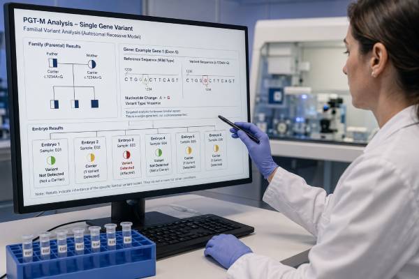
Preimplantation genetic testing for monogenic disease, or PGT-M, tests IVF embryos for a specific condition caused by a variant in one gene. It can be used when an intended parent has a dominant condition, both partners carry the same recessive condition, one partner carries an X-linked condition, or a family has another clearly defined inherited risk. Unlike broad carrier screening, PGT-M is designed around the exact family variant and inheritance pattern.
The process usually requires records confirming the pathogenic or likely pathogenic variant, DNA samples from the intended parents, and sometimes samples from an affected child or other relatives. A genetics laboratory builds a family-specific assay, often combining direct variant testing with linkage analysis of nearby DNA markers. Embryos are created through IVF, biopsied at the blastocyst stage, and frozen while testing is completed. Results may classify an embryo as affected, unaffected, a carrier, or inconclusive. PGT-M can greatly reduce the chance of an affected pregnancy, but it cannot guarantee an embryo for transfer or eliminate the need to discuss prenatal confirmation.
- PGT-M targets a known familial gene variant; it is not a general test for every inherited condition.
- Test development often begins before IVF and may take several weeks or longer.
- A recessive-condition carrier embryo is usually unaffected but can pass the variant to a future child.
- An unaffected result does not rule out unrelated genetic conditions, chromosome abnormalities, or birth defects.
- Allele dropout, recombination, contamination, and embryo mosaicism are managed with linked DNA markers and laboratory safeguards.
- CVS or amniocentesis should still be discussed after pregnancy because PGT-M has a small residual error risk.
Table of Contents
- Which Inherited Risks PGT-M Can Test
- Building a Family-Specific Test
- IVF, Biopsy, and Laboratory Analysis
- Affected, Carrier, Unaffected, and Inconclusive Results
- Accuracy, Limitations, and Special Situations
- Choosing Embryos and Confirming a Pregnancy
- Planning, Costs, and Questions for the Team
Which Inherited Risks PGT-M Can Test
PGT-M is appropriate only when the laboratory can define a testable genetic target. The starting point is usually a clinical laboratory report naming the gene, the exact DNA variant, its classification, and the condition associated with it. A family history by itself is rarely enough.
For an autosomal dominant condition, a parent with one disease-causing variant may have a 50% chance of passing it to each embryo. Examples include Huntington disease, Marfan syndrome, neurofibromatosis type 1, and many hereditary cancer syndromes. Penetrance and severity can vary, so an embryo that inherits the variant may not have the same course as the affected parent.
For an autosomal recessive condition, both intended genetic parents usually carry a pathogenic variant in the same gene. Each embryo then has a 25% chance of being affected, a 50% chance of being an unaffected carrier, and a 25% chance of inheriting neither familial variant. Cystic fibrosis, sickle cell disease, spinal muscular atrophy, and many metabolic disorders follow this pattern.
X-linked inheritance requires attention to fetal sex chromosomes and the condition’s biology. A carrier with an X-linked recessive variant may have sons who are affected and daughters who are carriers, though female carriers can sometimes have symptoms. Some X-linked dominant conditions affect all sexes but may differ in severity. PGT-M can test the variant directly rather than relying only on embryo sex.
Mitochondrial DNA conditions are more difficult because eggs can contain different proportions of altered mitochondrial DNA. PGT may estimate heteroplasmy—the percentage of mitochondrial DNA carrying the variant—but a threshold that predicts health is not available for every condition. The mutation load can also shift between embryo, placenta, and child. Specialized centers should manage these cases.
PGT-M may also be requested for adult-onset conditions, reduced-penetrance variants, or cancer predisposition. These uses are ethically accepted in many settings when counseling covers uncertainty, severity, available surveillance or treatment, and the future child’s interests. A variant associated with increased risk is not equivalent to a fully penetrant childhood disorder.
A variant of uncertain significance is generally not an appropriate sole target because its relationship to disease is unresolved. The laboratory may decline testing or require reclassification. A variant of uncertain significance should not be treated as if it were known to cause the family condition.
PGT-M cannot be designed reliably when the diagnosis is only suspected, several genes could explain the condition, or the familial variant has not been found. In that situation, updated diagnostic testing of an affected relative may be the most useful first step.
Building a Family-Specific Test
PGT-M often requires a preclinical workup before ovarian stimulation. The genetics laboratory reviews the original test report, family relationships, inheritance pattern, and available DNA samples. It decides whether direct testing alone is sufficient or whether linked markers are needed.
Direct variant analysis asks whether the embryo sample contains the familial DNA change. The challenge is that a blastocyst biopsy contains only a few cells. The laboratory must amplify the DNA, and one copy of a gene may occasionally fail to amplify. This is called allele dropout. If the disease-associated allele drops out, an affected embryo could appear unaffected; if the normal allele drops out, an unaffected embryo could appear affected.
Linkage analysis lowers this risk by tracking a group of nearby DNA markers, known as a haplotype, that travels with the familial variant. Instead of relying on one DNA position, the laboratory asks whether the embryo inherited the chromosome segment associated with the affected or unaffected family branch. Karyomapping and other genome-wide marker methods can create this haplotype framework.
Useful samples may include:
- Blood or saliva from both intended parents
- DNA from an affected child or pregnancy
- DNA from grandparents, siblings, or other informative relatives
- Stored prenatal, newborn, or diagnostic material
- Donor samples when a donor contributes egg or sperm and testing is needed for the inheritance model
A relative’s sample is not always necessary. Some laboratories can phase variants using parental samples, single-sperm testing, long-read methods, or embryos of known status. However, lack of an informative relative can lengthen development or make the case less certain.
The laboratory also confirms sample identity and examines whether nearby recombination could separate a marker from the variant. A crossover close to the gene may make an embryo result inconclusive. The farther a marker lies from the variant, the greater the chance that recombination changes the expected linkage.
Test development may take several weeks to several months. Rare genes, complex variants, repeat expansions, pseudogenes, deletions, de novo variants, and low-quality archival DNA can require extra work. Starting IVF before the assay is ready can lead to embryos being stored for longer than expected or to discovering that the requested test is not feasible.
The final pretest plan should state what result categories will be issued, whether carrier status will be disclosed, whether PGT-A will be added, and which prenatal test is recommended after transfer.
IVF, Biopsy, and Laboratory Analysis
PGT-M requires IVF even when the intended parents have no infertility diagnosis. Ovarian stimulation aims to produce multiple eggs because attrition occurs at every stage. Not every egg is mature, not every mature egg fertilizes, and not every fertilized egg becomes a blastocyst suitable for biopsy.
Fertilization is commonly performed by intracytoplasmic sperm injection. Injecting one sperm into each mature egg can reduce the amount of extra sperm DNA surrounding the embryo, which helps limit contamination in some PGT-M workflows. The need for ICSI should still be discussed because clinic practices differ.
At the blastocyst stage, an embryologist removes several trophectoderm cells. The embryo is vitrified, and the biopsy is sent to the genetics laboratory under a strict chain of identity. The laboratory amplifies DNA and applies the family-specific assay. Results may take several days to a few weeks after the biopsy arrives.
PGT-M and PGT-A can be performed on the same biopsy. This may identify embryos unaffected by the monogenic condition that also have no detected whole-chromosome aneuploidy. Adding PGT-A can help rank embryos in some situations, particularly with older eggs or a larger cohort, but it may also create mosaic or segmental findings and reduce the number labeled for transfer. It should not be added automatically without separate counseling.
A simple probability calculation can underestimate attrition. In a recessive condition, 75% of embryos are expected to be unaffected on average, but that does not mean three of four retrieved eggs will be transferable. Some eggs will not form blastocysts, some unaffected embryos will be aneuploid, and random variation can produce a cycle with no unaffected embryo.
Consider a carrier couple with four blastocysts. The inheritance probabilities allow several possible outcomes: all four could be unaffected, one could be affected, or all four could be affected. Each embryo is an independent event. The expected percentages become more reliable across many embryos, not in a single small cohort.
Biopsy and freezing are generally well tolerated by good-quality blastocysts in experienced laboratories, but embryo damage or failure to survive warming can occur. Patients should ask for the clinic’s own post-biopsy survival and frozen-transfer results.
Affected, Carrier, Unaffected, and Inconclusive Results
The report should connect each embryo identifier to the targeted condition, inheritance status, and any additional chromosome result. Common categories include affected, unaffected noncarrier, unaffected carrier, at risk, inconclusive, recombinant, or no result.
| Result | Typical interpretation | Transfer implication |
|---|---|---|
| Unaffected noncarrier | The embryo did not inherit the tested familial variant or disease haplotype. | Usually eligible, subject to embryo quality and any chromosome testing. |
| Unaffected carrier | The embryo carries one recessive variant but is not expected to have the recessive disease. | Often eligible; counseling covers future reproductive implications. |
| Affected | The embryo inherited the genotype expected to cause the targeted condition. | Usually not selected, though policies and patient choices vary for reduced-penetrance or mild conditions. |
| At risk | The embryo inherited a predisposition variant whose future expression is uncertain. | Requires condition-specific counseling rather than a simple affected/unaffected label. |
| Inconclusive or recombinant | The assay could not resolve inheritance because of DNA quality, recombination, or conflicting markers. | Rebiopsy, transfer without a definitive result, or nonuse may be discussed. |
| No result | Insufficient reliable DNA data were obtained. | The embryo is not proven affected; options depend on clinic policy and embryo condition. |
Carrier embryos deserve clear explanation. For most autosomal recessive disorders, a person with one pathogenic variant remains healthy. Excluding carrier embryos can sharply reduce the transferable pool without preventing disease in that child. Some families still prefer noncarrier embryos when several equally suitable choices exist.
For dominant or X-linked conditions, the word “carrier” may be misleading because inheriting one variant can cause disease. The report should use terminology matched to the inheritance pattern.
Reduced penetrance complicates “affected” labels. An embryo with a BRCA1 or BRCA2 pathogenic variant has increased future cancer risk, not a certainty of childhood illness. An embryo with a familial cardiomyopathy variant may never develop disease or may have variable severity. Consent should establish how these results will be described before embryos are created.
If PGT-A is added, an embryo may be unaffected for the gene condition but aneuploid or mosaic. The clinic should provide both result layers rather than collapsing them into “normal” or “abnormal.”
Accuracy, Limitations, and Special Situations
PGT-M is highly accurate when the familial variant is confirmed, the assay is well designed, and linkage is informative. It still has a residual error risk. Misdiagnosis can result from allele dropout, contamination, recombination, embryo mosaicism, sample mix-up, or a laboratory limitation that was not recognized before testing.
The test is targeted. An embryo reported unaffected for cystic fibrosis has not been cleared of every other genetic condition. De novo variants can arise, unrelated recessive conditions may be present, and structural birth defects can occur without a detectable genetic cause.
Complex variants may need specialized methods. Repeat expansions, such as those causing Huntington disease or fragile X syndrome, can be difficult to amplify directly from embryo DNA. Linkage may provide the primary result. Large deletions, duplications, pseudogene regions, and variants in highly similar gene families can also require custom validation.
A de novo variant in an affected child creates phasing challenges because neither parent’s blood shows the variant. PGT-M may still be possible, but the laboratory must determine which parental chromosome carries the mutation in the germline. Germline mosaicism means the recurrence risk can be higher than a negative parental blood test suggests.
Non-disclosure PGT-M is sometimes requested for an adult-onset condition when an at-risk parent does not want to learn whether they carry the familial variant. The laboratory and clinic may design a process that avoids revealing the parent’s status, but treatment logistics can inadvertently disclose information. These cases need careful ethics, genetics, and psychological planning.
Human leukocyte antigen matching may be combined with PGT-M when a family hopes to have an unaffected child whose cord blood could help an affected sibling. The chance of finding an embryo that is unaffected, HLA-compatible, chromosomally suitable, and developmentally viable can be low. The future child’s welfare and the uncertainty of treatment benefit require independent ethical review in many settings.
Some conditions are not technically or clinically suitable. PGT-M cannot guarantee phenotype when disease depends heavily on environment, multiple genes, methylation, or uncertain variant interpretation. Polygenic embryo scores are not a substitute for validated single-gene testing.
Choosing Embryos and Confirming a Pregnancy
Embryo selection should follow the priorities agreed before testing. If several unaffected embryos are available, the clinic may rank them by chromosome status, blastocyst development, morphology, and maternal age at retrieval. Single-embryo transfer usually minimizes twin pregnancy risk.
Families may face choices they did not expect. There may be only carrier embryos, only embryos with an uncertain chromosome result, or no embryo with a conclusive PGT-M result. A prewritten ranking plan can reduce pressure. It should address whether the family would transfer an unaffected carrier, a mosaic embryo, or an embryo with no PGT-M result.
Prenatal confirmation should be offered after pregnancy. Chorionic villus sampling can test placental tissue in the first trimester, while amniocentesis tests amniotic-fluid cells later. The prenatal laboratory needs the exact familial variant and should not rely only on the phrase “PGT pregnancy.”
Cell-free DNA does not confirm most single-gene PGT-M results. Commercial single-gene cfDNA tests are not equivalent to diagnostic testing and may not cover the familial variant. A targeted diagnostic assay on CVS or amniotic fluid is the usual confirmatory method.
Ultrasound remains necessary because an unaffected PGT-M result does not exclude structural anomalies. Newborn testing may be considered if prenatal diagnosis was declined, especially when early treatment would matter.
Patients should keep copies of the familial variant report, PGT-M workup, embryo result, and transfer record. These documents may be needed years later for the child’s care or for relatives considering testing.
Planning, Costs, and Questions for the Team
PGT-M costs include more than the embryo analysis. Families may pay for genetic counseling, custom assay development, relatives’ sample collection, IVF medications, egg retrieval, embryo biopsy, freezing, storage, and frozen transfer. Some laboratories charge a fixed setup fee plus a per-embryo fee. Coverage varies widely and may separate medical genetics benefits from infertility benefits.
Time is another cost. Assay development, insurance authorization, coordinating relatives, and repeated retrievals can add months. People with declining ovarian reserve may need the genetics and fertility teams to work in parallel, sometimes freezing eggs or embryos while the assay is finalized.
Questions to ask include:
- Is the familial variant classified as pathogenic or likely pathogenic, and has the diagnosis been confirmed in the right relative?
- Will the assay use direct testing, linkage, or both?
- Which relatives’ samples are needed, and what happens if they are unavailable?
- Can the laboratory distinguish unaffected carriers from noncarriers?
- How will recombination or allele dropout be recognized?
- Is PGT-A being offered separately, and how might it change the transferable embryo count?
- What are the expected affected, carrier, and unaffected proportions for this inheritance pattern?
- How often does the laboratory issue inconclusive results for similar cases?
- What is the clinic’s policy on carrier, mosaic, or no-result embryos?
- Which prenatal diagnostic test will confirm the result?
PGT-M offers a way to reduce a defined inherited risk before pregnancy begins, but its success depends on more than genetic accuracy. A usable embryo must also develop, survive biopsy and warming, implant, and continue through pregnancy. The clearest counseling combines the inheritance probabilities with age-specific IVF expectations so families understand both the genetic and reproductive parts of the process.
References
- Indications and management of preimplantation genetic testing for monogenic conditions: a committee opinion (2023) 2023 (Position Statement)
- Genetic counseling for pre-implantation genetic testing of monogenic disorders (PGT-M) 2023 (Review)
- The Role of Preimplantation Genetic Testing for Monogenic Disorders (PGT-M) in Hemoglobinopathies 2025 (Review)
- Clinical outcomes following preimplantation genetic testing for monogenic conditions: a systematic review 2025 (Systematic Review)
- Use of preimplantation genetic testing for monogenic adult-onset conditions: an Ethics Committee opinion (2024) 2024 (Position Statement)
- Prenatal diagnosis following preimplantation genetic testing for monogenic conditions: a single-centre retrospective study 2025 (Review)
Disclaimer
This article is educational and does not replace genetic counseling, IVF care, or laboratory-specific advice. PGT-M feasibility and accuracy depend on the exact familial variant, inheritance pattern, available relatives, assay design, and embryo cohort. Embryo transfer and prenatal-testing decisions should be made with the reproductive and genetics teams that hold the complete records.





