
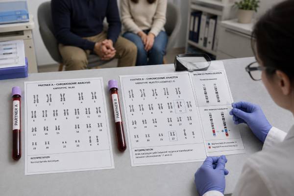
A parental karyotype test examines the number and visible structure of chromosomes in one or both partners. It may help explain recurrent miscarriage, severe male-factor infertility, or a pregnancy affected by an unbalanced chromosome rearrangement. Most people who carry a balanced translocation or inversion are healthy because they have the expected amount of genetic material, arranged differently. The reproductive risk appears when eggs or sperm receive an unbalanced chromosome combination, which can prevent implantation, cause miscarriage, or lead to a child with a chromosome condition.
The test usually requires a blood sample and does not need fasting. Results often take about 2 to 4 weeks because the laboratory must grow cells before viewing the chromosomes. A normal result cannot rule out every genetic cause of infertility or pregnancy loss, and an abnormal result does not mean a healthy pregnancy is impossible. Genetic counseling is especially important because risks differ greatly by the chromosomes involved, the breakpoints, the carrier’s sex, reproductive history, and maternal age.
- A parental karyotype usually checks 46 chromosomes for extra, missing, or visibly rearranged material.
- Balanced translocations and inversions may cause no health problems in the carrier but can raise reproductive risks.
- Testing is most informative after recurrent pregnancy loss, severe sperm abnormalities, or an unbalanced fetal or miscarriage result.
- A normal karyotype does not exclude small DNA changes, single-gene conditions, or all causes of infertility.
- No fasting or medication changes are usually needed; the sample is generally blood drawn into a heparin tube.
- An abnormal result should be interpreted with a genetic counselor before choosing natural conception, prenatal diagnosis, donor gametes, or IVF with PGT-SR.
Table of Contents
- What the Test Examines
- When Parental Karyotyping Is Considered
- How the Test Is Performed
- Understanding Normal and Abnormal Results
- Reproductive Risks of Balanced Rearrangements
- Options After an Abnormal Result
- Limits, Costs, and Questions to Ask
What the Test Examines
A karyotype is an organized image of chromosomes from dividing cells. Humans usually have 46 chromosomes: 22 pairs of autosomes and one pair of sex chromosomes. A laboratory stains the chromosomes to create light and dark bands, then evaluates their number, size, shape, and banding pattern. This type of chromosome analysis can identify changes large enough to be seen under a microscope.
A parental karyotype can detect several broad types of findings:
- Numerical chromosome changes: an extra or missing chromosome, such as 47,XXY or 45,X in some or all tested cells.
- Reciprocal translocations: segments from two different chromosomes have exchanged places.
- Robertsonian translocations: two acrocentric chromosomes—usually chromosomes 13, 14, 15, 21, or 22—have joined near their centromeres.
- Inversions: a chromosome segment has broken, reversed direction, and rejoined.
- Large deletions or duplications: visible chromosome material is missing or present in extra copies.
- Marker chromosomes or other complex rearrangements: extra or reorganized material cannot always be identified fully by standard banding alone.
- Mosaicism: two or more chromosome cell lines are present, provided the abnormal line appears in enough tested blood cells to be detected.
“Balanced” means the person appears to have no major net gain or loss of chromosome material. For example, someone with a balanced reciprocal translocation may have all the expected genes, but parts of two chromosomes have traded places. The carrier is often healthy, although an apparently balanced rearrangement can occasionally disrupt a gene or involve a small imbalance below karyotype resolution.
“Unbalanced” means genetic material is missing or duplicated. The health effects depend on which segments and genes are involved and how much material changed. Unbalanced rearrangements are more likely to cause developmental differences, congenital anomalies, pregnancy loss, or failure of an embryo to implant.
Karyotyping does not read individual DNA letters. It cannot reliably detect most single-gene variants, small copy-number changes, or subtle chromosome rearrangements. A normal result therefore answers a focused question: no large numerical or structural chromosome abnormality was visible in the cells examined.
When Parental Karyotyping Is Considered
Parental karyotyping is usually selected for a specific clinical reason. Current recurrent pregnancy loss guidance does not support automatic karyotyping for every couple. Instead, clinicians consider the chance of finding a meaningful rearrangement and whether the result would change counseling or care.
Testing may be particularly useful when:
- Two or more pregnancies have ended in loss and the couple’s history suggests a chromosome rearrangement.
- Testing of miscarriage tissue found an unbalanced structural chromosome change rather than a common random trisomy.
- A prior fetus or child had multiple congenital anomalies, developmental disability, or an unbalanced translocation.
- A family member carries a balanced translocation, inversion, or other chromosome rearrangement.
- One partner has azoospermia, very low sperm concentration, small testes, or other signs of a sex chromosome abnormality.
- There is unexplained infertility combined with a suggestive personal or family history.
- An assisted-reproduction team needs to clarify whether preimplantation genetic testing for structural rearrangements would apply.
In recurrent miscarriage, testing the pregnancy tissue can sometimes guide parental testing more efficiently. A products of conception genetic test may show that a loss was caused by a random whole-chromosome error, an unbalanced inherited rearrangement, or no detectable chromosome imbalance. If the tissue contains an unbalanced translocation, both partners are commonly offered karyotyping to determine whether either carries the balanced form.
Karyotyping is also part of selected male infertility evaluations. Men with nonobstructive azoospermia or severe oligospermia have a higher rate of chromosome abnormalities than men with normal sperm counts. Depending on the clinical picture, a clinician may order a karyotype alongside hormone tests, Y-chromosome microdeletion analysis, or CFTR testing. The exact threshold and test combination should follow current specialty guidance and the individual semen-analysis findings.
A history of miscarriage alone does not prove that a parent carries a rearrangement. Most early miscarriages result from sporadic chromosome errors in the embryo, and the chance rises with maternal age. Uterine, endocrine, immune, and other factors may also contribute. A complete recurrent pregnancy loss genetic evaluation places a parental karyotype in that broader context.
How the Test Is Performed
The usual sample is peripheral blood from each person being tested. The collection itself is similar to any routine blood draw, but the laboratory commonly needs a sodium-heparin tube because living white blood cells must be cultured. Fasting is not usually required, and ordinary medications rarely interfere. Patients should follow the ordering laboratory’s instructions because specimen type, tube color, shipping temperature, and timing can differ.
In the laboratory, technicians stimulate lymphocytes to divide, culture them for about 72 hours, and stop cell division during metaphase, when chromosomes are condensed and easiest to see. The cells are placed on slides and treated with a banding method, usually G-banding. A cytogenetic technologist examines multiple metaphase cells, prepares representative karyograms, and a qualified specialist reviews the result.
Typical turnaround is about 2 to 4 weeks. It may take longer if cells grow slowly, the finding is complex, extra cells must be counted for suspected mosaicism, or follow-up methods are needed. A failed culture is uncommon with a properly handled blood sample, but recollection may be necessary if too few dividing cells are available.
The report uses International System for Human Cytogenomic Nomenclature, often called ISCN. The notation can look cryptic. Common examples include:
| Example | Plain-language meaning |
|---|---|
| 46,XX | Forty-six chromosomes with an XX sex chromosome pattern; no visible abnormality reported. |
| 46,XY | Forty-six chromosomes with an XY sex chromosome pattern; no visible abnormality reported. |
| 46,XX,t(2;8)(q21;q24) | An XX individual with a reciprocal translocation between chromosomes 2 and 8 at the listed bands. |
| 45,XY,rob(13;14)(q10;q10) | An XY individual with a balanced Robertsonian translocation involving chromosomes 13 and 14; 45 chromosomes is expected for this rearrangement. |
| 46,XX,inv(9)(p12q13) | An XX individual with an inversion in chromosome 9 between the specified breakpoints. |
| mos 45,X/46,XX | Two cell lines were detected: one missing an X chromosome and one with an XX pattern. |
The report may recommend fluorescence in situ hybridization, chromosomal microarray, or another test if the chromosome finding needs clarification. Relatives may also be offered targeted karyotyping when a familial rearrangement is identified.
Understanding Normal and Abnormal Results
A normal parental karyotype is usually written as 46,XX or 46,XY. It means the laboratory did not see a chromosome-number abnormality or structural rearrangement at the test’s resolution in the cells examined. It does not mean fertility is normal, miscarriage cannot recur, or every genetic risk has been excluded.
A normal result does not reliably detect:
- Single-gene variants or carrier status for most inherited disorders
- Small deletions and duplications below the microscope’s resolution
- Low-level mosaicism absent or rare in blood
- Epigenetic conditions, mitochondrial DNA changes, or most repeat expansions
- Sperm or egg aneuploidy that occurs during gamete formation despite a normal blood karyotype
- Chromosome abnormalities that arose only in a particular embryo
An abnormal result may be balanced, unbalanced, numerical, mosaic, or uncertain in reproductive significance. The most common clinically important parental findings in recurrent pregnancy loss are balanced reciprocal and Robertsonian translocations. Some inversions also affect reproductive risk, although many common heterochromatic variants are considered benign or of limited clinical importance.
A report may use wording such as “normal variant,” “chromosomal polymorphism,” or “heteromorphism.” Examples include variation in satellite size or heterochromatin length. Older studies sometimes linked these findings to infertility or miscarriage, but many are now regarded as common variants without proven clinical effect. Interpretation should follow the laboratory’s classification and current genetic counseling rather than assumptions based on the notation alone.
A numerical finding such as 47,XXY can explain impaired sperm production and may have broader health implications. Mosaic 45,X/46,XX can be associated with reduced ovarian reserve, infertility, or Turner syndrome features, although effects vary with the tissues involved and proportion of abnormal cells. These results may lead to endocrine, reproductive, or medical follow-up beyond pregnancy planning.
The word “balanced” should not be mistaken for “zero risk.” It generally predicts that the carrier has no large net gain or loss of DNA, but reproductive outcomes depend on how the rearranged chromosomes separate during egg or sperm formation. The same general type of translocation can create very different risks in different families.
Reproductive Risks of Balanced Rearrangements
During meiosis, chromosome pairs must align and separate so each egg or sperm receives one copy of each chromosome region. A balanced rearrangement changes how chromosomes pair. Several segregation patterns are possible, producing gametes that are normal, balanced like the parent, or unbalanced.
A pregnancy may therefore have one of several outcomes:
- The embryo has a normal chromosome arrangement.
- The embryo inherits the same balanced rearrangement and is usually healthy like the carrier parent.
- The embryo has an unbalanced duplication and deletion, which may lead to implantation failure or miscarriage.
- The pregnancy continues with a fetal chromosome condition whose features depend on the specific imbalance.
There is no universal percentage that applies to all translocation carriers. Risk estimates can vary from low single digits to much higher levels. Important factors include:
- Which chromosomes are involved
- The precise breakpoints and size of possible duplicated or deleted segments
- Whether the carrier is the egg-producing or sperm-producing partner
- The type of rearrangement—reciprocal, Robertsonian, inversion, or complex
- The carrier’s prior pregnancies and any tested miscarriage results
- Maternal age, which adds an independent risk of sporadic aneuploidy
- Whether the calculation concerns miscarriage, an affected live birth, or any unbalanced conception
A Robertsonian translocation involving chromosomes 13 and 14 has different implications from one involving chromosomes 14 and 21. A person carrying rob(14;21), for example, may have a risk of a pregnancy with translocation Down syndrome, while rob(13;14) more often contributes to miscarriage because many unbalanced combinations are not viable. Rarely, a Robertsonian rearrangement raises concerns about uniparental disomy, in which both copies of a chromosome come from one parent; whether testing is relevant depends on the chromosomes and fetal result.
Natural conception can still lead to a healthy child. Studies of couples with recurrent loss and abnormal parental karyotypes show that cumulative live birth is often achievable, although miscarriage risk may remain higher. The most useful counseling separates the chance per recognized pregnancy from the cumulative chance over repeated attempts and accounts for the emotional and medical burden of additional losses.
Options After an Abnormal Result
An abnormal parental karyotype should lead to individualized counseling, not a single automatic treatment. A genetics professional can translate the notation, estimate the range of reproductive risks, discuss uncertainty, and identify whether relatives may also be carriers.
Common reproductive paths include:
Natural conception with prenatal screening or diagnosis
Some couples continue trying naturally. Prenatal screening, including cell-free DNA or serum screening, may estimate risk for selected whole-chromosome conditions, but screening cannot reliably exclude every unbalanced rearrangement. Diagnostic testing through chorionic villus sampling or amniocentesis can examine fetal chromosomes directly. The best method may be karyotype, chromosomal microarray, targeted testing for the familial rearrangement, or a combination. A prenatal genetic testing discussion should clarify what each test can and cannot detect.
IVF with PGT-SR
Preimplantation genetic testing for structural rearrangements evaluates embryos created through IVF for chromosome imbalances related to a known parental rearrangement. PGT-SR may reduce the chance of transferring an embryo with an unbalanced chromosome result, but it does not guarantee a transferable embryo, implantation, pregnancy, or live birth.
The number of embryos available is strongly affected by ovarian response and age. Some cycles produce no embryo classified as suitable for transfer. Depending on the platform and family-specific design, PGT-SR may identify unbalanced embryos but may not always distinguish a chromosomally normal embryo from one carrying the same balanced rearrangement as the parent. Prenatal diagnostic testing is commonly offered after pregnancy because embryo testing samples only a few placental-lineage cells and has technical limits.
Donor eggs, donor sperm, or donor embryos
Using a gamete from a donor without the rearrangement can avoid passing on that specific parental chromosome risk. It introduces separate medical, emotional, legal, and family considerations. Donors still undergo screening, but no reproductive option removes every genetic or pregnancy risk.
Adoption or choosing not to pursue pregnancy
These are valid family-building or life choices. Counseling should be nondirective and should respect personal values, finances, cultural beliefs, tolerance for treatment, and experience of prior loss.
No option is inherently right for every carrier. A couple may reasonably choose natural conception after understanding the risks, while another may prefer IVF with PGT-SR to reduce the likelihood of another chromosome-related miscarriage. Access, cost, age, fertility diagnosis, and the expected number of embryos often shape the choice as much as the karyotype itself.
Limits, Costs, and Questions to Ask
Parental karyotyping has a narrower scope than many people expect. Its strength is detecting large chromosome changes across the genome. Its weakness is limited resolution. A chromosomal microarray can detect smaller gains and losses, while sequencing can identify many single-gene variants, but those methods do not replace every function of a karyotype. Microarray, for example, usually cannot identify a balanced translocation because there is no net gain or loss of DNA.
The clinical value of testing depends on the pretest probability. In an unselected couple with no suggestive history, the chance of finding a meaningful balanced rearrangement is low. In a couple whose miscarriage tissue had an unbalanced translocation, the result may immediately change recurrence counseling and reproductive planning. This is why risk-based testing can be more useful than ordering the test simply because conception has taken longer than expected.
Costs vary by country, laboratory, insurance plan, and whether one or both partners are tested. Coverage may require documentation of recurrent losses, severe male infertility, an abnormal pregnancy result, or a genetics consultation. Ask for an estimate that separates the laboratory charge from clinician, counseling, blood-draw, and follow-up fees.
Before testing, useful questions include:
- What finding in our history makes a parental karyotype appropriate?
- Should both partners be tested at the same time, or should testing start with one person?
- Would testing miscarriage tissue first provide more direct information?
- What resolution does the laboratory report, and how many cells are examined?
- How will a normal result change the rest of the evaluation?
- If a rearrangement is found, can the laboratory or counselor provide a family-specific risk estimate?
- Would relatives benefit from testing?
- Which prenatal test would diagnose the specific familial rearrangement?
- Can PGT-SR distinguish normal embryos from balanced-carrier embryos in this case?
- What costs and waiting times apply to karyotyping, counseling, IVF, embryo testing, and prenatal diagnosis?
Seek prompt clinical advice if a pregnancy is ongoing and a parent is known to carry a rearrangement, because timing affects access to chorionic villus sampling and amniocentesis. The result is not an emergency in the usual sense, but early referral preserves more testing and counseling options.
A parental karyotype can turn an unexplained reproductive history into a defined chromosome mechanism, but it may also be normal or reveal a finding with variable significance. Its greatest value comes from ordering it for a clear reason and interpreting it alongside pregnancy history, semen findings, fetal or miscarriage testing, maternal age, and the couple’s reproductive priorities.
References
- Guideline on the management of recurrent pregnancy loss 2023 (Guideline)
- The Value of Parental Karyotyping in Recurrent Pregnancy Loss Lies in Individual Risk Assessments 2024 (Review)
- Systematic review of subsequent pregnancy outcomes in couples with parental abnormal chromosomal karyotypes and recurrent pregnancy loss 2022 (Systematic Review)
- A Systematic Review of Reproductive Counseling in Cases of Parental Balanced Translocation Carriers 2022 (Systematic Review)
- Updates to Male Infertility: AUA/ASRM Guideline (2024) 2024 (Guideline)
Disclaimer
This information is educational and cannot replace individualized care from a reproductive specialist, medical geneticist, or genetic counselor. Karyotype findings and reproductive risks depend on the exact chromosome rearrangement, family history, laboratory method, and pregnancy context. Do not make treatment or pregnancy-testing decisions from a report without professional interpretation.





