
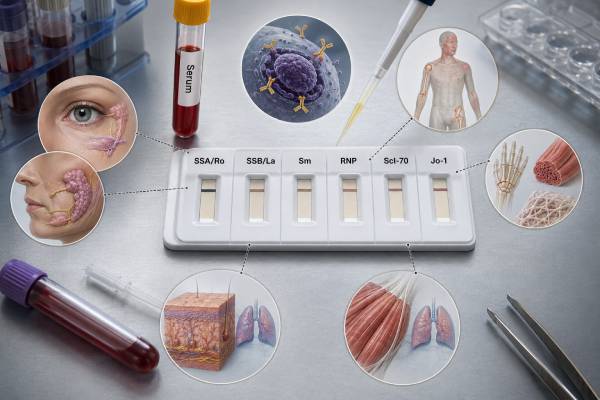
An ENA 6 panel checks for six autoantibodies that are commonly associated with systemic connective tissue diseases: SSA/Ro, SSB/La, Smith, RNP, Scl-70, and Jo-1. The panel is usually ordered after an antinuclear antibody result or a clinical pattern raises concern for lupus, Sjögren disease, mixed connective tissue disease, systemic sclerosis, or inflammatory myositis. Each antibody points toward a different cluster of possibilities, but none can diagnose a disease in isolation. The strength of the result, the test method, the ANA pattern, and the person’s symptoms all change its meaning. A weak isolated positive can be incidental or assay-related, while a strong disease-matched antibody may guide organ screening and specialist care. A negative ENA 6 result also does not close the door on autoimmunity because many relevant antibodies are not included and some patients remain seronegative. The report is best viewed as a six-part clue set rather than a single positive-or-negative verdict.
- The classic ENA 6 panel includes SSA/Ro, SSB/La, Sm, RNP, Scl-70, and Jo-1.
- SSA/Ro and SSB/La often support Sjögren disease or lupus, while Sm is strongly associated with lupus.
- RNP may support mixed connective tissue disease; Scl-70 and Jo-1 raise specific lung-disease considerations.
- Low-positive results may require confirmation: assay methods and laboratory cutoffs can produce discordant findings.
- No fasting is usually required: interpretation matters more than special preparation.
Table of Contents
- How the ENA 6 Panel Works
- SSA/Ro and SSB/La Results
- Sm and RNP Results
- Scl-70 and Jo-1 Results
- Reading Antibody Combinations and Strength
- Who Is Most Likely to Benefit From Testing
- Negative Results and Panel Limitations
- Preparation, Laboratory Methods, and Follow-Up
How the ENA 6 Panel Works
ENA means extractable nuclear antigen. The term describes a historical group of salt-soluble cellular proteins that became useful targets for autoimmune antibody testing. Modern laboratories usually test purified, recombinant, or synthetic antigens with automated immunoassays, but the familiar ENA name remains.
The six-antibody panel is narrower than an extended ENA or systemic autoimmune rheumatic disease panel. Its advantage is focus: it covers several of the most established antibody-disease associations without producing dozens of low-level findings. Its limitation is equally important: it cannot detect every antibody linked to lupus, systemic sclerosis, myositis, Sjögren disease, or overlap syndromes.
The panel generally includes:
| Reported antibody | Alternate name or target | Main disease associations |
|---|---|---|
| SSA/Ro | Ro60 and/or Ro52, depending on assay | Sjögren disease, lupus, cutaneous lupus, several overlap conditions |
| SSB/La | La | Sjögren disease and lupus, usually with SSA |
| Sm | Smith antigen | Systemic lupus erythematosus |
| RNP | Usually U1-ribonucleoprotein | Mixed connective tissue disease, lupus, overlap disease |
| Scl-70 | Topoisomerase I | Systemic sclerosis |
| Jo-1 | Histidyl-tRNA synthetase | Antisynthetase syndrome and inflammatory myositis |
The panel is often used after a positive ANA by IFA test. ANA immunofluorescence shows whether antinuclear antibodies are present and may reveal a pattern, while ENA testing identifies specific antigen targets. The two approaches answer related but different questions.
Why ANA and ENA can disagree
An ANA and an ENA panel can disagree without either result being automatically wrong. Indirect immunofluorescence exposes a broad range of antigens inside cultured cells, whereas an ENA assay presents a selected set of manufactured targets. A weak ANA may have no matching specificity among the six ENA antigens. Conversely, some SSA or Jo-1 antibodies may be detected on a targeted assay even when the nuclear ANA screen is negative or shows mainly cytoplasmic staining.
The sequence also matters. A laboratory may perform ENA testing only when ANA exceeds a certain titer, while a clinician may order an individual antibody directly because the symptoms are highly specific. That is especially relevant for suspected Sjögren disease, subacute cutaneous lupus, antisynthetase syndrome, or antibody-related pregnancy risk.
Discordance should prompt a method check rather than a reflex diagnosis. The clinician can compare the ANA titer and pattern, review which antigens the ENA platform contains, and decide whether a focused test or second method is worthwhile. Repeating every autoimmune test without a clinical reason usually creates more noise.
A result can be reported as negative or positive, as a numerical index, or in units defined by the manufacturer. There is no universal scale. A value of 2.0 may be strongly positive on one platform and meaningless on another. The laboratory’s own cutoff and method are essential parts of the result.
SSA/Ro and SSB/La Results
SSA/Ro is the broadest and most clinically varied antibody in the ENA 6 panel. It is common in Sjögren disease and lupus, but it can also occur in subacute cutaneous lupus, neonatal lupus risk, systemic sclerosis, inflammatory myositis, autoimmune liver disease, and some people without a classifiable systemic illness.
SSA is not one single antigen
The Ro system includes at least two major targets, Ro60 and Ro52. Some ENA 6 assays combine them under one SSA result. Others detect one better than the other, and some laboratories report them separately. Ro52 positivity has broad overlap across autoimmune conditions and can carry special relevance in myositis or interstitial lung disease. Ro60 has a more traditional association with Sjögren disease and lupus.
A combined SSA-positive result therefore does not always reveal which Ro component is driving the test. When the clinical question involves lung disease, myositis, pregnancy, or a conflicting ANA pattern, separate Ro52 and Ro60 testing may add useful detail.
Sjögren disease and dryness symptoms
Anti-SSA is an important classification marker for Sjögren disease, but dryness alone is not enough to interpret it. Medications, menopause, diabetes, dehydration, contact lenses, sleep disorders, and local gland disease can all cause dry eyes or dry mouth. A convincing evaluation may include Schirmer tear testing, ocular staining, salivary flow measurement, gland imaging, or minor salivary gland biopsy.
Anti-SSB usually appears with SSA. Isolated SSB without SSA has much less diagnostic value than the combination. A clinician may recheck an isolated low SSB result, review the ANA pattern, and look for objective gland dysfunction before assigning significance. A focused anti-SSA/Ro antibody assessment may be helpful when the laboratory’s combined result is unclear.
Pregnancy-related meaning
SSA and SSB antibodies can cross the placenta. Most pregnancies with these antibodies do not develop antibody-related fetal complications, but a small proportion are affected by neonatal lupus manifestations, including congenital heart block. Risk is higher when there was a previously affected pregnancy.
A pregnant person with known SSA or SSB positivity should inform the obstetric and rheumatology teams early. Management may include medication review and fetal cardiac surveillance during the gestational period when conduction disease is most likely to appear. The antibody result is not a reason to avoid pregnancy, but it is a reason for coordinated care.
Skin and systemic lupus clues
SSA can support lupus when it appears with photosensitive rash, oral ulcers, inflammatory arthritis, blood-count abnormalities, kidney findings, or other classification features. It is especially associated with subacute cutaneous lupus, which often causes annular or scaly photosensitive lesions. Some patients with SSA-associated disease can have a negative ANA by certain methods, so strong clinical suspicion may justify direct SSA testing even when the initial screen is not clearly positive.
Sm and RNP Results
Sm and RNP antibodies target related ribonucleoprotein complexes, so they are often detected together. Their relative strength and the clinical phenotype help separate lupus from mixed connective tissue disease and overlap syndromes.
Anti-Sm and lupus
Anti-Sm is relatively specific for systemic lupus erythematosus. A confirmed positive result in a patient with compatible features carries substantial diagnostic weight. Its sensitivity is limited, however, which means many people with lupus are Sm-negative. A negative Sm result should never be used to rule out lupus.
Sm is not a dependable disease-activity marker. It can remain positive whether lupus is quiet or active. Monitoring usually relies more on symptoms, examination, urinalysis, kidney function, blood counts, complement, and sometimes anti-dsDNA. A positive anti-Smith antibody supports diagnostic reasoning but does not show whether the kidneys, skin, joints, brain, heart, or lungs are currently inflamed.
Weak Sm results deserve method-aware interpretation. Multiplex and line assays can occasionally report low-level reactivity that is not confirmed with another technique. A strong result with a matching ANA pattern and lupus phenotype is more persuasive than an isolated borderline result in someone without objective autoimmune findings.
Anti-RNP and mixed connective tissue disease
RNP is found in lupus, systemic sclerosis overlap, inflammatory myositis overlap, and mixed connective tissue disease. Mixed connective tissue disease usually involves high anti-U1-RNP levels plus a recognizable clinical combination such as Raynaud phenomenon, puffy hands, inflammatory arthritis, myositis, esophageal dysfunction, or lung involvement.
RNP positivity alone does not establish mixed connective tissue disease. Low or moderate RNP values are common in lupus and can appear in undifferentiated connective tissue disease. The diagnosis depends on the pattern over time, not just the lab label.
People with strong RNP-associated disease may need assessment for pulmonary hypertension, interstitial lung disease, muscle inflammation, or swallowing problems depending on symptoms. Shortness of breath, reduced exercise tolerance, chest discomfort, new weakness, or persistent swallowing difficulty should not be attributed to a blood test without direct evaluation.
Sm/RNP combined results
Some laboratories report Sm and RNP separately; others also provide a combined Sm/RNP marker. A combined positive may reflect antibodies to shared proteins and may not equal a true Sm-specific positive. The individual component results are more useful when deciding how strongly the pattern supports lupus or MCTD.
When both Sm and RNP are positive, clinicians ask which is stronger, whether ANA is strongly positive, and whether the person’s features are predominantly lupus-like, MCTD-like, or overlapping. The name of the disease may remain uncertain early on, and follow-up can reveal the stable phenotype more clearly than a one-time panel.
Scl-70 and Jo-1 Results
Scl-70 and Jo-1 are included because they identify disease patterns that may require prompt lung evaluation. They are also two results where assay limitations can have major consequences.
Scl-70 and systemic sclerosis
Scl-70 is another name for anti-topoisomerase I antibody. It is associated with systemic sclerosis, particularly diffuse skin involvement and interstitial lung disease. A convincing result is most meaningful when it occurs with Raynaud phenomenon, puffy or tightening fingers, fingertip ulcers, abnormal nailfold capillaries, reflux or swallowing problems, or other systemic sclerosis findings.
Commercial Scl-70 assays can produce false-positive results, especially low positives found through broad multiplex panels. Referral-center studies have shown that some patients with positive commercial results test negative by more specific immunodiffusion methods and never develop classifiable systemic sclerosis. Because the label can trigger anxiety and extensive imaging, confirmation is reasonable when the result is weak or the clinical picture does not fit.
A true positive does not predict an individual outcome with certainty. It indicates a risk pattern, not a guaranteed course. Clinicians may use pulmonary function tests and chest imaging to screen for interstitial lung disease and monitor over time.
Jo-1 and antisynthetase syndrome
Jo-1 is an antibody to histidyl-tRNA synthetase. It is the most common of several antisynthetase antibodies and can occur with interstitial lung disease, inflammatory muscle disease, inflammatory arthritis, fever, Raynaud phenomenon, or mechanic’s hands—rough, cracked skin along the sides of the fingers.
The lung disease may appear before obvious muscle weakness. A person with Jo-1 positivity and unexplained cough, breathlessness, or reduced oxygen level needs direct pulmonary assessment rather than reassurance based on normal muscle enzymes. Conversely, a weak isolated Jo-1 result without muscle, lung, joint, or skin features may need confirmation.
A negative Jo-1 does not exclude antisynthetase syndrome. Other antibodies, including PL-7, PL-12, EJ, OJ, KS, and Zo, are not part of the classic ENA 6 panel. A broader myositis antibody panel may be appropriate when interstitial lung disease or proximal weakness is a central concern.
Reading Antibody Combinations and Strength
The ENA 6 panel becomes more useful when results are read as a pattern instead of six independent yes-or-no answers.
| Result pattern | Possible interpretation | Important caution |
|---|---|---|
| SSA positive, SSB positive | Supports Sjögren disease or lupus in the right phenotype | Objective dryness or systemic features are still needed |
| Sm strong positive, RNP positive | Can strongly support lupus | Activity and organ involvement require separate tests |
| RNP dominant, Sm negative | May fit MCTD or overlap disease | RNP level alone does not diagnose MCTD |
| Scl-70 isolated low positive | Could represent early systemic sclerosis or a false positive | Confirm when ANA pattern and symptoms do not match |
| Jo-1 positive | Raises concern for antisynthetase syndrome | Assess lungs even if weakness is mild |
| All six negative | No detected antibody in this limited panel | Does not exclude seronegative disease or antibodies outside the panel |
Result strength matters, but not in a simple linear way. A number twice the cutoff is not necessarily twice as clinically important. The relationship between antibody concentration and disease likelihood differs by antibody and assay. Strong, reproducible, disease-matched results carry more weight than weak isolated findings.
The ANA pattern can provide a consistency check. A classic speckled pattern may fit SSA, Sm, or RNP. A pattern associated with topoisomerase I can support Scl-70. A cytoplasmic pattern may accompany Jo-1 even though the panel is called “nuclear antigen” testing. When the ENA result and ANA pattern sharply conflict, the laboratory method and possibility of confirmation deserve attention.
Combinations may also reflect one autoimmune syndrome rather than several simultaneous diseases. SSA plus Jo-1 may describe a myositis-lung phenotype with Ro52 positivity. Sm plus RNP may occur within lupus. The report should not be converted mechanically into one diagnosis per positive antibody.
Who Is Most Likely to Benefit From Testing
The panel is most useful when symptoms suggest a systemic autoimmune rheumatic disease. Testing people with only nonspecific fatigue, diffuse pain, or a family history but no objective features increases incidental findings and can produce more confusion than clarity.
Features that may justify ENA 6 testing include:
- Persistent inflammatory joint swelling
- Photosensitive or characteristic autoimmune rash
- Raynaud phenomenon with puffy fingers, ulcers, or capillary changes
- Objective dry eyes or dry mouth
- Proximal muscle weakness, elevated creatine kinase, or characteristic rash
- Unexplained interstitial lung disease
- Protein or blood in urine with concern for immune kidney disease
- Cytopenias, serositis, or recurrent oral ulcers in a lupus-like pattern
- A positive ANA with a clinically relevant titer and pattern
Testing can be ordered as a reflex after ANA or as selected individual antibodies. Direct SSA testing may be reasonable despite a negative ANA when Sjögren disease, subacute cutaneous lupus, or pregnancy-related risk is strongly suspected. Direct Jo-1 or broader myositis testing may be appropriate in interstitial lung disease even when the classic nuclear ANA is negative.
Children, pregnant patients, and people with severe lung or kidney findings may need more focused evaluation than a standard adult screening sequence. The panel should be chosen for the clinical question, not simply because it is available.
Negative Results and Panel Limitations
An all-negative ENA 6 panel means the assay did not detect these six antibody specificities above its thresholds. It does not mean the immune system is normal or that all connective tissue diseases are excluded.
Important antibodies missing from the classic panel may include anti-dsDNA, centromere, RNA polymerase III, PM/Scl, fibrillarin, MDA5, TIF1-gamma, NXP2, Mi-2, SRP, HMGCR, chromatin, ribosomal P, and many others. The correct next panel depends on the organ pattern.
A negative result may occur because:
- The disease is genuinely seronegative
- The relevant antibody is not included
- The level is below the assay cutoff
- The method does not display the target in a form the antibody recognizes
- Testing occurred early in disease
- Immunosuppressive treatment or biological variation affected detectability
Repeating the same panel immediately usually adds little. Repeat testing is more useful after a meaningful change in symptoms, when a borderline result needs confirmation, or when another method is chosen to resolve discordance.
The panel is also poor for routine activity monitoring. Sm, RNP, SSA, SSB, Scl-70, and Jo-1 often persist despite clinical improvement. Organ-specific follow-up—such as urine protein for lupus nephritis, pulmonary function for systemic sclerosis or antisynthetase syndrome, and muscle strength or enzymes for myositis—provides more actionable information.
Preparation, Laboratory Methods, and Follow-Up
The test uses venous blood and usually requires no fasting. Patients should provide a current medication and supplement list. Prescribed drugs should not be stopped solely for testing unless the clinician gives specific instructions. High-dose biotin can interfere with some immunoassay platforms, so the ordering team or laboratory should advise whether it needs to be paused.
Laboratories may use ELISA, chemiluminescence, multiplex bead assays, line immunoassays, immunoblotting, immunodiffusion, or combinations of methods. Each platform has different sensitivity, specificity, antigen design, and cutoffs. This explains why results from two laboratories may not match exactly.
After a positive result, follow-up should be antibody-specific:
- SSA/SSB: assess dryness objectively, review pregnancy status, and evaluate skin, blood-count, kidney, nerve, or lung features as indicated.
- Sm: complete a lupus-focused assessment, including urine, kidney function, blood counts, complement, and anti-dsDNA when appropriate.
- RNP: look for Raynaud phenomenon, swollen hands, inflammatory arthritis, muscle disease, swallowing problems, and pulmonary complications.
- Scl-70: confirm a weak discordant result and consider lung screening, nailfold examination, and systemic sclerosis evaluation.
- Jo-1: assess respiratory symptoms, lung imaging or function, muscle strength, muscle enzymes, joints, and skin findings.
Urgent evaluation is warranted for rapidly worsening shortness of breath, low oxygen levels, chest pain, new severe muscle weakness, dark urine after muscle symptoms, significant swelling with reduced urine, or neurologic symptoms. The ENA result can direct attention, but urgent care decisions should follow the person’s condition rather than the antibody number.
A rheumatologist often provides the most integrated interpretation, especially when several antibodies are positive, the ANA and ENA disagree, or symptoms cross more than one disease category. Pulmonology, nephrology, dermatology, maternal-fetal medicine, or neurology may be needed when a specific organ risk is present.
References
- Prevalence and clinical significance of anti-SSA antibody in the Chinese health screening population 2024
- False positive anti-Topoisomerase I (Scl-70) antibody results in clinical practice: a case series from a scleroderma referral center 2022 (Review)
- Myositis interstitial lung disease and autoantibodies 2023 (Review)
- A Review of Myositis-Associated Interstitial Lung Disease 2024 (Review)
- ENA Panel 2024 (Official Page)
- ENA test 2025 (Official Page)
Disclaimer
ENA 6 results vary by assay and must be interpreted with symptoms, examination findings, ANA testing, and organ-specific studies. A positive antibody does not confirm a diagnosis, and a negative panel does not exclude autoimmune disease; seek urgent care for severe breathing, kidney, neurologic, or muscle symptoms.





