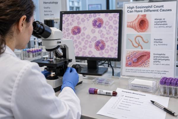
An eosinophil count measures a type of white blood cell involved in allergic inflammation, defense against certain parasites, and several immune disorders. The result is usually reported as an absolute eosinophil count, or AEC, within a complete blood count with differential. A high value is common in asthma, eczema, medication reactions, and some parasitic infections, but it can also point to eosinophilic granulomatosis with polyangiitis (EGPA), a blood disorder, or another condition that can injure organs.
The number alone does not establish a diagnosis. Its meaning depends on how high it is, whether it persists, what symptoms are present, recent medications and travel, and whether the lungs, heart, nerves, skin, kidneys, or digestive tract appear affected. Mild elevations often have a straightforward cause. Persistent counts of 1,500 cells/µL or more deserve a structured evaluation, especially when symptoms or abnormal organ tests are present.
- A normal absolute eosinophil count is generally below 500 cells/µL, although the laboratory’s reference interval should guide interpretation.
- Mild eosinophilia is 500–1,499 cells/µL; moderate is 1,500–4,999 cells/µL; severe is 5,000 cells/µL or higher.
- Allergy, asthma, eczema, and medication reactions are frequent causes, while parasite risk depends strongly on travel, residence, food, soil, and freshwater exposures.
- EGPA becomes more concerning when high eosinophils occur with adult-onset asthma, sinus disease, neuropathy, lung infiltrates, purpura, or heart involvement.
- Seek prompt medical assessment for chest pain, breathing difficulty, fainting, new weakness or numbness, coughing blood, reduced urine, or a widespread rash with fever.
Table of Contents
- What the eosinophil count measures
- Eosinophil ranges and result interpretation
- Common causes of high eosinophils
- When high eosinophils suggest EGPA
- How the test is done and when to repeat it
- Follow-up testing for eosinophilia
- Urgent warning signs and next steps
What the eosinophil count measures
The eosinophil count shows how many eosinophils are circulating in a microliter of blood. Eosinophils are made in bone marrow and spend only a short time in the bloodstream before moving into tissues. They contain inflammatory proteins that can help respond to parasites but can also damage tissue when eosinophils become excessive or activated in the wrong setting.
Most laboratories report eosinophils in two ways:
- Absolute eosinophil count: the actual number of eosinophils per microliter, often written as cells/µL or ×10⁹/L.
- Eosinophil percentage: the percentage of all white blood cells that are eosinophils.
The absolute count is more useful. A percentage can look high when the total white blood cell count is low, even if the true eosinophil number is normal. For example, 8% eosinophils may appear abnormal, but if the total white blood cell count is only 4,000 cells/µL, the AEC is 320 cells/µL and remains within a typical range.
When a laboratory does not list the AEC, it can be estimated:
AEC = total white blood cell count × eosinophil percentage as a decimal
If the white blood cell count is 10,000 cells/µL and eosinophils are 12%, the AEC is 1,200 cells/µL. A broader white blood cell count interpretation can help explain why the percentage and absolute number do not always move together.
Blood eosinophils are only a sample of eosinophil activity. Most eosinophils live in tissues, so a normal blood result does not fully exclude an eosinophilic disorder. Corticosteroids can also lower the count quickly, sometimes within hours, and may mask an elevation that was present before treatment.
Eosinophil ranges and result interpretation
Eosinophilia generally means an AEC of at least 500 cells/µL. The usual categories help clinicians decide how quickly and broadly to investigate, but the severity of illness does not always match the number.
| Absolute eosinophil count | Common description | Typical interpretation |
|---|---|---|
| Below 500 cells/µL | Usually normal | Interpret with the laboratory range and clinical context |
| 500–1,499 cells/µL | Mild eosinophilia | Often allergy, asthma, eczema, medication effect, or a limited infection |
| 1,500–4,999 cells/µL | Moderate eosinophilia | Requires closer assessment if persistent or accompanied by symptoms |
| 5,000 cells/µL or higher | Severe eosinophilia | Raises concern for significant reactive disease, a clonal blood disorder, or organ-threatening eosinophilic disease |
Hypereosinophilia usually refers to an AEC of 1,500 cells/µL or higher. Hypereosinophilic syndrome requires more than a high count: eosinophil-related organ damage must be present, and the cause must be assessed. Older definitions emphasized persistence for six months, but clinicians should not delay evaluation or treatment when the heart, lungs, nervous system, or another organ may be at risk.
One result can be temporary. Eosinophils vary during the day, can rise after a recent exposure, and may fall after corticosteroids or other treatment. The trend is often more informative than a single measurement. A value that remains elevated on repeat testing carries more weight than an isolated mild increase that returns to normal.
A low eosinophil count usually has little clinical importance. Acute stress, infection, and corticosteroids can suppress eosinophils. Unlike neutrophils or lymphocytes, an eosinophil count near zero usually does not indicate immune deficiency by itself.
Common causes of high eosinophils
High eosinophils are most often reactive, meaning another condition is stimulating normal eosinophils rather than the bone marrow producing an abnormal clone. The history often narrows the possibilities faster than broad laboratory screening.
Allergy, asthma, and skin disease
Allergic rhinitis, asthma, atopic dermatitis, and some food-related disorders commonly cause mild or moderate eosinophilia. The count does not identify the allergen and does not prove that symptoms are allergic. A person with seasonal nasal symptoms may have a modest rise, while another person with severe asthma may have a normal count because of corticosteroid treatment or natural variation.
Total IgE may rise alongside eosinophils, but neither marker is specific. A total IgE blood test can add context, while a specific IgE test addresses sensitization to selected allergens. Results still need to match the person’s actual reaction history.
Eosinophilic diseases can affect a single organ, such as eosinophilic esophagitis, eosinophilic pneumonia, or eosinophilic gastrointestinal disease. In these disorders, tissue biopsy or imaging may matter more than the blood count.
Medication reactions
Almost any medication can be relevant, including antibiotics, anti-seizure drugs, nonsteroidal anti-inflammatory drugs, allopurinol, and some supplements. A mild increase may occur without symptoms, but medication-related eosinophilia can also be part of a severe systemic reaction.
Drug reaction with eosinophilia and systemic symptoms, often called DRESS, commonly develops two to eight weeks after starting a medication. Warning features include fever, a widespread rash, facial swelling, swollen lymph nodes, and liver, kidney, lung, or heart abnormalities. Stopping a suspected medication should be directed by a clinician because abrupt withdrawal can be unsafe for some drugs, and alternative causes still need consideration.
Parasites and travel-related exposure
Eosinophilia is most closely associated with tissue-invasive helminths, or parasitic worms. Relevant exposures include residence or travel in endemic regions, walking barefoot on contaminated soil, freshwater contact, insect bites, and eating raw or undercooked freshwater fish, crab, crayfish, snails, or meat.
The timing matters. Eosinophils may rise while larvae migrate through tissue, before eggs appear in stool. A negative stool ova-and-parasite examination therefore does not exclude all infections. Strongyloides often requires serologic testing, and infection can persist for decades. This is especially important before high-dose corticosteroids or other immune-suppressing treatment because unrecognized Strongyloides can become life-threatening.
Not every parasite causes eosinophilia, and a high count does not prove a parasite is present. Testing should follow the geographic and exposure history rather than relying on a generic parasite panel.
Other immune, endocrine, and blood disorders
Autoimmune and inflammatory conditions, including EGPA, can elevate eosinophils. Adrenal insufficiency can also cause an increase because the body lacks the normal eosinophil-suppressing effect of cortisol.
Cancer-related causes include certain lymphomas and myeloid or lymphoid neoplasms with molecular abnormalities involving genes such as PDGFRA, PDGFRB, FGFR1, or JAK2. Clues include very high or persistent counts, anemia, abnormal platelets, immature blood cells, an enlarged spleen, unexplained weight loss, night sweats, or elevated vitamin B12 and tryptase. These findings warrant hematology assessment rather than assuming the result reflects allergy.
When high eosinophils suggest EGPA
EGPA is a rare inflammatory disease that combines eosinophilic inflammation with small- to medium-vessel vasculitis. High eosinophils are an important clue, but they are not sufficient for diagnosis. The pattern of symptoms is what makes EGPA stand out from routine allergic disease.
EGPA often develops in adults with asthma, chronic sinus inflammation, or nasal polyps. Over time, inflammation may affect the lungs, skin, peripheral nerves, heart, digestive tract, or kidneys. Features that should raise suspicion include:
- Asthma that began in adulthood or has become difficult to control
- Recurrent sinusitis or nasal polyps
- Migrating or patchy lung opacities on imaging
- New foot drop, wrist drop, numbness, burning pain, or asymmetric weakness
- Purpura, tender skin nodules, or other unexplained rashes
- Chest pain, shortness of breath, palpitations, fainting, or signs of heart failure
- Abdominal pain, gastrointestinal bleeding, or unexplained weight loss
- Blood or protein in urine, although severe kidney disease is less common than in some other ANCA-associated vasculitides
The 2022 classification criteria give substantial weight to a maximum eosinophil count of at least 1,000 cells/µL, obstructive airway disease, and nasal polyps. However, classification criteria are designed mainly for research after a clinician has already established that small- or medium-vessel vasculitis is present. They should not be used as a self-diagnosis checklist.
ANCA is positive in only a minority of people with EGPA. When present, it is often directed against myeloperoxidase. A negative ANCA test therefore does not exclude EGPA. ANCA-positive disease is more often linked with classic vasculitic features such as neuropathy and kidney involvement, while ANCA-negative disease may show more eosinophilic tissue and cardiac involvement, though there is substantial overlap.
A focused EGPA blood test panel can organize useful markers, but diagnosis still depends on symptoms, imaging, organ testing, and sometimes biopsy. Eosinophils may also fall after corticosteroid use, so clinicians often review older blood counts to find the untreated peak.
How the test is done and when to repeat it
An eosinophil count is obtained from a standard blood draw, usually as part of a complete blood count with differential. No fasting is normally required. The collection itself takes a few minutes, and common effects are brief discomfort, bruising, or lightheadedness.
Preparation is mainly about providing accurate context:
- Bring a complete list of prescription drugs, over-the-counter medicines, vitamins, and supplements.
- Note any recent corticosteroid tablets, injections, inhalers, nasal sprays, or topical treatments.
- Record new rashes, wheezing, sinus symptoms, digestive symptoms, fever, weight change, numbness, or weakness.
- Describe travel and residence over several years, not just the most recent trip.
- Mention raw or undercooked foods, freshwater exposure, soil contact, and animal exposure.
Do not stop corticosteroids or other prescribed medicines solely to obtain a higher or more “accurate” eosinophil count. The risk of stopping treatment may outweigh the value of the test. Clinicians can often interpret the result by reviewing the dose, timing, previous counts, and clinical picture.
A repeat count is commonly appropriate when mild eosinophilia is unexpected and the person is otherwise well. The interval may range from days to several weeks depending on the number, symptoms, and likely trigger. More urgent assessment is appropriate when the AEC is 1,500 cells/µL or higher, rises rapidly, persists, or accompanies signs of organ involvement.
Trend the absolute number rather than only the percentage. It is also useful to compare the result with hemoglobin, platelets, total white blood cells, liver enzymes, kidney function, and the blood smear.
Follow-up testing for eosinophilia
Follow-up should be targeted. Ordering every available test at once can produce false-positive results and distract from the most likely cause.
A typical first evaluation includes a repeat complete blood count with differential, review of a peripheral blood smear, metabolic and liver panels, medication review, and a focused examination. Additional testing depends on the pattern:
| Clinical clue | Possible follow-up |
|---|---|
| Asthma, rhinitis, eczema, or food-related symptoms | Allergy assessment, total or specific IgE when clinically relevant, pulmonary testing |
| Travel, migration, soil, freshwater, or raw-food exposure | Exposure-specific serology, stool testing, infectious disease or tropical medicine review |
| Asthma plus neuropathy, lung infiltrates, purpura, or systemic symptoms | ANCA with MPO and PR3 antibodies, urinalysis, kidney function, chest imaging, organ-directed studies |
| Chest symptoms or marked persistent eosinophilia | ECG, troponin, echocardiography, and sometimes cardiac MRI |
| Abnormal smear, enlarged spleen, cytopenias, or unexplained severe elevation | Hematology evaluation, serum tryptase and vitamin B12, molecular testing, possible bone marrow examination |
For suspected vasculitis, inflammatory markers such as C-reactive protein and erythrocyte sedimentation rate can show inflammation but cannot identify EGPA by themselves. Urinalysis is particularly useful because blood or protein may reveal kidney inflammation before obvious symptoms develop.
When Strongyloides exposure is plausible, clinicians may test before starting systemic corticosteroids. Stool testing can help for some parasites, but the correct number and type of specimens vary. A specialist may recommend serology or other assays based on the location and timing of exposure.
Biopsy is not required in every case. When needed, the best tissue is usually an affected and safely accessible site, such as skin, nerve, muscle, lung, gastrointestinal tract, or kidney. The goal is to show the type of inflammation or vasculitis and exclude mimics, not simply to prove that eosinophils exist.
Urgent warning signs and next steps
The count should be interpreted with symptoms, not in isolation. A person with an AEC of 900 cells/µL and new neurologic weakness may need faster assessment than someone with an AEC of 2,000 cells/µL, no symptoms, and a clear short-lived medication trigger.
Seek urgent medical care for:
- Chest pain, severe shortness of breath, fainting, or a new irregular heartbeat
- Sudden weakness, foot or wrist drop, loss of sensation, confusion, or severe headache
- Coughing blood, blue lips, or rapidly worsening wheeze
- A widespread rash with fever, facial swelling, mouth sores, or skin peeling
- Severe abdominal pain, vomiting blood, or black stools
- Markedly reduced urine, visible blood in urine, or sudden swelling
- Persistent high fever, rapid deterioration, or signs of shock
For a nonurgent abnormal result, start with three questions: How high is the absolute count? Is it persistent? Are there symptoms or other abnormal tests? Then review medications and exposure history before assuming allergy or ordering broad panels.
A practical follow-up plan often includes repeating the count, checking for organ involvement, and referring according to the dominant clues. Allergy or pulmonary specialists may help when asthma and atopy lead the picture. Infectious disease or tropical medicine is useful for complex exposure risk. Rheumatology often coordinates suspected EGPA, while hematology evaluates severe, unexplained, or potentially clonal eosinophilia.
The most important point is that eosinophilia is a sign, not a final diagnosis. Mild elevations are common and often manageable, but persistent hypereosinophilia or any evidence of heart, nerve, lung, kidney, skin, or gastrointestinal injury deserves timely evaluation.
References
- World Health Organization and International Consensus Classification of eosinophilic disorders: 2024 update on diagnosis, risk stratification, and management 2024 (Review)
- The multidisciplinary approach to eosinophilia 2023 (Review)
- The challenge of diagnosing and classifying eosinophilia and eosinophil disorders: A review 2024 (Review)
- 2022 American College of Rheumatology/European Alliance of Associations for Rheumatology Classification Criteria for Eosinophilic Granulomatosis with Polyangiitis 2022 (Guideline)
- Post-Travel Parasitic Disease Including Evaluation of Eosinophilia 2025 (Guideline)
- Hypereosinophilia: clinical and therapeutic approach in 2025 2025 (Review)
Disclaimer
An eosinophil count cannot diagnose allergy, a parasite, EGPA, or a blood disorder by itself. Results should be interpreted by a qualified clinician using symptoms, medications, exposure history, repeat testing, and organ-specific evaluation. Seek urgent care for breathing difficulty, chest pain, fainting, new neurologic symptoms, coughing blood, or a severe rash with fever.