The erythrocyte sedimentation rate, or ESR, is a blood test that measures how far red blood cells settle in a vertical tube during one hour. Inflammation changes plasma proteins and encourages red cells to form stacks or clusters that sink faster, producing a higher result. ESR is useful because it can support the presence of inflammatory activity and help monitor selected conditions, including polymyalgia rheumatica, giant cell arteritis, rheumatoid arthritis, infection, and some cancers. It is also easy to misinterpret. Age, anemia, pregnancy, kidney disease, immunoglobulin levels, red-cell shape, and laboratory technique can change ESR even when inflammation is absent or modest. A normal result does not rule out disease, and a high result does not reveal the cause. This article explains common ranges, why ESR rises or falls, how it compares with CRP, and how clinicians decide whether an abnormal value needs repeat testing or a broader evaluation.
- Indirect marker: ESR reflects red-cell settling influenced by plasma proteins, not a single inflammatory molecule.
- Age and blood counts matter: Older age, anemia, pregnancy, and red-cell characteristics can shift the result.
- Units are mm/hr: The number is the millimeters of clear plasma above settled cells after one hour.
- Very high needs context: ESR above 100 mm/hr often warrants timely investigation but is not a diagnosis.
- CRP complements ESR: The two markers change at different speeds and are affected by different factors.
Table of Contents
- How the ESR Test Works
- Normal Ranges by Age and Sex
- Inflammatory Causes of High ESR
- Noninflammatory Reasons ESR Can Rise
- Very High and Low ESR Results
- ESR Versus CRP and Other Tests
- Testing, Follow-Up, and Practical Interpretation
How the ESR Test Works
Red blood cells normally remain suspended in plasma because their surfaces repel one another. During inflammation, liver production of fibrinogen and other proteins rises, and immunoglobulins may also increase. These proteins reduce the repulsive forces between cells and promote rouleaux, formations that resemble stacks of coins. Larger aggregates settle more quickly than individual cells.
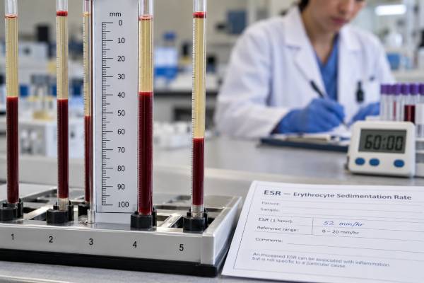
In the traditional Westergren method, anticoagulated blood is placed in a long, narrow, vertical tube. After one hour, the laboratory measures the distance from the top of the red-cell column to the top of the plasma. A result of 30 mm/hr means the cells descended 30 millimeters during that period.
ESR is therefore a physical phenomenon rather than a direct protein measurement. Several elements contribute:
- Concentrations of fibrinogen and immunoglobulins
- Number of red blood cells and the hematocrit
- Cell size, shape, and flexibility
- Plasma viscosity and other protein changes
- Tube dimensions, angle, vibration, temperature, and processing delay
The Westergren approach remains the reference method, although many laboratories use automated or modified systems. Alternative instruments must be validated against the reference method because they may behave differently, especially at high values or in samples with unusual red-cell properties.
ESR often rises more slowly than CRP after an acute inflammatory trigger and may stay elevated longer during recovery. Its persistence can be useful in chronic disorders but can also make it difficult to tell whether inflammation is current or recently resolved.
The test is commonly ordered when symptoms suggest a systemic inflammatory condition or when a clinician is following a known disease. It should not be used as a general screening test without a clinical question. Minor elevations are common and nonspecific, especially in older adults.
Normal Ranges by Age and Sex
There is no universal ESR normal range. Laboratories use different methods and reference populations. The upper limit tends to increase with age and is often higher in females. Children and newborns have their own intervals.
Commonly cited adult ranges using a Westergren-type method are approximately:
| Group | Illustrative range | Important note |
|---|---|---|
| Men younger than 50 | 0–15 mm/hr | Some laboratories use an upper limit of 20 |
| Women younger than 50 | 0–20 mm/hr | Hormonal and hematologic factors can affect the result |
| Men 50 and older | 0–20 mm/hr | Age-adjusted laboratory ranges may be higher |
| Women 50 and older | 0–30 mm/hr | Exact limits vary substantially with age and method |
These values are examples, not replacements for the range on a report. Some laboratories provide detailed age bands extending into older age, while others use broad categories. A result labeled high by one laboratory may fall within another laboratory’s age-adjusted interval.
Formulas are sometimes used to estimate an age-adjusted upper limit, such as age divided by two for men and age plus ten divided by two for women. These formulas are rough clinical aids, not validated reference intervals for every patient. They can also normalize values that still deserve attention when symptoms are concerning.
ESR is reported in millimeters per hour, usually written mm/hr or mm/h. It is not directly convertible to CRP, which is reported as a protein concentration. An ESR of 40 mm/hr and a CRP of 40 mg/L are unrelated numerical scales.
A borderline elevation may reflect normal variation, especially if the person feels well and the complete blood count is normal. The significance rises when the value is clearly above the age-appropriate range, persists on repeat testing, or accompanies symptoms and other abnormalities.
Inflammatory Causes of High ESR
A high ESR often results from increased fibrinogen, immunoglobulins, or other inflammation-related proteins. The test cannot identify the source, so clinicians organize causes by clinical pattern.
Autoimmune and rheumatic disease can produce sustained elevations. ESR is especially useful in polymyalgia rheumatica and giant cell arteritis, where a high value supports the diagnosis and may help monitor treatment. Rheumatoid arthritis, vasculitis, and some connective-tissue diseases can also raise it. However, normal ESR does not completely exclude these disorders.
Infection is another major category. Bone and joint infection, endocarditis, tuberculosis, deep abscesses, and other prolonged infections may create marked elevations. Acute infections can raise ESR, but the slower response means CRP is often more informative early in the illness.
Inflammatory tissue injury from surgery, trauma, infarction, or chronic organ inflammation may increase ESR. The result can stay high after symptoms begin improving because elevated plasma proteins and altered red-cell dynamics take time to normalize.
Malignancy may raise ESR through inflammation, anemia, increased immunoglobulins, tissue injury, or infection. Multiple myeloma and some lymphomas can produce particularly high values because of abnormal immunoglobulin concentrations. ESR is not a cancer screening test, and most people with an elevated result do not have cancer.
Kidney disease can be associated with inflammation, anemia, and altered proteins, all of which tend to raise ESR. The value may remain chronically elevated and be less useful for detecting a new problem unless compared with that person’s baseline.
The degree of elevation does not map neatly to severity. A patient with active lupus may have a high ESR but modest CRP, while a patient with severe bacterial infection may show large increases in both. A person with localized inflammation may have a normal ESR. Clinical context always outranks a generic cutoff.
Noninflammatory Reasons ESR Can Rise
ESR is strongly affected by the blood itself. An elevated result can occur without a major inflammatory process.
Anemia is a common reason. When fewer red cells occupy the tube, they may settle faster. Macrocytosis, in which cells are larger than usual, can also increase sedimentation. This is why ESR should be interpreted with hemoglobin, hematocrit, and red-cell indices.
Pregnancy raises fibrinogen and changes red-cell concentration, so ESR can increase substantially, particularly later in pregnancy. Nonpregnant reference intervals should not be applied.
Older age is associated with higher typical values. Age-related changes in plasma proteins, anemia prevalence, and chronic conditions all contribute.
High immunoglobulins can speed sedimentation even when the source is not a conventional inflammatory flare. Monoclonal proteins in plasma-cell disorders are a notable example. Polyclonal immunoglobulin increases in chronic liver disease or infection can have a similar effect.
Kidney disease, obesity, and some endocrine states may cause modest elevations through mixed inflammatory and noninflammatory mechanisms. Menstruation has also been reported to influence results in some people.
Technical problems can falsely elevate ESR. A tube that is not perfectly vertical allows cells to fall along the side more quickly. Vibration, excessive room temperature, an incorrect blood-to-anticoagulant ratio, or delayed testing can change the measurement. A difficult collection may affect sample quality.
These influences explain why an isolated ESR of 25 or 35 mm/hr cannot be interpreted without a complete blood count and clinical history. The white blood cell count may reveal infection or hematologic clues, while hemoglobin can show whether anemia is likely inflating the rate.
Very High and Low ESR Results
An ESR above 100 mm/hr is often called an extreme elevation. Studies have found that most extreme values are associated with a significant underlying condition, commonly infection, autoimmune or inflammatory disease, kidney disease, or malignancy. This level generally deserves timely evaluation, especially when it is new.
An extreme result is not an emergency solely because of the number. Urgency depends on symptoms and vital signs. Fever with low blood pressure, shortness of breath, severe headache with visual change, neurologic symptoms, or rapidly worsening illness requires urgent care. An asymptomatic person with a chronic baseline near 100 mm/hr needs a different approach.
Very high ESR can be especially important in suspected giant cell arteritis. New headache, scalp tenderness, jaw pain while chewing, visual disturbance, or unexplained constitutional symptoms in an older adult require immediate clinical assessment. Treatment may begin before confirmatory testing because delayed therapy can risk permanent vision loss.
Low ESR is usually not clinically concerning, but it can occur when red cells do not form aggregates or when blood is unusually concentrated. Possible associations include:
- Polycythemia or high hematocrit
- Sickle-shaped or markedly abnormal red cells
- Hereditary spherocytosis
- Very high white blood cell counts
- Low fibrinogen
- Severe heart failure in some settings
A low result does not guarantee absence of inflammation. Sickle cell disease, for example, may prevent normal rouleaux formation even during an inflammatory event. Low fibrinogen from severe liver disease or consumption can also keep ESR low while the patient is seriously ill.
The key lesson is that both extremes can be shaped by red-cell and protein biology. ESR should never be interpreted as a direct “inflammation score” without considering those factors.
ESR Versus CRP and Other Tests
ESR and CRP are often ordered together because their differences can add context. CRP is a defined protein made by the liver in response to inflammatory cytokines. ESR is a physical measurement influenced by many proteins and red-cell features.
| Feature | ESR | CRP |
|---|---|---|
| Response speed | Usually slower to rise and fall | Often rises and falls relatively quickly |
| Influenced by age and anemia | Strongly | Much less |
| Influenced by immunoglobulins | Strongly | Indirectly |
| Useful in selected chronic diseases | Yes, especially PMR and giant cell arteritis | Yes, and often better for acute changes |
| Specific for a disease | No | No |
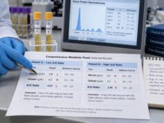
A high ESR with normal CRP may occur with anemia, older age, pregnancy, kidney disease, or monoclonal immunoglobulins. It can also occur when inflammation is resolving or in selected autoimmune patterns. A high CRP with normal ESR may fit very early inflammation, a rapid acute response, or factors that prevent red-cell aggregation.
The CRP blood test is generally more responsive for acute infection and treatment change. ESR may remain helpful when disease-specific evidence supports its use or when long-term trends are established.
Other companion tests depend on the suspected cause. A complete blood count can identify anemia, leukocytosis, thrombocytosis, or abnormal cells. Ferritin, fibrinogen, kidney and liver tests, cultures, autoantibodies, protein electrophoresis, and imaging may be appropriate. Ordering all tests indiscriminately can create misleading incidental results; the symptom pattern should guide selection.
Testing, Follow-Up, and Practical Interpretation
ESR requires a venous blood sample and usually no fasting. If other tests are ordered at the same time, fasting instructions may apply. Tell the healthcare professional about pregnancy, anemia, known blood disorders, recent infection, and medications. No medicine should be stopped merely to prepare for an ESR unless specifically directed.
A practical interpretation follows several steps:
- Use the laboratory’s range. Check age, sex, method, and units.
- Review the complete blood count. Anemia or abnormal cell shape may explain part of the elevation.
- Match the result to symptoms. Joint stiffness, fever, weight loss, headache, infection signs, or kidney symptoms alter the differential.
- Compare CRP and previous ESR values. Discordance and trends can be more informative than one number.
- Investigate persistent or extreme elevation. The workup should be targeted, not a blind search.
A small unexplained elevation in a well person may be repeated after a reasonable interval rather than prompting extensive testing. A marked rise with symptoms deserves faster evaluation. When ESR is being used to monitor a known condition, the patient’s baseline and clinical response are more useful than a universal target.
ESR should not be lowered directly. Antibiotics, steroids, and immune-suppressing medicines carry risks and must be used only when an underlying diagnosis supports them. Lifestyle measures may improve chronic health but cannot replace investigation of a substantial unexplained value.
Seek prompt care for visual symptoms, severe new headache, chest pain, difficulty breathing, confusion, high fever with deterioration, or focal neurologic deficits. These symptoms can be urgent even if ESR is normal.
The ESR test is best viewed as a broad lens. It can show that the protein and cellular environment of blood favors faster settling, often because of inflammation. It cannot show where the problem is or what treatment is needed. The answer comes from combining the rate with the person.
Monitoring deserves a final caution. When ESR is already elevated because of anemia, kidney disease, pregnancy, or a monoclonal protein, it may not return to the standard range even after the inflammatory condition improves. Clinicians may instead look for a meaningful change from the person’s own baseline. Small fluctuations should not automatically trigger treatment changes, especially when symptoms and organ-specific findings are stable. Conversely, treatment can improve pain or fever before ESR falls because fibrinogen and immunoglobulin patterns resolve slowly.
Medication effects are usually indirect. Corticosteroids and disease-modifying drugs may lower ESR by suppressing inflammation, while therapies that change hemoglobin or plasma proteins may alter the measurement through other pathways. There is no standard list of medicines that must be withheld before testing. The safest approach is to provide a complete medication and supplement list and let the clinician interpret the result in context. When a result is unexpected, repeating the test with a properly collected sample and reviewing the blood count can be more useful than immediately ordering extensive imaging. In children, reference ranges and causes differ further, so pediatric interpretation should use age-appropriate standards. The same applies after childbirth and during recovery from major surgery, when temporary physiological changes can keep ESR elevated beyond the period of obvious symptoms and should be followed according to the underlying clinical situation carefully over time by experienced clinicians.
References
– Erythrocyte Sedimentation Rate (ESR) 2024 – Sed Rate (Erythrocyte Sedimentation Rate) 2025 – Erythrocyte Sedimentation Rate 2023 – Sedimentation Rate (ESR): What It Is & Normal Range 2024 – Performance Evaluation of Alternate ESR Measurement on the BC-780 Automated Hematology Analyzer 2024 – ICSH Recommendations for Modified and Alternate Methods of Measuring the Erythrocyte Sedimentation Rate 2017
Disclaimer
This article is for general education and cannot diagnose the cause of an abnormal ESR. Reference intervals and methods differ, and age, pregnancy, anemia, blood-cell shape, and plasma proteins may change the result independently of active inflammation. Discuss persistent, extreme, or symptom-associated results with a qualified healthcare professional.